ISSN 0102-695X DOI 10.1590/S0102-695X2012005000135 Received 4 Jun 2012 Accepted 24 Sep 2012 Available online 22 Nov 2012 Revista Brasileira de Farmacognosia Brazilian Journal of Pharmacognosy 23(2): 249-258, Mar./Apr. 2013
Inl uence of rosmarinic acid and
Salvia
ofi cinalis
extracts on melanogenesis of
B16F10 cells
Karina B. Oliveira,
1Érika Palú,
2Almeriane M. Weffort-Santos,
2Brás H. Oliveira
* 31Programa de Pós-graduação em Ciências Farmacêuticas, Universidade Federal
do Paraná, Brazil,
2Departamento de Patologia Médica, Setor de Ciências da Saúde, Universidade
Federal do Paraná, Brazil,
3Departamento de Química, Setor de Ciências Exatas, Universidade Federal do
Paraná, Brazil.
Abstract: Melanin is a photoprotective skin pigment, and pathologies characterized by hypo or hyperpigmentation are common. New compounds that regulate melanogenesis are, therefore, opportune, and many natural products with this property, as polyphenols, have been described. Salvia ofi cinalis L., Lamiaceae, is a widely used food spice that contains high amounts of phenol derivates, including rosmarinic acid. The aim of this work was to evaluate the contribution of rosmarinic acid in the melanogenic activity of sage extracts. Fluid and aqueous extracts of sage and purii ed rosmarinic acid were assayed for B16F10 cytotoxicity and, then, evaluated on melanin production and tyrosinase activity. While sage extracts showed a concentration-dependent ability to signii cantly increase melanin production without necessarily changing the enzymatic activity, rosmarinic acid showed a dual behavior on melanogenesis, increasing melanin biosynthesis and tyrosinase activity at low concentrations and decreasing it at higher levels. Rosmarinic acid may collaborate with sage extracts activity on melanogenesis, although other compounds may be involved. This is the i rst time that a dual action of rosmarinic acid on melanogenesis is reported, which may be useful in further studies for therapeutic formulations to treat skin pigmentation disorders.
Keywords:
melanin melanogenesis rosmarinic acid
Salvia officinalis
tyrosinase
Introduction
Salvia ofi cinalis L., Lamiaceae (sage) is a well-known vegetal specie largely used for food spicing. It presents many pharmacological properties, most of them associated with its polyphenol content (Lu & Foo, 2002; Bauer et al., 2012; Ben Taarit et al., 2012; Generalic et al., 2012). Rosmarinic acid (RA – 1) is the major phenol compound of sage, also found in many other plants used as seasoning (Petersen & Simmonds, 2003). Relevant biological activities such as neuroprotective (Iuvone et al., 2006; De Oliveira et al., 2012), antioxidant (Zheng & Wang, 2001; Perez-Tortosa et al., 2012), anticancer (Karmokar et al., 2012; Sharmila & Manoharan, 2012),
anti-inl ammatory (Sahu et al., 1999; Al-Musayeib et al.,
2011), and immunomodulatory (Kang et al., 2003; Costa et al., 2012) have been attributed to RA. Photoprotection and melanogenic properties have also been described, in which positive effects on human keratinocyte cells after UVA (Psotova et al., 2006) and UVB exposures
(Vostalova et al., 2010) were observed, resulting in an increased cell viability and reduced DNA damage. The effects of RA on mouse melanoma cells (Lee et al., 2007) and in the reduction of photo-induced skin alterations in mice (Sanchez-Campillo et al., 2009) have also been shown.
HO
HO
OH O
H O O
OH OH
1
Melanin pigmentation of the skin has important cosmetic and therapeutic roles, which can be compromised in various dermatological conditions. Hyperpigmentation disorders, such as freckles, lentigines, melasma or age spots, can be the result of a misregulated production or accumulation of melanin by melanocytes (Virador
et al., 1999). Hypomelanosis, on the other hand, is caused by lower number of melanocytes, interference on melanin biosynthesis or by a decrease in tyrosinase availability (Wulf et al., 2004). The treatment of these conditions is based on melanin biosynthesis, which initial steps involve the enzyme tyrosinase, the key element that catalyses the hydroxylation of L-tyrosine to 3,4-dihydroxyphenylalanine (L-DOPA), and from that to dopaquinone. After several steps melanin is produced as a complex mixture of pigments (Kim & Uyama, 2005; Chang, 2009). Within this context, compounds that interfere with this biosynthetic pathway, increasing or decreasing the amount of melanin formed, may be potential candidates to treat pigmentary disorders.
Some agents used to treat skin
hyperpigmentation, like hydroquinone, can cause side effects such as dermatitis and skin irritation (Maeda & Fukuda, 1996). Other disorders, such as vitiligo, have no effective cure and limited treatment options (Huggins et al., 2012). The search for natural products for treatment of skin pigmentation disorders is, therefore, opportune. Many inhibitors and enhancers of melanin biosynthesis have been described and some are obtained from natural sources. Kojic acid, for example, is a well-known tyrosinase inhibitor produced by Aspergillus and Penicillium fungi (Smit et al., 2009). Some plant polyphenols, on the other hand, have been described as melanin inducers (Kim & Uyama, 2005).
Considering that S. oficinalis contains high amounts of RA among other polyphenols, the aim of this work was to study the melanogenic activity promoted by RA, and its contribution in the melanogenic effects
promoted by a luid extract and an aqueous extract prepared
from dried sage leaves.
Materials and Methods
Plant material
Dried and crushed S. oficinalis leaves were purchased in a local market (Curitiba, Brazil), and
identiied by macro and microscopic analysis (Wichtl,
2004), and by comparison with an authentic sample of the Laboratory of Pharmacognosy, Department of Pharmacy, Federal University of Paraná, Brazil. A specimen voucher (300-A) of the plant has been deposited at the Herboteca Carlos Stellfeld, Federal University of Paraná, Brazil. For isolation of RA, a dry extract of aerials parts of sage, containing around 6% (w/w) of
RA according to the supplier label and conirmed by chromatographic quantiication, was purchased from
Apsen Pharmaceuticals.
Instrumentation and chemicals
1H and 13C NMR spectra were acquired on a
Bruker Avance 200 spectrometer (Rheinstetten, Germany) using tetramethylsilane (TMS, Sigma Aldrich) as internal standard. A Varian 920 series HPLC system (Palo Alto, CA) with quaternary pump, autosampler, column oven and diode array detector was used for the chromatographic analyses. The absorbances were measured in a Shimadzu Model 1800 UV-VIS spectrophotometer. A Harrison Research Chromatotron Model 7924T (Palo Alto, CA) was used for preparative chromatography. HPLC grade solvents were purchased from Merck (São Paulo, Brazil). All other solvents and reagents used were of analytical grade.
Preparation of extracts
Aqueous extract (AE) of sage was obtained
by extraction of crushed dried leaves (10 g, <710 μm)
with water at 80 °C (100 mL) for 15 min. The mixture
was iltered (Whatman nº 1), and the extract volume
completed to 100 mL. Fluid extract (FE) was prepared by successive percolations of crushed dried leaves of
sage (20 g, <710 μm) with 56% ethanol (v/v) for three
days, in the dark, at room temperature (Farm Bras, 2002). The extracts were combined and concentrated to 20 mL.
Samples were sterilized by iltration through a 0.22 μm membrane ilter and stored at -25 °C till use.
Rosmarinic acid isolation and characterization
Isolation of RA from sage was based in a
previous report with modiications (Christ & Kesselring,
1982). Sample of 10 g of dry extract of sage was dissolved in water (300 mL) at 80 °C with magnetic stirring. The
solution was iltered (Whatman nº 1), acidiied to pH
2.5 and centrifuged. The supernatant was extracted with diethyl ether (5 x 100 mL), the organic fractions were combined, and the solvent evaporated. The resultant solid was fractionated by Chromatotron, in a silica gel with gypsum (Aldrich, 346446) rotor. Elution was performed with ethyl acetate/hexane/acetic acid (55:35:10 v/v/v) and all obtained fractions were analyzed by TLC. Those containing RA were combined, the solvent evaporated and the residue stored under nitrogen. The isolated solid was characterized as RA by spectroscopic methods (UV, 1H and 13C NMR). For the biological assays, RA stock solution
(10-2 M in RPMI full medium) was sterilized by iltration
through a 0.22 μm membrane ilter and stored at -25 °C.
Rosmarinic acid content of FE and AE
Inluence of rosmarinic acid and Salvia oficinalis extracts on melanogenesis of B16F10 cells
Karina B. Oliveira et al.
with some modiications (Wang et al., 2004). Briely,
samples of AE and FE were diluted to 1:50 in methanol 50% (v/v), and injected into a silica C18 column (Agilent
Eclipse C18, 150 mm x 4.6 mm x 5 μm) at 25 °C. Elution
was performed with methanol (eluent A) and 0.1% phosphoric acid in water (eluent B) in a step gradient mode (45% of A in B, 0-5 min, and 80% of A in B, 5-10
min). The low rate was 1.0 mL/min and detection at 330
nm. The concentration of RA was determined by external
standard calibration using a ive-point analytical curve.
Cell cultures and viability
B16F10 and McCoy cell lines were maintained under sterile conditions at 37 °C in RPMI 1640 medium (Himedia Laboratories; Mumbai, India) supplemented with 10% heat-inactivated fetal bovine serum, 2 mM L-glutamine, 100 IU/mL penicillin G, and 50 μg/mL streptomycin sulfate (RPMI full medium). The cell lines were subcultured twice a week at 105 cells/mL, and the
number of viable cells used in the experiments, determined by the trypan blue exclusion test, always exceeded 95%.
Cell proliferation assay and cytotoxicity evaluation
B16F10 and McCoy cells were seeded in 24-well culture plates in RPMI full medium, and allowed
to attach overnight at 37 °C in a fully humidiied, 5%
CO2 incubator. Test solutions (RA, AE and FE) were
then added to semi-conluent cultures to give inal RA concentrations ranging from 0.001 to 1,000 μM, and the
cultures re-incubated at the same conditions for 24 h. The medium was then removed, and the cells were properly treated with neutral red (NR) dye, following the NR assay (Borenfreund & Puerner, 1985). Absorbance of the treated cells was measured at 540 nm, and viability was
quantiied as the percentage (%) of living cells relative to
the control. Data was expressed as mean (%) ±SD from a triplicate.
Preparation of cell lysates
In order to test RA, FE and AE on melanin synthesis and tyrosinase activity, B16F10 cells were cultured in 25 cm2 culture lasks in full RPMI medium
containing 10 mM of NH4Cl as lysosomotropic agent (Saeki & Oikawa, 1983), in the presence of FE, AE or
RA, with or without 500 μM L-tyrosine, for 24 h at 37
°C, in a fully humidiied, 5% CO2 incubator. Treatment
of the cells with 400 mM kojic acid dissolved in DMSO was used as a positive control for melanogenesis inhibition. B16F10 cells kept under the same conditions but without any treatment were used as a control of the experiments. After treatment, the cells were washed twice with cold phosphate buffered saline (PBS), and
detached by short incubation with 0.02% EDTA at 37 °C. After centrifugation at 800 g for 5 min, the cell pellets were sonicated on ice for 1 h with 0.1 M Tris-HCl pH 7.2
lysis buffer (200 μL). The cell homogenates were used for melanin quantiication and protein determination by
the Bradford method (Bradford, 1976). Their respective supernatants (cell lysates), obtained after centrifugation of the homogenates at 10,000 g for 10 min at 4 °C, were used for tyrosinase activity.
Melanin production in B16F10 cells
Aliquots of 100 μL of the homogenates, obtained
from lysed cells pretreated with RA, AE and FE, were added to 900 µL of 1 M NaOH and incubated overnight at 37 °C. Melanin concentration was determined spectrophotometrically at 470 nm, using an external calibration curve (10-100 µg/mL) prepared with melanin from Sepia oficinalis (Sigma, M-2649). Melanin content was calculated relative to the control, following the formula [(concentration of melanin per µg of protein of treated cells/concentration of melanin per µg of protein of control) x100], and expressed as mean intracellular melanin per ug of total protein (%) ±SD (Tsuboi et al., 1998; Heo et al., 2009).
Cellular tyrosinase activity
Tyrosinase was assayed spectrophotometrically according to a published method (Sarkar et al., 2006) with
some modiications. Briely, the reaction mixture (1 mL), composed of cell lysates (100 μg of protein) from
pre-treated B16F10 cells with FE, AE, or RA, added of 0.1% L-DOPA in 0.1 M NaH2PO4 pH 6.8, was kept at 37 °C for 1 h. Absorbance was then measured at 470 nm. Tyrosinase activity was calculated as a percentage (%) relative to the control, according to the equation [(absorbance of pre-treated cells lysate/absorbance of control cell lysate) x100], and was expressed as mean (%) ±SD.
Free tyrosinase assay
Solutions (1 mL) were prepared with cell lysates
from control B16F10 cells (100 μg of protein) as a source
the substrate alone. Results were expressed as mean (%) ±SD (Nagata et al., 2004; An et al., 2008).
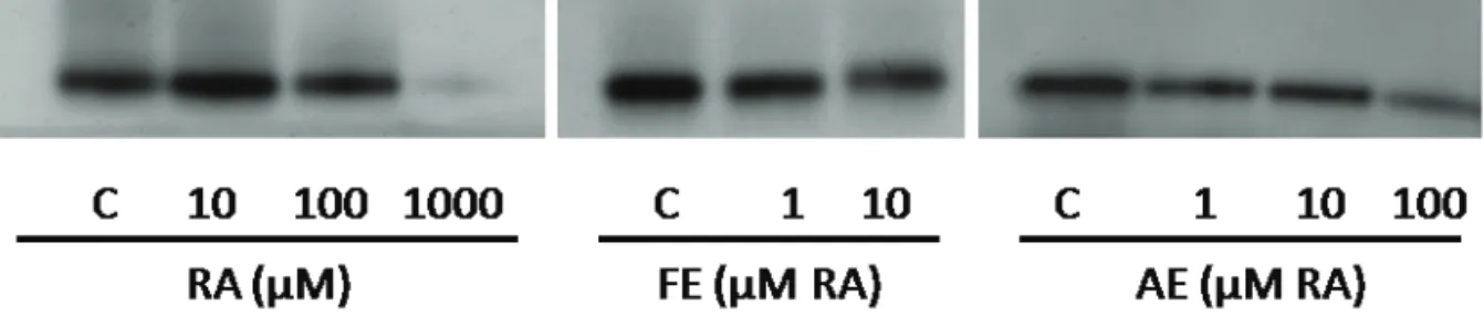
Zymography
Lysates of B16F10 cells (100 μg of protein)
pre-treated with RA, FE and AE were mixed with appropriate volume of Blue/Orange 6X Loading Dye (Promega, Madison, WI, USA) and resolved in a 8% SDS-polyacrylamide gel at 200 mV for 50 min. After electrophoresis, the gel was washed in 0.1 M NaH2PO4 pH 6.8, transferred to a staining solution containing 5 mM L-DOPA, and kept at 37 °C for 5 h in the dark, after which the dark bands of tyrosinase could be visualized and photo documented (Sato & Toriyama, 2009).
Statistical analyses
Student’s t-test or analyses of variance (ANOVA)
followed by Tukey HSD -α = 0.05 whereas necessary
were carried out using the Graph Pad-Prism 5 software.
Differences were considered signii cant at p≤0.05.
Results and Discussion
Isolated RA
The amount of purii ed RA obtained from 10 g
of sage dry extract was 4.9±0.85 % (w/w). Its spectral
proi le, and the 1H and 13C NMR data were all consistent
with the literature data for RA, coni rming the identity
of the compound (Kelley et al., 1975; 1976; Wang et al., 2004; Xu et al., 2008).
Rosmarinic acid content in extracts
The concentration of RA in FE (5.88 mg/mL) was about 10 times higher than in AE (0.51 mg/mL) as expected. From these results and considering the dry weights of FE (156.1 mg/mL) and AE (14.3 mg/mL), proper dilutions were made in order to obtain solutions with standardized RA concentrations for bioassays, allowing the comparison of the effects.
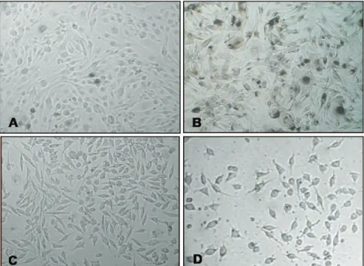
Cell phenotype alterations induced by extracts and RA
After treating B16F10 cells with varying concentrations of RA, FE, and AE, the effects on their morphology evaluated by phase-contrast microscopy
were evident, leading to profound modii cations in the
cell pigmentation patterns. They changed from the
characteristic spindle-like morphology of i broblasts,
interspersed by spherical-shaped cells (Figure 1A)
to irregular, l attened shape, with long dendrites-like
protrusions around a cytoplasm darkened by the presence of melanin (Figure 1B). These phenotypic changes were far more evident when L-tyrosine was added to the cultures, and were all suggestive of melanogenesis induction (Fernandes et al., 2004).
The treatment of McCoy cells with non-toxic
Inluence of rosmarinic acid and Salvia oficinalis extracts on melanogenesis of B16F10 cells
Karina B. Oliveira et al.
concentrations of tests did not induce any morphological alterations (Figure 1C). On the other hand, classical toxic effects such as cytoplasm vacuolization, retraction of dendrites, and cell detachment were easily observed for both cell lines, particularly in experiments with
the extracts at 1,000 μM of RA (Figure 1D). When the
incubation time was longer (>48 h), the culture dishes
of B16F10 and McCoy cells were fully conluent and
signals of deterioration were evident, as detached and
loating cells in the culture medium. These observations
led us to perform all experiments within 24 h, in order to avoid misinterpretation of results obtained from stressed, unhealthy cells (Virador et al., 1999).
Cell toxicity of sage extracts and RA
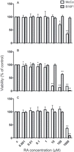
Different from McCoy cells, the B16F10 cell line is very sensitive to environmental changes, easily acquiring characteristics of melanogenic activation under stress. During the screening of prospective bioactive compounds for modulation of skin pigmentation using this cell line, it is therefore important to consider possible adverse effects on their survival and proliferation status, in order to avoid misinterpretation of results that could be compromised by unhappily cultured cells. Therefore, quantitative determination of cell viability after treatment with sage extracts and RA for 24 h was carried out using the NR technique. The results for B16F10 cells (Figure 2) were consistent with the phenotypic observations described earlier. RA did not interfere with their viability up to 1,000 µM (Figure 2A), contrasting with a previous report (Yoshida et al., 2005). However,
RA was signiicantly deleterious for McCoy cells at 100 and 1,000 μM, reducing their viability to 70 and 35%, respectively. FE also reduced signiicantly the viability
of McCoy cells in all concentrations tested (Figure 2B),
being more pronounced in the 10-1,000 μM range of
RA content. For B16F10 cells, the viability was reduced
only at FE concentrations higher than 100 μM of RA. The results of AE showed that only at 1,000 μM of RA
the viability of both cell lines was compromised (Figure 2C).
With these results, it become evident that the
McCoy ibroblastic cells were more sensitive than
B16F10 melanoma cells to the toxic effects of RA treatment, which is expected and in accordance with the characteristic drug resistance of tumor cells in contrast with normal cells (Rumjanek et al., 2001).
Comparing the results obtained for FE, AE and RA, a clear indication that FE has other compounds apart from RA that may be responsible for the toxicity observed raised. Also, the distinct results obtained for FE and AE treatments may be explained by their different production ways, and consequently, by their different chemical composition. In fact, the chromatograms of the
extracts showed distinct proiles and less polar substances,
such as luteolin, were found in higher concentrations in FE (data not shown). Hence, it is likely that compounds present only in FE may account for at least some of the more pronounced toxic effects observed.
Figure 2. Effects of rosmarinic acid (RA) (A), sage luid (B) and aqueous extracts (C) on McCoy and B16F10 cell lines viability. For conditions see Material and Methods. Each bar represents the percentage of living cells relative to control, expressed as mean±SD from four independent experiments, each one performed in triplicate (*p<0,05; **p<0.0001).
Inluence of RA and sage extracts on melanin production
Inhibition of tyrosinase gene expression or maturation and direct inactivation of the enzyme can be used as tools for controlling melanogenesis (Virador et al., 1999). Using one or both approaches, the
melanogenic activity will, therefore, relect on melanin
the melanogenic pathways, they were used in this work as a model to evaluate the effects of sage extracts and RA on melanogenesis. In order to avoid interferences on melanogenesis caused by the reduction of cell number
inluenced by the testing substances, only non-toxic
concentrations of FE, AE, and RA, as well as of kojic acid, L-tyrosine, DMSO and NH4Cl, used alone or in combinations, were used in the experiments.
Since cellular melanin contents relect turnover,
accumulation, and distribution of melanin associated
with cellular proliferation, semi-conluent B16F10
cultures were treated with sage extracts and RA for 24 h, with or without L-tyrosine, and both melanin content and tyrosinase activity were determined. FE was the only sample that, in the absence of L-tyrosine, showed little ability to interfere positively with the melanin content of B16F10 cells (Figure 3A), in contrast with L-tyrosine alone, which increased more than three times the melanin content compared to control population. These results for L-tyrosine were expected since it is a substrate for tyrosinase activity, empowering melanin synthesis (Kongshoj et al., 2007). However, the discrepancy of the effects with extracts and RA led us to question whether a low availability of substrate for melanin production could be obscuring the results.
To address this hypothesis, a new set of experiments with addition of L-tyrosine (500 μM) was carried out, having in mind that L-tyrosine not only stimulates melanin production but also can be used as a substrate for tyrosinase action. Under this circumstance, a new spectrum of activities was developed and a strikingly
signiicant inluence of FE, AE, and RA on pigment
production in the presence of L-tyrosine was observed (Figure 3B). Both sage extracts had a
concentration-dependent ability to signiicantly stimulate melanogenesis in B16F10 cells. It was even more pronounced at 10 μM
of RA, when FE and AE increased in 117% and 90%, respectively, the melanin content. Furthermore, the well known melanogenic inhibitory effect of kojic acid (Chang, 2009) could then be revealed, decreasing in more than 50% the melanin content.
While the melanin assay results showed that both S. oficinalis extracts have signiicantly stimulated
melanin production in B16F10 cells at 1 and 10 μM, the effect of puriied RA on pigment production was intriguing, acting signiicantly as a stimulator of melanin formation at 10 μM, and as an inhibitor at 1,000 μM. Increased melanin production by RA in the 1-100 μM
range using a similar cell system has been reported (Wulf et al., 2004; Lee et al., 2007; Sanchez-Campillo et al., 2009). However, to the best of our knowledge, this is
the irst time that a signiicant inhibitory melanogenic
activity for RA is described.
Interestingly is also the results indicating that FE and AE were even better than RA alone on melanin
production when L-tyrosine was present, suggesting that RA is one but not the only one of the compounds present in these mixtures contributing with these effects. Therefore, it is reasonable to keep in mind that FE and AE may have substances that would serve as alternative substrates for tyrosinase activity, leading to dramatic changes in melanin production. Moreover, since FE induced a higher melanin biosynthesis than AE, it is possible that stimulating agents of melanogenesis are in higher concentrations in FE.
Figure 3. Inluence of rosmarinic acid (RA), sage luid (FE) and aqueous extracts (AE) on melanin production by B16F10 cells. B16F10 cells were treated with the indicated concentrations of the tests for 24 h at 37 °C, without (A) or with (B) L-tyrosine (LTYR). For conditions see Material and Methods. Each bar represents the mean percentage±SD of melanin content related to control (n=5-8; *p<0.05; **p<0.01; ***p<0.0001). KA: kojic acid.
Cellular and free tyrosinase activity
Tyrosinase is an enzyme widely distributed in nature, and considered the key enzyme of melanogenesis in animals (Jimenez-Atienzar et al., 2005; Kim &
Uyama, 2005). The inluence of substances and extracts
Inl uence of rosmarinic acid and Salvia ofi cinalis extracts on melanogenesis of B16F10 cells
Karina B. Oliveira et al.
used as a free enzyme source (An et al., 2008) and added of RA, FE, and AE for 60 min in a L-DOPA solution; then, the enzyme activity was measured (Figure 4B).
As expected and in contrast with the stimulant effects observed for L-tyrosine, kojic acid signii cantly reduced both cellular (Figure 4A) and free (Figure 4B) tyrosinase activities, which is in accordance with the results obtained for melanin content (Figure 3B), and also with its well documented characteristic as a tyrosinase inhibitor. RA stimulated the cellular tyrosinase activity
but only at 10 μM, increasing it at 50% when compared to controls. This signii cant effect was coni rmed by
the greater intensity of the tyrosinase band observed in the zymogram (Figure 5). However, at that same concentration, RA did not alter the free tyrosinase activity (Figure 4B), suggesting that the main mechanism of action involved is related to the enzyme synthesis instead to its activity. At higher concentrations, RA led to an
opposite effect, with a signii cant dose-related decrease
in tyrosinase activity in both systems, recapitulating the
trend proi le of pigment formation observed for similar
concentrations (Figure 3B).
These contrasting results led us to propose that RA, depending on its concentration, has a dual effect on melanogenesis of B16F10 cells. Within this hypothesis, RA, at low concentrations, would act as a positive modulator of melanogenesis, interfering on tyrosinase synthesis that is followed by a rapid increase in its activity and visualized by melanin production. This action would explain the increased melanin content and cellular tyrosinase activity observed in our experiments
at 10 μM. Moreover, this suggestion is consistent with
the reported data showing that RA increases tyrosinase expression by activating the PKA pathway (Lee et al., 2007).
On the other hand, as a strong enzymatic inhibition has been observed for RA at higher but non-cytotoxic concentrations, it is possible that RA, as other polyphenols, acts as a competitive inhibitor of the tyrosinase binding site, leading to the formation of o-quinone derivates and blocking the enzymatic oxidation of L-DOPA to o-dopaquinone. Moreover, RA is a potent antioxidant, and as such, at high concentrations, it may also act reducing the o-dopaquinone present in
the reaction mixture to L-DOPA, limiting the formation of dopachrome and, consequently, of melanin (Chang, 2009). Within this context, the exposure of B16F10 cells to higher concentrations of RA would trigger several processes related to oxidation, simultaneously acting as a negative modulator of melanogenesis.
Figure 4. Inl uence of rosmarinic acid (RA), sage l uid (FE) and aqueous extracts (AE) on cellular (A) and free tyrosinase activities (B). For conditions see Material and Methods. Each bar represents the mean percentage ± SD of tyrosinase activity related to control (n= 5-8; *p<0.05; **p<0.01; ***p<0.0001). KA: kojic acid; LTYR: L-tyrosine.
The interpretation of the results obtained for FE and AE may be even more complex due to the wide variety of substances that are present in those extracts. If they do exist, they may have different ways of acting on melanogenesis, interfering with various/different steps of tyrosinase synthesis and activity, and possibly acting synergistically. FE did not change cellular and
free tyrosinase activity, except at 10 μM of RA, when a signiicant decrease in enzyme activity by 20% was
observed. AE showed similar behavior, and only at 100
μM of RA it reduced signiicantly the cellular tyrosinase
activity by 60%. Thus, the concentrations of FE and AE that induced an increase in melanin synthesis (Figure 3B) did not affect free or cellular tyrosinase activities in the same way.
Together, the results suggest the involvement of substances other than RA in sage extracts with different mechanisms on the biosynthesis of tyrosinase and/or on its activity. It is known that the S. oficinalis aqueous and hydroalcoholic extracts have polyphenols as the major
chemical constituents, particularly lavonoids, mostly
represented by luteolin and its derivate luteolin-7-O -glucuronide, and the caffeic acid derivates, including RA (Fecka & Turek, 2007). Previous report has shown that luteolin inhibits cAMP synthesis, an important factor for tyrosinase biosynthesis (Choi et al., 2008).
Also, at higher concentrations, this lavonoid was able
of inhibiting tyrosinase activity in a reversible and non-competitive manner (Xie et al., 2003; An et al., 2008; Choi et al., 2008). Consequently, compounds like luteolin present in the extracts may indirectly reduce tyrosinase biosynthesis and its activity, eventually blocking the stimuli evoked by other substances such as RA, for example, upon enzyme transcription and action. Furthermore, the higher production of melanin induced by sage extracts could lead to higher amounts of oxidants such as H2O2 in the medium. As an inhibitory action of H2O2 on melanogenesis, mediated by a reduction of the intracellular concentration of the melanogenic enzymes achieved partially at the transcriptional level, has been described (Jimenez-Cervantes et al., 2001; Wood et al., 2004), it may contribute to amplify the oxidant potential of the system enhancing the tyrosinase inhibition (Jimenez-Cervantes et al., 2001; Wood et al., 2004).
It is worthy of note that FE and AE extracts were produced by different methods, and differences in their chemical composition and biological activities are expected to occur, as shown in this work, emphasizing the need of standardized plant drug processing for therapeutic purposes.
In conclusion, the results of the present study suggest that sage extracts and RA interfere in melanogenesis at cellular melanin levels depending on their concentration in the medium, but not necessarily changing the activity of cellular and free tyrosinase. RA and other phenolic substances may collaborate with the effects of sage extracts in melanin production and tyrosinase activation. RA showed a dual behavior on melanogenesis, acting as stimulator of melanin synthesis and cellular tyrosinase activity at low concentrations, and as an inhibitor at high levels. Salvia oficinalis and RA can be considered as potential therapeutic agents for
treating diseases related to skin pigmentation. Further studies of the mechanisms involved in their action on melanogenesis and about their potency as pigmenting agents are, thus, encouraged.
Acknowledgement
The inancial support of CAPES/Ministry of
Education, Brazil, to KBO is greatly acknowledged.
Authors contributions
KBO (PhD student) has contributed to the plant
material identiication, has ran the lab work concerning
to the plant extracts' preparation; rosmarinic acid
isolation, identiication and evaluation along with most
of the cellular tests as well as analysing the data, drafting, and reviewing the manuscript. EK contributed to the cytotoxicity tests. AMWS contributed to the cellular tests' design, analysis of the data, supervision of the cellular lab work, and critically reviewing the manuscript. BHO
supervised the plant material identiication; the isolation, and identiication of rosmarinic acid, the data analyses
as well as the review of the manuscript. All authors have
read the inal manuscript and approved its submission.
References
Al-Musayeib N, Perveen S, Fatima I, Nasir M, Hussain A 2011. Antioxidant, anti-glycation and anti-inlammatory activities of phenolic constituents from Cordia sinensis. Molecules 16: 10214-10226.
An SM, Kim HJ, Kim JE, Boo YC 2008. Flavonoids, taxifolin and luteolin attenuate cellular melanogenesis despite increasing tyrosinase protein levels. Phytother Res 22: 1200-1207.
Bauer J, Kuehnl S, Rollinger JM, Scherer O, Northoff H, Stuppner H, Werz O, Koeberle A 2012. Carnosol and carnosic acids from Salvia oficinalis inhibit microsomal prostaglandin E-2 synthase-1. J Pharmacol Exp Ther 342: 169-176.
Ben Taarit M, Msaada K, Hosni K, Marzouk B 2012. Physiological changes, phenolic content and antioxidant activity of Salvia oficinalis L. grown under saline conditions. J Sci Food Agric 92: 1614-1619.
Borenfreund E, Puerner JA 1985. Toxicity determined in vitro by morphological alterations and neutral red absorption. Toxicol Lett 24: 119-124.
Bradford MM 1976. A rapid and sensitive method for the quantitation of microgram quantities of protein utilizing the principle of protein-dye binding. Anal Biochem 72: 248-254.
Chang TS 2009. An updated review of tyrosinase inhibitors. Int J Mol Sci 10: 2440-2475.
Inluence of rosmarinic acid and Salvia oficinalis extracts on melanogenesis of B16F10 cells
Karina B. Oliveira et al.
of luteolin related to the inhibition of cAMP pathway in alpha-MSH-stimulated B16 melanoma cells. Arch Pharm Res 31: 1166-1171.
Christ B, Kesselring K 1982. Process for isolating rosmarinic acid from plants. A. Nattermann & Cie GmbH 06/242657, apud Chemical Abstracts 53: 19955d. Costa RS, Carneiro TCB, Cerqueira-Lima AT, Queiroz NV,
Alcantara-Neves NM, Pontes-de-Carvalho LC, Velozo ED, Oliveira EJ, Figueiredo CA 2012. Ocimum gratissimum Linn. and rosmarinic acid, attenuate eosinophilic airway inlammation in an experimental model of respiratory allergy to Blomia tropicalis. Int Immunopharmacol 13: 126-134.
De Oliveira NCD, Sarmento MS, Nunes EA, Porto CM, Rosa DP, Bona SR, Rodrigues G, Marroni NP, Pereira P, Picada JN, Ferraz ABF, Thiesen FV, Da Silva J 2012. Rosmarinic acid as a protective agent against genotoxicity of ethanol in mice. Food Chem Toxicol 50: 1208-1214.
Farmacopeia Brasileira 2002. 4th ed., parte II. São Paulo: Atheneu.
Fecka I, Turek S 2007. Determination of water-soluble polyphenolic compounds in commercial herbal teas from Lamiaceae: peppermint, melissa, and sage. J Agr Food Chem 55: 10908-10917.
Fernandes SS, Arcuri R, Morgado-Diaz JA, Benchimol M 2004. Increase of melanogenesis by retinoic acid: an ultrastructural and morphometric study. Tissue Cell 36: 95-105.
Generalic I, Skroza D, Surjak J, Mozina SS, Ljubenkov I, Katalinic A, Simat V, Katalinic V 2012. Seasonal variations of phenolic compounds and biological properties in sage (Salvia oficinalis L.). Chem Biodivers 9: 441-457.
Heo SJ, Ko SC, Cha SH, Kang DH, Park HS, Choi YU, Kim D, Jung WK, Jeon YJ 2009. Effect of phlorotannins isolated from Ecklonia cava on melanogenesis and their protective effect against photo-oxidative stress induced by UV-B radiation. Toxicol In Vitro 23: 1123-1130. Huggins RH, Henderson MD, Mulekar SV, Ozog DM, Kerr HA,
Jabobsen G, Lim HW, Hamzavi IH 2012. Melanocyte-keratinocyte transplantation procedure in the treatment of vitiligo: the experience of an academic medical center in the United States. J Am Acad Dermatol 66: 785-793. Iuvone T, De Filippis D, Esposito G, D'Amico A, Izzo AA 2006.
The spice sage and its active ingredient rosmarinic acid protect PC12 cells from amyloid-beta peptide-induced neurotoxicity. J Pharmacol Exp Ther 317: 1143-1149. Jimenez-Atienzar M, Escribano J, Cabanes J, Gandia-Herrero
F, Garcia-Carmona F 2005. Oxidation of the lavonoid eriodictyol by tyrosinase. Plant Physiol Biochem 43: 866-873.
Jimenez-Cervantes C, Martinez-Esparza M, Perez C, Daum N, Solano F, Garcia-Borron JC 2001. Inhibition of melanogenesis in response to oxidative stress:
transient downregulation of melanocyte differentiation markers and possible involvement of microphthalmia transcription factor. J Cell Sci 114: 2335-2344.
Kang MA, Yun SY, Won J 2003. Rosmarinic acid inhibits Ca2+-dependent pathways of T-cell antigen receptor-mediated signaling by inhibiting the PLC-gamma 1 and Itk activity. Blood 101: 3534-3542.
Karmokar A, Marczylo TH, Cai H, Steward WP, Gescher AJ, Brown K 2012. Dietary intake of rosmarinic acid by ApcMin mice, a model of colorectal carcinogenesis: levels of parent agent in the target tissue and effect on adenoma development. Mol Nutr Food Res 56: 775-783.
Kelley CJ, Harruff RC, Carmack M 1976. The polyphenolic acids of Lithospermum ruderale. II. Carbon-13 nuclear magnetic resonance of lithospermic and rosmarinic acids. J Org Chem 41: 449-455.
Kelley CJ, Mahajan JR, Brooks LC, Neubert LA, Breneman WR, Carmack M 1975. Polyphenolic acids of Lithospermum ruderale Dougl. ex Lehm. (Boraginaceae). 1. Isolation and structure determination of lithospermic acid. J Org Chem 40: 1804-1815.
Kim YJ, Uyama H 2005. Tyrosinase inhibitors from natural and synthetic sources: structure, inhibition mechanism and perspective for the future. Cell Mol Life Sci 62: 1707-1723.
Kongshoj B, Mikkelsen ND, Kobayasi T, Lerche CM, Wulf HC 2007. Ammonium chloride and L-tyrosine enhance melanogenesis in vitro but not in vivo even in combination with ultraviolet radiation. Photodermatol Photoimmunol Photomed 23: 197-202.
Lee J, Kim YS, Park D 2007. Rosmarinic acid induces melanogenesis through protein kinase A activation signaling. Biochem Pharmacol 74: 960-968.
Lu YR, Foo LY 2002. Polyphenolics of Salvia - a review. Phytochemistry 59: 117-140.
Maeda K, Fukuda M 1996. Arbutin: mechanism of its depigmenting action in human melanocyte culture. J Pharmacol Exp Ther 276: 765-769.
Nagata H, Takekoshi S, Takeyama R, Homma T, Yoshiyuki Osamura R 2004. Quercetin enhances melanogenesis by increasing the activity and synthesis of tyrosinase in human melanoma cells and in normal human melanocytes. Pigment Cell Res 17: 66-73.
Perez-Tortosa V, Lopez-Orenes A, Martinez-Perez A, Ferrer MA, Calderon AA 2012. Antioxidant activity and rosmarinic acid changes in salicylic acid-treated Thymus membranaceus shoots. Food Chem 130: 362-369. Petersen M, Simmonds MS 2003. Rosmarinic acid.
Phytochemistry 62: 121-125.
Psotova J, Svobodova A, Kolarova H, Walterova D 2006. Photoprotective properties of Prunella vulgaris and rosmarinic acid on human keratinocytes. J Photochem Photobiol B 84: 167-174.
MC, Marques-Santos LF, Maia RC, Capella MA 2001. Multidrug resistance in tumour cells: characterization of the multidrug resistant cell line K562-Lucena 1. An Acad Bras Cienc 73: 57-69.
Saeki H, Oikawa A 1983. Stimulation of tyrosinase activity of cultured melanoma cells by lysosomotropic agents. J Cell Physiol 116: 93-97.
Sahu A, Rawal N, Pangburn MK 1999. Inhibition of complement by covalent attachment of rosmarinic acid to activated C3b. Biochem Pharmacol 57: 1439-1446.
Sanchez-Campillo M, Gabaldon JA, Castillo J, Benavente-Garcia O, Del Bano MJ, Alcaraz M, Vicente V, Alvarez N, Lozano JA 2009. Rosmarinic acid, a photo-protective agent against UV and other ionizing radiations. Food Chem Toxicol 47: 386-392.
Sarkar C, Singh SK, Mandal SK, Saha B, Bera R, Ratha J, Datta PK, Bhadra R 2006. Human placental protein/peptides stimulate melanin synthesis by enhancing tyrosinase gene expression. Mol Cell Biochem 285: 133-142. Sato K, Toriyama M 2009. Effect of pyrroloquinoline quinone
(PQQ) on melanogenic protein expression in murine B16 melanoma. J Dermatol Sci 53: 140-145.
Sharmila R, Manoharan S 2012. Anti-tumor activity of rosmarinic acid in 7,12-dimethylbenz(a)anthracene (DMBA) induced skin carcinogenesis in Swiss albino mice. Indian J Exp Biol 50: 187-194.
Smit N, Vicanova J, Pavel S 2009. The hunt for natural skin whitening agents. Int J Mol Sci 10: 5326-5349. Tsuboi T, Kondoh H, Hiratsuka J, Mishima Y 1998. Enhanced
melanogenesis induced by tyrosinase gene-transfer increases boron-uptake and killing effect of boron neutron capture therapy for amelanotic melanoma. Pigment Cell Res 11: 275-282.
Virador VM, Kobayashi N, Matsunaga J, Hearing VJ 1999. A standardized protocol for assessing regulators of pigmentation. Anal Biochem 270: 207-219.
Vostalova J, Zdarilova A, Svobodova A 2010. Prunella vulgaris extract and rosmarinic acid prevent UVB-induced DNA damage and oxidative stress in HaCaT keratinocytes.
Arch Dermatol Res 302: 171-181.
Wang HF, Provan GJ, Helliwell K 2004. Determination of rosmarinic acid and caffeic acid in aromatic herbs by HPLC. Food Chem 87: 307-311.
Wichtl M 2004. Herbal Drugs and Phytopharmaceuticals. Stuttgart: Medpharm, Scientiic Publishers, p. 538-545. Wood JM, Chavan B, Hafeez I, Schallreuter KU 2004.
Regulation of tyrosinase by tetrahydropteridines and H2O2. Biochem Biophys Res Commun 325: 1412-1417. Wulf HC, Sandby-Moller J, Kobayasi T, Gniadecki R 2004.
Skin aging and natural photoprotection. Micron 35: 185-191.
Xie LP, Chen QY, Huang H, Wang HZ, Zhang RQ 2003. Inhibitory effects of some lavonoids on the activity of mushroom tyrosinase. Biochemistry-Moscow 68: 487-491.
Xu J-Z, Shen J, Cheng Y-Y, Qu H-B 2008. Simultaneous detection of seven phenolic acids in Danshen injection using HPLC with ultraviolet detector. J Zhejiang University Science B 9: 728-733.
Yoshida M, Fuchigami M, Nagao T, Okabe H, Matsunaga K, Takata J, Karube Y, Tsuchihashi R, Kinjo J, Mihashi K, Fujioka T 2005. Antiproliferative constituents from Umbelliferae plants VII. Active triterpenes and rosmarinic acid from Centella asiatica. Biol Pharm Bull 28: 173-175.
Zheng W, Wang SY 2001. Antioxidant activity and phenolic compounds in selected herbs. J Agric Food Chem 49: 5165-5170.
*Correspondence
Brás Heleno de Oliveira
Laboratório de Química de Produtos Naturais, Departamento de Química, Universidade Federal do Paraná, Centro Politécnico, Jardim das Américas, 81531-990 Curitiba-PR, Brazil