*e-mail: [email protected]
Radiation Induced Synthesis of In
2O
3Nanoparticles - Part 1: Synthesis of In
2O
3Nanoparticles by Sol-gel Method Using Un-irradiated and
γ
-Irradiated Indium Acetate
Ajayb Al-Resheedia, Norah Saad Alhokbanya, Refaat Mohammed Mahfouzb*
aDepartment of Chemistry, College of Science, King Saud University – KSU, Riyadh 11451, P.O. Box
2455, Kingdom of Saudi Arabia
bChemistry Department, Faculty of Science, Assiut University – AUN, Assiut 71516, Egypt
Received: March 31, 2013; Revised: November 13, 2013
Pure phase of In2O3 nanoparticles were synthesized by sol-gel method in non-aqueous medium using un-irradiated and γ-irradiated indium acetate precursors. The as-prepared In2O3 nanoparticles were characterized by XRD, FT-IR, SEM, TEM and TG techniques. The results showed that pre-irradiation to indium acetate with 102 KGy γ-ray absorbed dose has signiicant effects on the shape and size of the as-synthesized In2O3 nanoparticles. In case of un-irradiated precursor, the SEM images show agglomerated and disordered spherical nanoparticles. In γ-irradiated case the particles showed coral-like structure. The as-synthesized In2O3 nanoparticles prepared using γ-irradiated precursor showed higher thermal stability compared with those prepared using un-irradiated one. The results were discussed in view of the role of γ-irradiation on the morphology and thermal behavior of In2O3 nanoparticles.
Keywords: -irra, γ, In2O3, sol-gel process
1. Introduction
In2O3 is an important n-type semi-conductor with a wide band gap of potential use in optoelectronic devices and gas sensing applications1-3. In
2O3 has two crystalline phases: Cubic In2O3 (c- In2O3) and hexagonal In2O3 (h- In2O3) with cubic phase as favorable phase for industrial applications4,5. Several routes such as chemical vapor deposition (CVD)6, sol-gel7, solution-phase growth8 and solid state decomposition9 have been employed for preparation of In2O3 nanoparticles. Pre-irradiation of the precursors with high energy charged particles (electrons, ions) as well as X-rays and γ-rays can aid in the synthesis of nanoparticles by sol-gel and by solid state thermal decomposition methods. Whereas the un-irradiated precursors are commonly used, the control provided by the broad range of dose and dose
rate might offer advantages in the ine-tuning of the size
and morphology of the nanoparticles10-13. Irradiation effects on the preparation of In2O3 nanoparticles were scanty investigated and to the best of our knowledge only one publication dealing with radiation induced synthesis of In2O3 nanoparticles is reported14.
In the present work, we started a comprehensive study on the radiation-induced synthesis of In2O3 nanoparticles by both sol-gel and solid-state thermal decomposition
methods. Several inluencing factors including variation of γ-ray absorbed dose, addition of surfactants, and variation of the used indium precursors were thoroughly investigated.
In our irst submission, we started with un-irradiated and γ-irradiated indium acetate with 102 KGy γ-ray absorbed dose as precursors for preparation of In2O3 nanoparticles by sol-gel method.
2. Experimental
2.1.
Synthesis of In
2O
3nanoparticles
All the chemicals used in this study were of analytical grade, purchased from Sigma-Aldrich and used without
further puriication
0.1 mole of both un-irradiated and 102 KGy absorbed dose of γ-irradiated anhydrous indium acetate (InAc) were
included in two separate round lasks at room temperature.
0.3 mole of anhydrous benzyl alcohol was added dropwise
to each lask with good stirring until white gels were formed.
100 mg of sodium dodecyl sulphate were added to each gel
with stirring for ≈5 min. The two gels were subjected to
thermal treatments at different calcination temperatures for different time intervals. At each calcination stage, the gel was investigated using FT-IR and TG techniques to follow the formation of In2O3 nanoparticles as inal product.
2.2.
Characterization
X-ray powder diffraction patterns (XRD) were recorded on siemens JDX-3530 X-ray diffractometer equipped with graphite monochromatized Cu Kα radiation (λ= 1.54178 Å). FT-IR measurements were recorded as KBr pellets in the range of 4400-400 cm–1 on Perkin- Elemer FT-IR spectrophotometer. TEM images were captured on JOEL JEM 2010 TEM. SEM images were recorded using HITACHIS-4800 SEM. TG measurements were recorded on Perkin-Elemer TG A7 thermogravimetric analyzer in the range of 30-1000 °C. The sample weight was 10.0 ± 0.1 mg with heating rate of 10 °C/min.
Radiation Induced Synthesis of In2O3 Nanoparticles - Part 1: Synthesis of In
2O3 Nanoparticles by Sol-gel Method Using Un-irradiated and γ-Irradiated Indium Acetate
γ-ray Cell 220 (Nordion INT-INC, Ontario, Canada). The exposure dose was attenuated to be 104KGy/h in order to save irradiation time. The source was calibrated against a Fricke ferrous sulphate dosimeter and the absorbed doses in the irradiated samples were calculated by applying appropriate corrections on the basis of photon mass
attenuation and the energy absorption coeficients for the
sample and the dosimeter solutions. After irradiation the samples were stored at room temperature for 24 h before analysis.
3. Results and Discussion
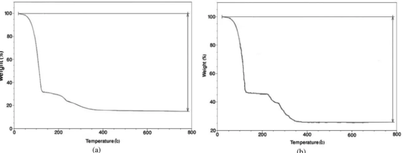
Figure 1 shows typical TG curves of the gel prepared using un-irradiated InAc precursor. Decomposition of the gel
proceeds through three major steps. The irst decomposition
step was in the range of 50-110 °C, the second weight loss step was in the range of 520-270 °C and the third weight loss was in the range of 280-350 °C. The total experimental
weight loss percentage (≈75%) was in good agreement with the percentage (74%) expected theoretically assuming
release of three benzyl alcohol molecules, with the formation
of In2O3 as inal solid residue. The decomposition of the
gel prepared using γ-irradiated InAc precursor proceeds by the same thermal behaviour as with the un-irradiated one.
Figure 2 shows FT-IR of the gels formed using un-irradiated and γ-irradiated InAc precursors before and after thermal oxidation treatments at 200 and at 400 °C calcinations temperatures for 5 hours respectively. The Figure 2 shows systematic decrease in the intensity of the absorption bands characteristic for benzyl alcohol molecules. Complete annealing of the gel and formation of In2O3 nanoparticles was achieved by calcination at
400 °C for 5 hours as shown in Figure 2.The igure show
the characteristic bands attributed to In-O phonon mode of vibration of cubic In2O3 nanoparticles in the range of 400-800 cm–1[15]. The chemisorbed H
2O and CO2 molecules on the surface of In2O3 could be easily detected by recording the bands appeared in the range of 3400-3500 cm–1 and 1700-1600 attributed to νOH and νC=O respectively.
XRD patterns of the as-prepared samples for both un-irradiated and γ-irradiated InAc with 102 KGy calcined at
200 °C for 5 hours show diffraction lines indexed as mixed phase of cubic and hexagonal In2O3 for both un-irradiated and γ-irradiated InAc16.
The diffraction lines for the samples calcined at 400 °C for 5 hours are all indexed to pure body centered cubic (bcc) structure of In2O3 (JCPDS Card No. 06-0416) as shown in Figure 3. No others impurity peaks were detected in the pattern which reveal the high phase purity of the sample. In addition, the EDS analyses also indicate only indium and oxygen as constituent’s elements. The broadening of the
obtained lines conirm nanocrystalline nature of the phase.
However, in case of γ-irradiation precursor, the XRD pattern shows more broadening in the width of diffraction lines. This broadening leads to a decrease in the lattice dimensions due to the radiation damage induced by γ-irradiation in the host
lattice of InAc precursor. This behavior was conirmed by
calculating the particle size of In2O3 nanoparticles by using of the Deby-Sherrer formula17.
λ =
β θB
0.89
cos
t (1)
where λ is the wavelength (in Angstrom), β is the broadening of the diffraction peaks (in radians) or full width at half maximum (FWHM), θB is the Bragg diffraction angle. The calculated average particle size of In2O3 nanoparticles was estimated to be in the ranges of (10-20) and (10-15) nm in case of un-irradiated and γ-irradiated precursors respectively.
SEM images of In2O3 nanoparticles obtained by using of un-irradiated and γ-irradiated InAc precursors are shown in Figure 4. Both images display the nanocrystalline nature
of the as-prepared powders, with signiicant differences in
the shape and size of the as-prepared In2O3 nanoparticles as a result of γ-irradiation. In case of un-irradiated precursor, the SEM images show agglomerated and disordered spherical nanoparticles. In γ-irradiated case the particles showed coral-like structure. TEM images of the as-prepared In2O3 nanoparticles showed also signiicant changes in the morphology of the particles prepared using un-irradiated and
γ-irradiated precursors. In case of γ-irradiation, in contrast to the agglomerated cubic In2O3 obtained using un-irradiated
Figure 1. Typical TG curves of the gels prepared using. (a) un-irradiated InAc; (b) γ-irradiated InAc (102 KGy).
precursors, the image obtained for γ-irradiated precursor showed blocks of agglomerated nanocubes (primary of nanocrystallites ) with big holes between these blocks These nanocubes exhibit different orientation compared with those prepared by un-irradiated precursor. The nanocubes in case of ɣ irradiated precursor are preferentially oriented
along 222 planes as was conirmed by XRD pattern of In2O3 obtained using γ-irradiated precursor. The average particle
size (≈10 nm) was estimated to be quite smaller than the value calculated using un-irradiated precursor (≈15 nm).
Thermal stability of In2O3 nanoparticles were examined to illustrate the role of γ-irradiation on the thermal stability of the as-prepared In2O3 nanoparticles. In general, the In2O3 nanoparticles prepared using γ-irradiated InAc precursor showed higher thermal stability compared with those prepared using un-irradiated InAc. The weight loss in the
temperature range of 50-600 °C was attributed to release of adsorbed water and volatile carbonate from the surface of In2O3 nanoparticles. Starting by 650 °C the TG curve for In2O3 nanoparticles prepared by un-irradiated InAC precursor show continuous weight loss due to degradation process. No detectable weight loss was recorded in the TG curves for In2O3 nanoparticles of γ-irradiated InAc up to 800 °C the results are shown in Figure 5.The stability of In2O3 nanoparticles prepared using un-irradiated precursor up to 800 °C clearly indicate that these particles can act as molten salts in the temperature range of 600 to 800 °C.This
is the irst report of the behavior of In2O3 as molten salt in the temperature range of 600 to 800 °C.
Figure 6 shows the RT-PL spectra of the as-synthesized In2O3 nanoparticles. While exciting the sample with 266 nm, a strong emission at 383 nm and a weak emission at 650 nm
Figure 2. FT-IR of the gels prepared using (a) un-irradiated and (b) γ-irradiated (102 KGy) InAc precursors. 1-without thermal treatment;
2- calcined at 200 °C for 5 hours; 3- calcined at 400 °C for 5 hours.
Radiation Induced Synthesis of In2O3 Nanoparticles - Part 1: Synthesis of In
2O3 Nanoparticles by Sol-gel Method Using Un-irradiated and γ-Irradiated Indium Acetate
were recorded. It is well known that bulk In2O3 cannot emit light, while its nanostructure can emit visible and UV light. The PL emission by nanocrystals in the visible range was mainly attributed to creation of oxygen vacancies. These oxygen vacancies induce the formation of new energy levels in the band gap which facilitate the electronic transitions between valence band and conduction bands18.
3.1.
Role of irradiation
Upon irradiation with Co-60 γ-ray source, the photoelectric effect is the main mode of interaction of
γ-ray with indium atom. In this way, stable molecules (M) are converted into solvated electrons (e) and high reactive free radicals (M+).In solid samples, the radiation effect is dominated by direct ionization of the material whereas for
Figure 4. SEM and TEM images of In2O3 nanoparticles obtained by using of un-irradiated (a, c) and γ-irradiated (b, d) InAc precursors.
Figure 5. Typical TG curves of the as-synthesized In2O3 nanoparticles. (a) un-irradiated and (b) γ-irradiated InAc precursors.
aqueous liquids the reaction with radical species such as OH- or solvated electrons is the dominant mechanism for damage to a solute. The solvated electrons are trapped in the interstitials and vacancies positions in the host lattice of indium acetate unit cell and not allowed to interact with anhydrous benzyl alcohol moiety. These crystal defects could lead to point defect (anion Frenkel defect where O2– is the interstitial ion), defect clusters and extended defects. As a result of these events, impurity and non-stoichiomertric centers are created. Accordingly some
kinds of modiications either in the morphology of the
as-synthesized nanoparticles or in the course of thermal decomposition behavior of γ-irradiated materials may be generated19. From crystallographic point of view, when unit cell is exposed to γ-irradiation , the atoms may be disturbed from the equilibrium positions. Accordingly, the scattering action changes so that the structure factor becomes different. The lattice parameters was calculated from 222 plane by using of the formula
+ +
=
2 2 2
2 2
1
d a
h k l
(2)
where d is the d-spacing parameter, and hkl are Miller indices (222) plane. The calculated parameter, a , in case of γ-irradiation
(10.05 Å) was estimated to be less than the estimated value (10.09 Å) using un-irradiated precursor. The two values are in good agreement with the reported value of 10.11 Å (international center for diffraction data card no. 04-015-4061)
4. Conclusion
In2O3 nanoparticles were prepared by sol-gel method in non-aqueous medium using anhydrous benzyl alcohol and indium acetate as precursor. Pre-irradiation of indium acetate with 102 KGy absorbed γ-ray dose led to signiicant changes in the morphology and thermal properties of the as-synthesized In2O3 nanoparticles. Further studies concerning the effects of varying γ-ray dose on the preparation of In2O3 nanoparticles and their applications as sensors are in progress in our laboratory.
Acknowledgments
This research project was supported by a grant from the research center of the center for the female scientiic and
medical colleges in King Saud University.
Figure 6. The RT-PL spectra of the as-synthesized In2O3 nanoparticles. (a) un-irradiated and (b) γ-irradiated InAc precursors.
References
1. Murali A, Barve A, Leppert VJ, Risbud SH, Kennedy IM and Lee H. Synthesis and Characterization of Indium Oxide Nanoparticles.Nano Letters. 2001; 1(6):287-289. http://dx.doi. org/10.1021/nl010013q
2. Li C, Zhang D, Liu X, Han S, Tang T, Han J and Zhou C. In2O3 nanowires as chemical sensors . Applied Physics Letters. 2003; 82:1613. http://dx.doi.org/10.1063/1.1559438
3. Xu JQ, Wang XH, Wang GQ, Han JJ and Sun YA. Solvothermal synthesis of In2O3 nanocrystal and its ethanol
sensing mechanism Electrochemical and Solid State Letters.
2006; 9(11):103-107. http://dx.doi.org/10.1149/1.2335943
4. Wang X, Zhang M, Liu J, Luo T and Qian Y. Shape- and phase-controlled synthesis of In2O3 with various morphologies
and their gas-sensing properties. Sensors and Actuators B: Chemical. 2009; 137(1):103-110. http://dx.doi.org/10.1016/j.
snb.2008.11.027
5. Oprea A, Gurlo A, Bârsan N and Weimar U. Transport and gas sensing properties of In2O3 nanocrystalline thick ilms:
A Hall effect based approach. Sensors and Actuators B: Chemical. 2009; 139(2):322-328. http://dx.doi.org/10.1016/j. snb.2009.03.002
Radiation Induced Synthesis of In2O3 Nanoparticles - Part 1: Synthesis of In
2O3 Nanoparticles by Sol-gel Method Using Un-irradiated and γ-Irradiated Indium Acetate
Growth. 2005; 276(3-4):471-477. http://dx.doi.org/10.1016/j.
jcrysgro.2004.11.394
7. Du J, Yang M, Nam Cha S, Rhen D, Kang M and Kang DJ. Indium Hydroxide and Indium Oxide Nanospheres, Nanoflowers, Microcubes, and Nanorods: Synthesis and Optical Properties. Crystal Growth & Design. 2008; 8(7):2312-2317. http://dx.doi.org/10.1021/cg701058v
8. Zhu P, Wu W, Zhou J and Zhang W. Preparation of size-controlled In2O3 nanoparticles. Applied Organometallic Chemistry. 2007; 21(10):909-912. http://dx.doi.org/10.1002/ aoc.1300
9. Dodd A. Synthesis of indium oxide nanoparticles by solid state chemical reaction. Journal of Nanoparticle Research. 2009; 11(8):2171-2177. http://dx.doi.org/10.1007/s11051-009-9707-x
10. Maurizot MS, Mostafavi M, Douki T and Belloni J, editors. Radiation chemistry from basics to applications in material and life sciences. Edp Sciences; 2008.
11. Chmieleki AG, editor. Emerging applications of radiation in nanotechnology. Vienna: IAEA; 2005. IAEA TECDDOC-1438. 12. Gerasimov GY. Radiation methods in nanotechnology. Journal of Engineering Physics and Thermophysics. 2011; 84(4):947-963. http://dx.doi.org/10.1007/s10891-011-0554-0
13. Chmielewski AB, Chmielewska DK, Michalik J and Sampa MH. Prospects and challenges in application of gamma, electron and ion beams in processing of nanomaterials. Nuclear Instruments and Methods in Physics Research. 2007; B265(1):339-346.
http://dx.doi.org/10.1016/j.nimb.2007.08.069
14. Mahfouz RM, Al-Ahmari Sh, Al-Fawaz A, Al-Othman Z, Kh-Warad KhI and Siddiqui MR. Kinetic Analysis for Non-isothermal Decomposition of Unirradiated and γ-Irradiated
Indium Acetyl Acetonate. Materials Research. 2011; 14(1):7-10. http://dx.doi.org/14(1):7-10.1590/S1516-14392011005000009
15. Nyquist RA and Kagel RO. Infrared Spectra of Inorganic Compounds. New York: Academic Pres; 1971. p. 208.
16. Fan Y, Wang S and Sun ZX. In2O3 hollow spheres: One-step
solvothermal synthesis and gas sensing properties. Materials Chemistry and Physics; 2012; 134(1):93-97. http://dx.doi.
org/10.1016/j.matchemphys.2012.02.034
17. Chandradass J, Dong SD, Chandradass J, Bae DS, Kim KH and Hyeonkim K. A simple method to prepare indium oxide
nanoparticles: Structural, microstructural and magnetic properties. Advanced Powder Technology. 2011; 22(3):370-374. http://dx.doi.org/10.1016/j.apt.2010.05.006
18. Cao BX, Zhao Y and Wu Z. Synthesis and characterization of In2S3 nanoparticles. Journal of Alloys and Compounds.
2009; 472(1-2):325-327. http://dx.doi.org/10.1016/j. jallcom.2008.04.047
19. Mahfouz RM, Monshi MAS and Abd El-Salam NM. Kinetics
of the thermal decomposition of γ-irradiated gadolinium acetate. Thermochimica Acta. 2002; 383(1-2):95-101. http:// dx.doi.org/10.1016/S0040-6031(01)00682-7