Liver mitochondrial dysfunction and
oxidative stress in the pathogenesis of
experimental nonalcoholic fatty liver
disease
1Departamento de Gastroenterologia (LIM 07), 2Departamento de Cirurgia (LIM 37),
3Departamento de Emergência (LIM 51), 4Departamento de Patologia(LIM 14),
Faculdade de Medicina, Universidade de São Paulo, São Paulo, SP, Brasil
C.P.M.S. Oliveira1,
A.M.M. Coelho2,
H.V. Barbeiro3, V.M.R. Lima1,
F. Soriano3, C. Ribeiro4,
N.A.T. Molan2, V.A.F. Alves4,
H.P. Souza3,
M.C.C. Machado2
and F.J. Carrilho1
Abstract
Oxidative stress and hepatic mitochondria play a role in the pathogen-esis of nonalcoholic fatty liver disease. The aim of the present study was to evaluate the role of hepatic mitochondrial dysfunction and oxidative stress in the pathogenesis of the disease. Fatty liver was induced in Wistar rats with a choline-deficient diet (CD; N = 7) or a high-fat diet enriched with PUFAs-ω-3 (H; N = 7) for 4 weeks. The control group (N = 7) was fed a standard diet. Liver mitochondrial oxidation and phosphorylation were measured polarographically and oxidative stress was estimated on the basis of malondialdehyde and glutathione concentrations. Moderate macrovacuolar liver steatosis was observed in the CD group and mild liver steatosis was observed in the periportal area in the H group. There was an increase in the oxygen consumption rate by liver mitochondria in respiratory state 4 (S4) and a decrease in respiratory control rate (RCR) in the CD group (S4: 32.70 ± 3.35; RCR: 2.55 ± 0.15 ng atoms of O2 min-1 mg protein-1) when compared to the H and control groups (S4: 23.09 ± 1.53, 17.04 ± 2.03, RCR: 3.15 ± 0.15, 3.68 ± 0.15 ng atoms of O2 min-1 mg protein-1, respectively), P < 0.05. Hepatic lipoperoxide concentrations were significantly increased and the concentration of reduced gluta-thione was significantly reduced in the CD group. A choline-deficient diet causes moderate steatosis with disruption of liver mitochondrial function and increased oxidative stress. These data suggest that lipid peroxidation products can impair the flow of electrons along the respiratory chain, causing overreduction of respiratory chain compo-nents and enhanced mitochondrial reactive oxygen species. These findings are important in the pathogenesis of nonalcoholic fatty liver disease.
Correspondence
C.P.M.S. Oliveira
Departamento de Gastroenterologia Faculdade de Medicina, USP Av. Dr. Enéas C. Aguiar, 255 Instituto Central, 9º andar, S. 9159 05403-000 São Paulo, SP Brasil
Fax: +55-11-3066-7301 E-mail: [email protected]
Publication supported by Programa de Apoio à Pós-Graduação-CAPES (PROAP).
Received May 4, 2005 Accepted September 22, 2005
Key words
•Hepatic mitochondrial dysfunction
•Oxidative stress
•Nonalcoholic fatty liver disease
•Choline-deficient diet
Introduction
Nonalcoholic fatty liver disease (NAFLD) involves fatty liver (hepatic steatosis) and nonalcoholic steatohepatitis (NASH) that progresses from hepatic steatosis with lobu-lar inflammation to ballooning degenera-tion, fibrosis, and eventually to cirrhosis (1,2). NAFLD is associated with several pre-disposing factors such as obesity, diabetes, dyslipidemia, jejunoileal bypass, drugs, and parenteral nutrition. However, the progres-sion to fibrosis and cirrhosis is not known. Some studies have shown that liver injury is mediated by oxidative stress (3,4), endotox-ins, cytokines (5,6), and hyperinsulinemia (7,8). Oxidative stress plays a central role in the pathogenesis of NASH. The increased production of reactive oxygen species (ROS) is known to cause lipid peroxidation, fol-lowed by an inflammatory response, and activation of stellate cells leading to fibro-genesis (9-11). Mitochondria are respon-sible for oxidative phosphorylation and fatty acid ß-oxidation and are the main source of cellular ROS. Therefore, dysfunction of liver mitochondria may play an important role in the induction of hepatic steatosis and NASH. The aim of the present study was to evaluate the role of liver mitochondrial dysfunction and oxidative stress in the pathogenesis of experimental fatty liver induced by different diets in an animal model.
Material and Methods
The study was designed in accordance with the Guide for the Care and Use of Labo-ratory Animals published by the US National Institutes of Health (NIH Publication No. 85-23, revised in 1996) and the Guidelines of Animal Experimentation of the University of São Paulo School of Medicine, São Paulo, SP, Brazil, for the care and use of laboratory ani-mals. Male Wistar rats weighing 300 to 350 g were housed in cages with a controlled 12-h light/dark cycle, receiving water ad libitum. Fatty liver was induced in these animals by two different diets: choline-deficient diet (CD; N = 7) or high-fat diet enriched with
polyun-saturated fatty acids (PUFAs-ω-3) (H; N =
7) for 4 weeks (Table 1). The control group (N = 7) was fed a standard diet. After 4 weeks, the rats were sacrificed under anes-thesia with 0.2 mg/kg ketamine hydrochlo-ride injected intraperitoneally and serum, plasma and liver samples were collected for biochemical analysis, histological examina-tion, oxidative stress analysis, and analysis of mitochondrial function.
Biochemical analysis
Serum alanine aminotransferase (AST), aspartate aminotransferase (ALT), choles-terol, and triglycerides were analyzed by standard methods.
Histological analysis
Fragments of liver tissue previously fixed in 10% formalin solution were processed and stained with hematoxylin-eosin and Masson trichrome. The following histologi-cal variables were assessed and scored from 0 to 3 by a blinded experienced pathologist: macro- and microvacuolar fatty change, fatty zonal distribution, foci of necrosis, portal and perivenular fibrosis, as well as the in-flammatory infiltrate and its zonal distribu-tion.
Table 1. Composition of the experimental diets used.
Choline-deficient diet High-fat diet Standard diet
Casein 140 (14%) 140 (14%) 140 (14%) Corn starch 620 (62%) 487 (48.7%) 620 (62%) Sucrose 100 (10%) - 100 (10%) Soybean oil 40 (4%) 70 (7%) 40 (4%) AIN-93M mineral mix 40 (4%) 40 (4%) 40 (4%) AIN-93M vitamin mix 12 (1.2%) 12 (1.2%) 12 (1.2%) L-cystine 5 (0.5%) 2 (0.2%) 2 (0.2%)
Fish oil - 200 (20%)
-Choline bitartrate - 3 (0.3%) 3 (0.3%) Fiber 46 (4.6%) 46 (4.6%) 46 (4.6%)
Oxidative stress analysis
The thiobarbituric acid method was used to quantify lipid peroxidation in tissues, measured as thiobarbituric acid-reactive
sub-stances (TBARS).Tissues (100 mg/mL) were
homogenized in 1.15% KCl buffer, and
cen-trifuged at 14,000 g for 20 min. The
super-natant was then stored at -70ºC. An aliquot
of the supernatantwas added to a reaction
mixture of 1.5mL 0.8% thiobarbituric acid,
200 µL 8.1% (v/v) SDS,1.5 mL 20% (v/v)
acetic acid, pH 3.5, and 600 µL distilled
H2O, and heated to 90ºC for 45 min. After
cooling to room temperature,the samples
were cleared by centrifugation at 10,000 g
for 10min, and their absorbance was
meas-ured at 532 nm using
1,1,3,3-tetramethox-ypropaneas an external standard. The
quan-tity of lipid peroxides is reportedas nmol
malondialdehyde (MDA) equivalents/mg
protein. Glutathione (GSH) assay: tissues
were homogenized (100 mg/mL) in 5% (v/ v) sulfosalicylic acid. The homogenates were
centrifuged at 10,000 g for 20 min, and an
aliquot of the clear supernatant (20 mL) was
combined with 0.3 M Na2HPO4 (160 mL)
and 0.04% 5,59-dithiobis-(2-nitrobenzoic acid) in 1% sodium citrate (20 mL). After 10-min incubation at room temperature, ab-sorbance was read at 405 nm in a Tecan microplate reader (Grödig, Salzburg, Aus-tria). GSH concentrations were calculated from a standard curve constructed with known concentrations of reduced GSH and are reported as µg GSH/mg protein.
Oxidation and phosphorylation of liver mitochondria
Liver mitochondria were prepared as pre-viously described (12). Mitochondrial oxy-gen consumption was measured polaro-graphically (12) using a Gilson 5/6H Oxy-graph (Gilson Medical Electronics, Inc., Middleton, WI, USA) in a closed reaction vessel fitted with a Clark oxygen electrode
(Yellow Springs Instruments Co., Yellow Springs, OH, USA) at 28ºC. The respiratory control rate, considered to be an index of oxidative and phosphorylative mitochondrial function, was calculated as the rate of oxy-gen consumption in the presence of ADP (state 3, S3) over the consumption in the absence of ADP (state 4, S4). Respiratory states S3 and S4 were measured and reported as ng atoms of O2 min-1 mg protein-1. Mito-chondria protein content was determined by the method of Lowry et al.
Statistical analysis
Data are reported as means ± SEM. Groups were compared by one-way analysis of variance (ANOVA), with the level of significance set at P < 0.05.
Results
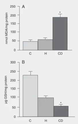
Oxida-tive stress was increased in animals fed the CD diet: hepatic lipoperoxide (TBARS) con-centrations were significantly increased and the concentration of reduced GSH was sig-nificantly lower in the CD group compared to the H and control groups (Figure 2A,B).
Discussion
NAFLD is ascribed to an imbalance be-tween the excessive uptake of free fatty acids by the liver with subsequent increase in TG synthesis, and the reduction of fatty acid oxidation and TG secretion (very low den-sity lipoproteins, VLDLs) in the liver. Cho-line is an essential nutrient for the secretion of TGs (VLDLs) by the liver, and its priva-tion impairs their transport, leading to fatty liver in the rat. A choline-deficient diet is a classic general model of NAFLD (13). On the other hand, lipogenesis is stimulated by a high carbohydrate diet, whereas it is inhib-ited by PUFAs and by fasting. The supply of PUFAs reduces the production and excre-tion of VLDL and decreases serum TG con-centration (14). Sekiya et al. (15) demon-strated that animal PUFAs (fish oil) amelio-rate hepatic steatosis through the suppres-sion of sterol regulatory element-binding protein-1 in ob/ob mice. In the present study, the group that received a high-fat diet en-riched with animal PUFA (fish oil) devel-oped only a mild liver steatosis in the peri-portal zone and had normal plasma levels of cholesterol and TGs. These data confirm
reports (14-16)that a high-fat diet enriched
with PUFAs-ω-3 (fish oil) has a protective
effect on the liver, causing only mild liver steatosis. In animals fed the choline-defi-cient diet, liver steatosis was more severe and TG levels were also increased. These differences suggest that a high carbohydrate diet with an increase of TGs or impairment of VLDL exportation could play an impor-tant role in the pathogenesis of NAFLD.
Mitochondria are involved in fatty acid ß-oxidation and in oxidative
phosphoryla-Figure 1. Initial and final weight of the animals. Data are reported as means ± SEM for 7 rats in each group as a function of di-ets. CD = choline-deficient diet; H = high-fat diet enriched with PUFAs-ω-3; C = standard diet (control).
Table 2. Oxidation and phosphorylation of liver mitochondria in experimental nonalco-holic fatty liver disease.
Group N RCR S3 S4 ADP/oxygen ratio
CD 9 2.55 ± 0.15* 81.39 ± 6.14 32.70 ± 3.35* 1.82 ± 0.07 H 7 3.15 ± 0.15 71.69 ± 4.10 23.09 ± 1.53 1.84 ± 0.03 C 6 3.68 ± 0.15 62.35 ± 7.17 17.04 ± 2.03 1.96 ± 0.01
Data are reported as means ± SEM ng atoms of O2 min-1 mg protein-1. RCR =
respiratory control rate; S3 (state 3) = oxygen consumption in the presence of ADP; S4 (state 4) = oxygen consumption in the absence of ADP; CD = choline-deficient diet; H = high-fat diet; C = standard diet (control).
*P < 0.05 for CD vs H and C (ANOVA).
tion and are an important cellular source of ROS. Therefore, mitochondrial dysfunction may play a central role in the accumulation of fat in the liver (“first hit”) and the exces-sive production of ROS by mitochondria may result in lipid peroxidation (“second hit”) (6). Drug-induced fatty liver has in-creased lipid peroxidation (17) and ob/ob mice have an increased ROS production (11). Our group has demonstrated in previous stud-ies that the presence of steatosis correlates with an increase in superoxide anion hydro-peroxide generation in animals fed a cho-line-deficient diet (18) and the use of vita-min C reduces liver steatosis and peroxida-tion (19). In the present study, lipid peroxi-dation was also increased and GSH was decreased in the CD group while the oxida-tive stress of animals fed a high-fat diet
enriched with PUFAs-ω-3 was similar to
that observed in controls. The lack of lipid peroxidation in the H group could be ex-plained by the fact that no steatosis devel-oped. Additionally, chronic impairment of mitochondrial ß-oxidation could cause mi-crovacuolar and/or mami-crovacuolar steatosis and an increase in ROS. Our results showed mitochondrial dysfunction with a marked succinate-dependent increase in basal respi-ration (S4) in CD animals, resulting in an average 92% increase of oxygen consump-tion when compared to the control group. This increased mitochondrial ROS forma-tion may further oxidize fat deposits, caus-ing a vicious cycle with more lipid peroxida-tion, more mitochondrial damage, and more ROS formation. ROS may oxidize fat depos-its, releasing lipid peroxidation products that damage mitochondrial DNA and proteins to partially block the flow of electrons along the respiratory chain, thus further increasing mitochondrial ROS formation. ROS may also deplete antioxidants and cause the
for-mation of tumor necrosis factor-α, two
ef-fects that may further impair the flow of
electrons and increase mitochondrial ROS formation (20).
PUFAs-ω-3 (fish oil) can be used by
cyclooxygenase to produce prostaglandins of lower potency, that will induce less in-flammation, or may reduce inflammation in
some other way. Since PUFA-ω-3
prosta-glandins are less potent they induce less ROS production, a fact that may explain our findings.
There was no difference in
mitochon-drial function between the H diet and
con-trol. The high-fat diet caused only mild liver steatosis but mitochondrial function was similar to that observed in controls. Accord-ing to this hypothesis, the increase in ROS production (“second hit”) may result from the damage of mitochondrial ß-oxidation (“first hit”), leading to oxidative stress. Our study suggests that the pathogenesis of fatty liver differed between the two diets: the CD diet led to more severe, predominantly mac-rovacuolar steatosis, increased mitochondrial dysfunction, increased oxidative stress, and higher plasma TG levels than the H diet. The choline-deficient rat model is important for assessing oxidative stress independent of obesity, whereas the high fat diet model more closely resembles a leptin-resistant model. On the other hand, a high-fat diet
enriched with polyunsaturated fat (ω-3)
slightly increases the arrival of fatty acids into the liver and, although producing mild steatosis, it preserves mitochondrial ß-oxi-dation and phosphorylation and the plasma triglyceride levels are similar to those in the control group.
The results of the present study suggest that the excessive arrival of fatty acid to the liver due to a high-fat diet enriched with
polyunsaturated fat (ω-3) may cause fatty
References
1. Matteoni CA, Younossi ZM, Gramlich T et al. (1999). Nonalcoholic fatty liver disease: a spectrum of clinical and pathological severity. Gastroenterology, 116: 1413-1419.
2. Falck-Ytter Y, Younossi ZM, Marchesini G et al. (2001). Clinical features and natural history of nonalcoholic steatosis syndromes. Seminars in Liver Disease, 21: 17-26.
3. Chitturi S & Farrell GC (2001). Ethiopathogenesis of nonalcoholic steatohepatitis. Seminars in Liver Disease, 21: 27-41.
4. Mehta K, Van Thiel DH, Shah N et al. (2002). Nonalcoholic fatty liver disease: pathogenesis and the role of antioxidants. Nutrition Re-views, 60: 289-293.
5. Tilg H & Diehl AM (2000). Cytokines in alcoholic and nonalcoholic steatohepatitis. New England Journal of Medicine, 343: 1467-1476. 6. Wigg AJ, Roberts-Thomson IC, Dymock RB et al. (2001). The role of small intestinal bacterial overgrowth, intestinal permeability, endo-toxaemia, and tumour necrosis factor alpha in the pathogenesis of non-alcoholic steatohepatitis. Gut, 48: 206-211.
7. Luyckx FH, Lefebvre PJ & Scheen AJ (2000). Non-alcoholic steato-hepatitis: association with obesity and insulin resistance, and influ-ence of weight loss. Diabetes Metabolism, 26: 98-106.
8. Scheen AJ & Luyckx FH (2003). Nonalcoholic steatohepatitis and insulin resistance: interface between gastroenterologists and endo-crinologists. Acta Clinica Belgica, 58: 81-91.
9. Yang S, Zhu H, Li Y et al. (2000). Mitochondrial adaptations to obesity-related oxidant stress. Archives of Biochemistry and Bio-physics, 378: 259-268.
10. Curzio M, Esterbauer H & Dianzani MU (1985). Chemotactic activity of hydroxyalkenals on rat neutrophils. International Journal of Tis-sue Reactions, 7: 137-142.
11. Lee KS, Buck M, Houglum K et al. (1995). Activation of hepatic stellate cells by TGF alpha and collagen type I is mediated by
oxidative stress through c-myb expression. Journal of Clinical In-vestigation, 96: 2461-2468.
12. Estabrook RW (1967). Mitochondrial respiratory control and the polarographic measurement of ADP:O ratio. In:Estabrook RW & Pullman ME (Editors), Methods in Enzymology. Academic Press, New York, 41-47.
13. Teramoto K, Bowers JL, Khettry U et al. (1993). A rat fatty liver transplant model. Transplantation, 55: 737-741.
14. Willumsen N, Skorve J & Hexeberg S (1993). The hypotriglyc-eridemic effect of eicosapentaenoic acid in rats is reflected in in-creased mitochondrial fatty acid oxidation followed by dimished lipogenesis. Lipids, 28: 683-690.
15. Sekiya M, Yahagi N & Matsuzaka T (2003). Polyunsaturated fatty acids ameliorate hepatic steatosis in obese mice by SREBP-1 sup-pression. Hepatology, 38: 1529-1539.
16. Clarke SD & Jump DB (1994). Dietary polyunsaturated fatty acid regulation of gene transcription. Annual Review of Nutrition, 14: 83-98.
17. Letteron P, Fromenty B, Terris B et al. (1996). Acute and chronic hepatic steatosis leads to in vivo lipid peroxidation in mice. Journal of Hepatology, 24: 200-208.
18. Oliveira CPMS, Gayotto LC, Tatai C et al. (2002). Oxidative stress in the pathogenesis of nonalcoholic fatty liver disease, in rats fed with a choline-deficient diet. Journal of Cellular and Molecular Medicine, 6: 399-406.
19. Oliveira CPMS, Gayotto LC, Tatai C et al. (2003). Vitamin C and vitamin E in prevention of nonalcoholic fatty liver disease (NAFLD) in choline deficient diet fed rats. Nutrition Journal, 2: 9.