ISSN 0100-879X
BIOMEDICAL SCIENCES
AND
CLINICAL INVESTIGATION
www.bjournal.com.br
www.bjournal.com.br
Volume 45 (5) 376-472 May 2012
Braz J Med Biol Res, May 2012, Volume 45(5) 444-449
doi:
10.1590/S0100-879X2012007500032
Time course of the hemodynamic responses to aortic depressor
nerve stimulation in conscious spontaneously hypertensive rats
M.T. Durand, A.L. Mota, A.R. Barale, J.A. Castania, R. Fazan Jr. and H.C. Salgado
Institutional Sponsors
The Brazilian Journal of Medical and Biological Research is partially financed by
Faculdade de Medicina de Ribeirão Preto Campus
Ribeirão Preto
Explore High - Performance MS Orbitrap Technology In Proteomics & Metabolomics
Time course of the hemodynamic responses
to aortic depressor nerve stimulation
in conscious spontaneously hypertensive rats
M.T. Durand
1, A.L. Mota
1, A.R. Barale
2, J.A. Castania
1, R. Fazan Jr.
1and H.C. Salgado
11Departamento de Fisiologia, Faculdade de Medicina de Ribeirão Preto, Universidade de São Paulo, Ribeirão Preto, SP, Brasil 2Instituto de Ciências Biomédicas, Universidade Federal de Uberlândia, Uberlândia, MG, Brasil
Abstract
The time to reach the maximum response of arterial pressure, heart rate and vascular resistance (hindquarter and mesenteric) was measured in conscious male spontaneously hypertensive (SHR) and normotensive control rats (NCR; Wistar; 18-22 weeks) subjected to electrical stimulation of the aortic depressor nerve (ADN) under thiopental anesthesia. The parameters of stimula-tion were 1 mA intensity and 2 ms pulse length applied for 5 s, using frequencies of 10, 30, and 90 Hz. The time to reach the hemodynamic responses at different frequencies of ADN stimulation was similar for SHR (N = 15) and NCR (N = 14); hypoten-sion = NCR (4194 ± 336 to 3695 ± 463 ms) vs SHR (3475 ± 354 to 4494 ± 300 ms); bradycardia = NCR (1618 ± 152 to 1358 ± 185 ms) vs SHR (1911 ± 323 to 1852 ± 431 ms), and the fall in hindquarter vascular resistance = NCR (6054 ± 486 to 6550 ± 847 ms) vs SHR (4849 ± 918 to 4926 ± 646 ms); mesenteric = NCR (5574 ± 790 to 5752 ± 539 ms) vs SHR (5638 ± 648 to 6777 ± 624 ms). In addition, ADN stimulation produced baroreflex responses characterized by a faster cardiac effect followed by a vascular effect, which together contributed to the decrease in arterial pressure. Therefore, the results indicate that there is no alteration in the conduction of the electrical impulse after the site of baroreceptor mechanical transduction in the baroreflex pathway (central and/or efferent) in conscious SHR compared to NCR.
Key words: Electrical stimulation; Arterial pressure; Aortic depressor nerve; Baroreflex; Spontaneously hypertensive rats; Vascular tone
Introduction
Correspondence: M.T. Durand, Departamento de Fisiologia, Faculdade de Medicina de Ribeirão Preto, USP, Av. Bandeirantes, 3900, 14049-900 Ribeirão Preto, SP, Brasil. Fax: +55-16-3633-0017. E-mail: [email protected]
Received October 27, 2011. Accepted February 28, 2012. Available online March 16, 2012. Published May 7, 2012. It has been well demonstrated that the arterial
barore-flex acts, in particular, to buffer acute changes in arterial
pressure by reciprocal modulation of the sympathetic and parasympathetic activities that control heart rate (HR) and vascular resistance (1,2). Remarkable attenuation of the
baroreflex control of HR (3-6), of baroreceptor afferent sen
-sitivity (7-11) and of baroreflex control of sympathetic nerve
activity (12-14) in spontaneously hypertensive rats (SHR) has been thoroughly documented in the literature.
Studies evaluating the hemodynamic responses caused by electrical stimulation of the baroreceptors in anesthetized rats demonstrated that the hemodynamic responses were
reduced in SHR, suggesting an alteration of the baroreflex
circuitry (15,16). Nevertheless, in a recent study, Salgado
et al. (17) demonstrated that electrical stimulation of the aortic depressor nerve (ADN) in conscious SHR produced equivalent or even greater depressor responses compared to normotensive rats, indicating that conscious SHR
exhib-ited a well-preserved baroreflex response to the electrical
stimulation of the ADN, probably because this maneuver bypasses the mechanical transduction of the barorecep-tors. Although Salgado et al. (17) reported a well-preserved
baroreflex function elicited by electrical activation of the ADN
in SHR, they did not compare the dynamic characteristics, i.e., the time course of the hemodynamic responses (arterial pressure, HR, and vascular resistance) to electrical stimula-tion of SHR and normotensive control rats (NCR).
Electrical stimulation of the aortic baroreceptors 445
the hemodynamic responses could indicate a derangement
of the conduction of the electrical impulse in the baroreflex
pathway in SHR. Therefore, in the current investigation, we determined the time elapsed to reach maximum hypoten-sion, bradycardia and the decrease in vascular resistance caused by electrical stimulation of the ADN in SHR and NCR.
Material and Methods
This is a retrospective analysis of previously published data (17). Male Wistar NCR and SHR 18-22 weeks of age (270-340 g) were used in the present study. The procedures were reviewed and approved by the Committee of Ethics in Animal Research of Faculdade de Medicina de Ribeirão Preto, Universidade de São Paulo (Protocol #040/2005).
The approach used to stimulate the left ADN and si-multaneously record the arterial pressure, HR and regional
blood flow has been described elsewhere (17-19). Briefly,
under thiopental sodium anesthesia (40 mg/kg; ip), a 4- to 6-mm long portion of the left ADN was carefully isolated below the junction with the superior laryngeal nerve and placed on a bipolar platinum electrode with an inter-electrode
distance of 2 mm. The correct identification of the nerve was confirmed by its typical pattern of discharge, which is
synchronous with the arterial pulse pressure. The ADN was covered with a silicone impression material (Super-Dent; Carlisle Laboratories, USA). To ensure complete polym-erization of the silicone impression material, the silicone that covered the ADN was left to stabilize for 30 min, and
the activity of the nerve was again recorded to confirm the
integrity of the signal. Once the integrity of the signal was
confirmed, the fine platinum wires of the electrodes were
exteriorized to the nape of the neck of the rats and soldered to a small plug connected to the electrical stimulator. While the animals were still under anesthesia the femoral artery was catheterized with polyethylene tubing (50 and PE-10,Intramedic Becton Dickinson, USA) in order to record arterial pressure. In addition, a laparotomy was performed
to place miniaturized Doppler flow probes (Iowa Doppler
Products, USA) around the superior mesenteric artery or the inferior abdominal aorta to measure the changes in blood
flow velocity and to calculate the mesenteric or hindquarter vascular resistance, respectively (18). Catheters and flow
probes were also exteriorized along with the electrodes to the nape of the neck and the surgical incision sites were closed using sutures. Twenty-four hours after the end of surgery, the rats were connected to the recording system, which consisted of a pressure transducer (P23Gb; Statham
Instruments, USA), a pulsed Doppler flowmeter (545C-4;
Department of Bioengineering, The University of Iowa, USA) and an electrical stimulator (EMG/EP, N200/A; BioMed, Hungary). The signals, i.e., the pulsatile arterial pressure, mean arterial pressure (MAP), and regional (mesenteric
or hindquarter) blood flow velocity, were fed to an IBM
personal computer equipped with a 12-bit analog-to-digital interface (CAD 12/36 Lynx Eletrônica, Brazil) and were continuously sampled (500 Hz). The HR was derived from the arterial pressure trace (pulse intervals), and the vascular resistances were calculated online as the ratio between the
MAP and mean blood flow velocity using computer software
(Advanced CODAS; Dataq Instruments, USA). Cardiovas-cular variables were recorded for at least 15 min before electrical stimulation of the ADN (1 mA, 2 ms pulse length, for 5 s) at 10, 30 and 90 Hz in a random sequence.
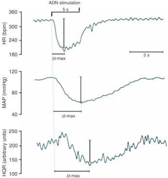
Each stimulus with a particular frequency was applied for a period of 5 s with intervals of at least 5 min. The differ-ence between the pre-stimulation baseline level of the MAP, HR, and vascular resistance (% of basal) and the maximum change in these variables elicited by each frequency of ADN
stimulation was quantified in milliseconds (Figure 1). The
results of the maximum changes of each variable (MAP, HR and vascular resistance) have been published in a previous study from our laboratory (17). The protocol was successfully carried out in 14 NCR and 15 SHR.
Data are reported as means ± SEM. The time elapsed for the cardiovascular variables to reach the maximum response due to ADN stimulation in SHR and NCR and pooled data was analyzed by repeated measures two-way ANOVA (groups: SHR vs NCR; pooled data: hemodynamic
variables MAP vs HR vs vascular resistance; repeated mea-sures: stimulus frequency). When ANOVA demonstrated
statistical significance, the Tukey multiple comparison post hoc test was used to test for differences between means. Baseline values for NCR vs SHR were compared by the unpaired Student t-test. Differences were considered to be
significant when P < 0.05.
Results
Basal hemodynamics
The basal MAP and HR of SHR (150 ± 5 mmHg and 393 ± 9 bpm, respectively) were higher than those of NCR (103
± 2 mmHg and 360 ± 5 bpm, respectively; P < 0.001).
Time course of the hemodynamic baroreflex
responses
The bradycardia, hypotension and fall in hindquarter vascular resistance elicited by the electrical stimulation of the ADN in NCR are illustrated in Figure 1. The time elapsed for the MAP and HR response to reach their maximum value during ADN stimulation was similar in SHR and NCR for all frequencies studied (Figure 2). The time elapsed to reach the maximum response of vascular resistance in the hindquarter and mesenteric bed was similar for the two strains (Figure 3). Moreover, there was no difference between the SHR and NCR for either of the frequencies examined (Figure 3).
When NCR and SHR data were pooled, the time elapsed
Figure 2. Time elapsed to reach (∆t-max) the maximum hypotensive (A) and bradycardic (B) response to electrical stimulation of the aortic depressor nerve in normotensive control rats (NCR) and spontaneously hypertensive rats (SHR) at different frequencies of stimulation (10, 30, 90 Hz). Data are reported as means ± SEM (mean arterial pressure = group: P = 0.706, frequency: P = 0.093, group vs frequency: P = 0.070; HR = group: P = 0.932, frequency: P = 0.843, group vs frequency: P = 0.137; two-way ANOVA).
Electrical stimulation of the aortic baroreceptors 447
to reach the maximum decrease in mesenteric and hind-quarter vascular resistance (10 Hz = 5529 ± 351; 30 Hz = 5520 ± 315; 90 Hz = 5999 ± 355 ms) was longer than the time elapsed to reach the maximum decrease in HR (10 Hz = 1780 ± 190; 30 Hz = 1764 ± 131; 90 Hz = 1634
± 255 ms; P < 0.001 vs MAP and vascular resistance) and MAP (10 Hz = 3794 ± 252; 30 Hz = 4486 ± 221; 90 Hz =
4142 ± 270 ms; P < 0.001 vs HR and vascular resistance). These data demonstrate that the electrical stimulation of the ADN elicits a faster cardiac effect, followed by a vascular effect.
Discussion
The present study demonstrates that there was no
significant difference in the time elapsed for the baroreflex
responses of MAP, HR, and vascular resistance to electrical stimulation of the ADN in SHR compared to NCR for any of the frequencies (10, 30, and 90 Hz) examined. In addition, when NCR and SHR data were pooled, it was also observed that the electrical stimulation of the ADN produced a prompt
cardiac effect followed by a vascular effect. These findings indicate that, as expected, the baroreflex acted immediately
upon the heart and later on the periphery (vessels) with a similar time course in SHR and NCR.
Previous reports have examined the time course of
baroreflex activation using carotid sinus nerve stimulation combined with recording of cardiac vagal fiber activity
(20,21). Kunze (20) demonstrated that the time elapsed
for cardiac vagal fiber activation following carotid sinus
nerve stimulation was 30-72 ms. In the present study, the bradycardic response occurred within 1.5 s after the start of ADN stimulation. Previous studies have demonstrated
that the reflex bradycardia evoked by increases in arterial
pressure is primarily mediated by rapid parasympathetic activation (22-25). Coleman (23) reported that the peak of
the reflex bradycardia following the hypertensive response
caused by phenylephrine was attained approximately 1 s after the maximum increase in arterial pressure. Thus, in the current study, the prompt bradycardia evoked by ADN stimulation in NCR and SHR is mainly attributable to rapid parasympathetic activation, which involves direct excitatory projection from the nucleus tractus solitarii to both the neurons of the nucleus ambiguous and the dorsal motor nucleus of the vagus (26,27).
On the other hand, the time elapsed to reach the maxi-mum hypotensive response and the maximaxi-mum decrease in vascular resistance in both groups was longer than the time to reach the bradycardic response. It has been demon-strated that these depressor responses are mainly caused
by the reflex withdrawal of sympathetic activity (19,28) and
a small contribution of other vasodilatory mechanisms, such
as the activation of sympathetic fibers, which release nitric
oxide (29). Thus, it is possible that the difference in the time elapsed to reach the bradycardic and vascular responses
in both groups of rats was due to the differential central processing of the baroreceptor input. It should also be noted
that Coleman (23) demonstrated that the reflex withdrawal
of sympathetic activity has a slower onset compared to parasympathetic activation.
Afferent baroreceptor sensitivity (7-10) and
baroreflex-mediated changes in HR (3,4) have been shown consistently
to be impaired in SHR. The decreased baroreflex sensitivity
for the control of the HR has been primarily attributed to a derangement of parasympathetic function (3,4,30-32). Nev-ertheless, in the present study, no difference was observed
between the time elapsed to reach reflex bradycardia in SHR and NCR, indicating that the baroreflex parasympathetic
pathway is preserved in SHR.
Moreover, it was also observed that the time elapsed to reach maximum hypotensive and vasodilatory re-sponses, mainly caused by withdrawal of the sympathetic vasoconstrictor tone, was similar in NCR and SHR. Several studies have evaluated the baroreflex control of sympathetic function by recording the renal sympa-thetic nerve activity in SHR (12-14,33,34). However, conflicting results have been reported. There are reports of decreased (12,14), normal (33,34), and increased (13) baroreflex control of sympathetic nerve activity in SHR. However, studies that evaluated the dynamic characteristics of baroreflex regulation of sympathetic activity using transfer functions reported no change in baroreflex control in SHR compared to Wistar-Kyoto rats (34,35). Thus, based on the results obtained in the current study, we propose that the transmission of the inhibitory baroreceptor input throughout the sympathetic pathway is not altered in SHR compared to NCR.
The electrical stimulation of afferent fibers bypasses
the site of baroreceptor mechanosensory transduction.
Therefore, the reflex response to ADN stimulation provides
information about the central processing of the afferent input and the properties of the central and efferent components
of the baroreflex. In a previous study from our laboratory, it
was demonstrated that electrical stimulation of the ADN of conscious SHR produced equivalent or greater depressor responses compared to NCR, suggesting that the central
and efferent components of the baroreflex were preserved
in SHR (17). These data suggested that there is no change in the transmission of the electrical impulse through the
central/efferent baroreflex pathway of SHR. Thus, if there is some alteration in the baroreflex of SHR, it could be in
the mechanosensory transduction of the baroreceptors, as reported previously (7-11).
In conclusion, electrical stimulation of the ADN dem-onstrated no differences in the time elapsed to reach the maximum responses of MAP, HR and vascular resistance in conscious SHR compared to NCR. In addition, the results also demonstrate that electrical stimulation of the ADN pro-duced a prompt cardiac effect followed by a vascular effect,
in both the SHR and NCR. Therefore, these data indicate that the central/efferent processing of the baroreceptor input, which is elicited by electrical stimulation of the ADN, is well preserved in SHR.
Acknowledgments
Research supported by FAPESP (#02/09406-5), CNPq (#134480/2006-6), and CAPES (#1681/07).
References
1. Krieger EM, Salgado HC, Michelini LC. Resetting of barore-ceptors. In: Guyton AC, Hall JE (Editors), Cardiovascular physiology IV, International review of physiology 26. Balti-more: University Park Press; 1982. p 119-146.
2. Thomas GD. Neural control of the circulation. Adv Physiol Educ 2011; 35: 28-32.
3. Head GA, Adams MA. Time course of changes in barorecep-tor reflex control of heart rate in conscious SHR and WKY: contribution of the cardiac vagus and sympathetic nerves. Clin Exp Pharmacol Physiol 1988; 15: 289-292.
4. Minami N, Head GA. Relationship between cardiovascular hypertrophy and cardiac baroreflex function in spontane -ously hypertensive and stroke-prone rats. J Hypertens 1993; 11: 523-533.
5. Xie HH, Shen FM, Zhang XF, Jiang YY, Su DF. Blood pres -sure variability, baroreflex sensitivity and organ damage in spontaneously hypertensive rats treated with various anti-hypertensive drugs. Eur J Pharmacol 2006; 543: 77-82. 6. Cisternas JR, Valenti VE, Alves TB, Ferreira C, Petenusso
M, Breda JR, et al. Cardiac baroreflex is already blunted in eight weeks old spontaneously hypertensive rats. Int Arch Med 2010; 3: 2.
7. Sapru HN, Wang SC. Modification of aortic baroreceptor re -setting in the spontaneously hypertensive rat. Am J Physiol 1976; 230: 664-674.
8. Andresen MC, Krauhs JM, Brown AM. Relationship of aortic wall and baroreceptor properties during development in normotensive and spontaneously hypertensive rats. Circ Res 1978; 43: 728-738.
9. Andresen MC, Yang M. Arterial baroreceptor resetting: contributions of chronic and acute processes. Clin Exp Pharmacol Physiol Suppl 1989; 15: 19-30.
10. Fazan PV, Junior FR, Salgado CH, Barreira AA. Morphology of aortic depressor nerve myelinated fibers in normotensive Wistar-Kyoto and spontaneously hypertensive rats. J Auton Nerv Syst 1999; 77: 133-139.
11. Brum PC, Da Silva GJ, Moreira ED, Ida F, Negrao CE, Krieger EM. Exercise training increases baroreceptor gain sensitivity in normal and hypertensive rats. Hypertension 2000; 36: 1018-1022.
12. Coote JH, Sato Y. Reflex regulation of sympathetic activity in the spontaneously hypertensive rat. Circ Res 1977; 40: 571-577.
13. Thoren P. Efferent renal nerve traffic in the spontaneously hypertensive rat. Clin Exp Hypertens A 1987; 9 (Suppl 1): 259-279.
14. DiBona GF, Jones SY, Sawin LL. Reflex effects on renal nerve activity characteristics in spontaneously hypertensive rats. Hypertension 1997; 30: 1089-1096.
15. Gonzalez ER, Krieger AJ, Sapru HN. Central resetting of baroreflex in the spontaneously hypertensive rat. Hyperten-sion 1983; 5: 346-352.
16. Gao XY, Huang XL, Wang HJ, Zhou LM, Xu Y, Wang W, et al. Depressor effect of closed-loop chip system in spontane-ously hypertensive rats. Auton Neurosci 2007; 137: 84-91. 17. Salgado HC, Barale AR, Castania JA, Machado BH,
Chap-leau MW, Fazan R Jr. Baroreflex responses to electrical stimulation of aortic depressor nerve in conscious SHR. Am J Physiol Heart Circ Physiol 2007; 292: H593-H600. 18. Haywood JR, Shaffer RA, Fastenow C, Fink GD, Brody
MJ. Regional blood flow measurement with pulsed Doppler flowmeter in conscious rat. Am J Physiol 1981; 241: H273-H278.
19. De Paula PM, Castania JA, Bonagamba LG, Salgado HC, Machado BH. Hemodynamic responses to electrical stimula-tion of the aortic depressor nerve in awake rats. Am J Physiol 1999; 277: R31-R38.
20. Kunze DL. Reflex discharge patterns of cardiac vagal effer -ent fibres. J Physiol 1972; 222: 1-15.
21. McAllen RM, Spyer KM. The baroreceptor input to cardiac vagal motoneurones. J Physiol 1978; 282: 365-374. 22. Glick G, Braunwald E. Relative roles of the sympathetic and
parasympathetic nervous systems in the reflex control of heart rate. Circ Res 1965; 16: 363-375.
23. Coleman TG. Arterial baroreflex control of heart rate in the conscious rat. Am J Physiol 1980; 238: H515-H520. 24. Head GA, McCarty R. Vagal and sympathetic components
of the heart rate range and gain of the baroreceptor-heart rate reflex in conscious rats. J Auton Nerv Syst 1987; 21: 203-213.
25. Stornetta RL, Guyenet PG, McCarty RC. Autonomic nervous system control of heart rate during baroreceptor activation in conscious and anesthetized rats. J Auton Nerv Syst 1987; 20: 121-127.
26. Spyer KM. Annual review prize lecture. Central nervous mechanisms contributing to cardiovascular control. J Physiol 1994; 474: 1-19.
27. Wang J, Irnaten M, Neff RA, Venkatesan P, Evans C, Loewy AD, et al. Synaptic and neurotransmitter activation of cardiac vagal neurons in the nucleus ambiguus. Ann N Y Acad Sci 2001; 940: 237-246.
28. Durand MT, Castania JA, Fazan R Jr, Salgado MC, Salgado HC. Hemodynamic responses to aortic depressor nerve stimulation in conscious L-NAME-induced hypertensive rats. Am J Physiol Regul Integr Comp Physiol 2011; 300: R418-R427.
29. Possas OS, Johnson AK, Lewis SJ. Role of nitrosyl factors in the hindlimb vasodilation elicited by baroreceptor afferent nerve stimulation. Am J Physiol Regul Integr Comp Physiol 2006; 290: R741-R748.
Electrical stimulation of the aortic baroreceptors 449
31. Minami N, Head GA. Cardiac vagal responsiveness during development in spontaneously hypertensive rats. Auton Neurosci 2000; 82: 115-122.
32. Corbett EK, Mary DA, McWilliam PN, Batten TF. Age-related loss of cardiac vagal preganglionic neurones in spontane-ously hypertensive rats. Exp Physiol 2007; 92: 1005-1013. 33. Lundin S, Ricksten SE, Thoren P. Renal sympathetic
activ-ity in spontaneously hypertensive rats and normotensive controls, as studied by three different methods. Acta Physiol Scand 1984; 120: 265-272.
34. Harada S, Imaizumi T, Ando S, Hirooka Y, Sunagawa K, Takeshita A. Arterial baroreflex dynamics in normotensive and spontaneously hypertensive rats. Am J Physiol 1992; 263: R524-R528.