ISSN 0100-879X
BIOMEDICAL SCIENCES
AND
CLINICAL INVESTIGATION
www.bjournal.com.br
www.bjournal.com.br
Volume 44 (11) 1070-1193 November 2011
Institutional Sponsors
The Brazilian Journal of Medical and Biological Research is partially financed by
Faculdade de Medicina de Ribeirão Preto Campus
Ribeirão Preto
Ex plor e H igh - Pe r for m a n ce M S Or bit r a p Te ch n ology I n Pr ot e om ics & M e t a bolom ics
analit icaw eb.com .br S C I E N T I F I C
Braz J Med Biol Res, November 2011, Volume 44(11) 1148-1155
doi: 10.1590/S0100-879X2011007500119
CPU0213, a novel endothelin type A and type B receptor antagonist,
protects against myocardial ischemia/reperfusion injury in rats
CPU0213, a novel endothelin type A and type B
receptor antagonist, protects against myocardial
ischemia/reperfusion injury in rats
Z.Y. Wang
1, W. Zhang
2, X.Z. Li
2, Y. Han
3, Y.P. Chen
1, Z. Liu
2,
L.P. Xie
2, Y. Ji
2and X. Lu
11Department of Geriatrics, the Second Affiliated Hospital, 2Key Laboratory of Human Functional Genomics,
Atherosclerosis Research Centre, 3Department of Geriatrics, the First Affiliated Hospital,
Nanjing Medical University, Nanjing, China
Abstract
The efficacy of endothelin receptor antagonists in protecting against myocardial ischemia/reperfusion (I/R) injury is controversial, and the mechanisms remain unclear. The aim of this study was to investigate the effects of CPU0123, a novel endothelin type A and type B receptor antagonist, on myocardial I/R injury and to explore the mechanisms involved. Male Sprague-Dawley rats weighing 200-250 g were randomized to three groups (6-7 per group): group 1, Sham; group 2, I/R + vehicle. Rats were sub-jected to in vivo myocardial I/R injury by ligation of the left anterior descending coronary artery and 0.5% sodium carboxymethyl cellulose (1 mL/kg) was injected intraperitoneally immediately prior to coronary occlusion. Group 3, I/R + CPU0213. Rats were subjected to identical surgical procedures and CPU0213 (30 mg/kg) was injected intraperitoneally immediately prior to coronary occlusion. Infarct size, cardiac function and biochemical changes were measured. CPU0213 pretreatment reduced infarct size as a percentage of the ischemic area by 44.5% (I/R + vehicle: 61.3 ± 3.2 vs I/R + CPU0213: 34.0 ± 5.5%, P < 0.05) and improved ejection fraction by 17.2% (I/R + vehicle: 58.4 ± 2.8 vs I/R + CPU0213: 68.5 ± 2.2%, P < 0.05) compared to vehicle-treated animals. This protection was associated with inhibition of myocardial inflammation and oxidative stress. Moreover, reduction in Akt (protein kinase B) and endothelial nitric oxide synthase (eNOS) phosphorylation induced by myocardial I/R injury was limited by CPU0213 (P < 0.05). These data suggest that CPU0123, a non-selective antagonist, has protective effects against myocardial I/R injury in rats, which may be related to the Akt/eNOS pathway.
Key words: Endothelin receptor antagonist; Myocardial ischemia/reperfusion injury; Akt; eNOS
Introduction
Correspondence: Y. Ji, Key Laboratory of Human Functional Genomics, Atherosclerosis Research Centre, Nanjing Medical Univer-sity, Nanjing 210029, China. Fax: + 86-25-8650-8960. E-mail: yongji@njmu.edu.cn and / X. Lu, Department of Geriatrics, the Second Affiliated Hospital, Nanjing Medical University, Nanjing 210011, China. Fax: +86-25-5850-9994. E-mail: luxiang66@njmu.edu.cn
Received March 9, 2011. Accepted August 16, 2011. Available online September 9, 2011. Published November 14, 2011.
Endothelin (ET) is a 21-amino acid peptide isolated from endothelial cells with powerful vasoconstrictive properties (1). There are three isoforms of ET (ET-1, ET-2, and ET-3). ET-1 is the most prominent isoform in the cardiovascular system, involved in endothelial dysfunction, vasomotor contraction,
leukocyte activation, and cellular proliferation (2).ET-1 exerts
its biological effect via endothelin type A (ETA) receptors
and endothelin type B (ETB) receptors. ETA receptors are
predominantly located on smooth muscle cells, where they
mediate vasoconstriction. ETB receptors are mainly found
on both endothelial and smooth muscle cells, where they mediate vasodilation or vasoconstriction (3,4).
There are numerous reports showing that plasma ET-1
levels are elevated during myocardial ischemia/reperfusion (I/R) injury (5,6). Both exogenous and endogenous ET-1 can potentiate myocardial damage produced by I/R injury (7), and ET-1 was associated with increased long-term mortality in a high-risk ST-elevation myocardial infarction population reperfused by primary percutaneous coronary intervention (PCI) (8). Thus, ET-1 is considered to play an important role in the pathophysiology of myocardial I/R injury, and therapy targeted at suppression of the ET system may prevent the development of myocardial I/R injury.
It is generally accepted that ETA receptor antagonists
ex-hibit protective effects against myocardial I/R injury (7,9,10).
ETA and ETB receptor antagonist protects against myocardial I/R injury 1149
ETB receptor antagonists remain controversial, since they
may block the beneficial effects of the ETB receptor (11,12).
Nevertheless, there is some evidence that non-selective
ETA/ETB receptor blockade may be superior to selective
ETA receptor blockade in certain conditions (13).
CPU0213 is a novel non-selective ETA/ETB receptor
antagonist (Figure 1). It has been reported to improve dia-betic cardiac insufficiency (14), and suppress ventricular fibrillation in cardiomyopathy (15). However, the effects of CPU0213 on myocardial I/R injury are unknown. In the pres-ent study, we evaluated whether CPU0123, a non-selective
ETA/ETB receptor antagonist, could ameliorate myocardial
damage induced by I/R, and tried to identify the potential signaling pathway involved.
Material and Methods
Animals
Male Sprague-Dawley rats weighing 200-250 g were used. All experiments were performed in accordance with the guidelines for the Principles of Laboratory Animal Care and the Guide for the Care and Use of Laboratory Animals published by the NIH (NIH Publication No. 85-23, revised 1996). The experimental protocols were approved by the Nanjing Medical University Committee on Animal Care.
Myocardial I/R model
Rats were anesthetized with 10% chloral hydrate. The heart was exposed through thoracotomy at the left fourth intercostal space, and myocardial ischemia was induced by ligation of the left anterior descending coronary artery (LAD) with 6.0 silk sutures. After 30 min of ischemia, the slipknot was released and the myocardium was reperfused for 6 h for measurement of some biochemical parameters, protein assay and cardiac function assessment, or for 24 h for infarct size evaluation. Rats were randomized to three groups: 1) Sham, animals were subjected to identical surgi-cal procedures, without coronary occlusion; 2) I/R + vehicle, 0.5% sodium carboxymethyl cellulose (Hercules, USA) was injected intraperitoneally immediately prior to coronary occlu-sion, and 3) I/R + CPU0213, 30 mg/kg CPU0213 was injected intraperitoneally immediately prior to coronary occlusion.
Determination of infarct size
Infarct size, infarct area (IA) and risk area (RA) were determined by triphenyltetrazolium chloride (TTC)/Evans blue double-staining method following 24 h of reperfusion. Briefly, the heart was quickly removed and perfused with 0.9% saline on a Langendorff apparatus. The LAD was reoccluded and 1 mL 1.5% Evans blue (Amresco, USA) was injected into the aorta and the coronary artery system to determine the non-ischemic area. The heart was then isolated and placed in a freezer at -20°C for 1 h. The frozen heart was cut into 1-2-mm thick parallel transverse slices and incubated in 1.5% TTC (Amresco) at 37°C for 15 min.
After fixation in 4% paraformaldehyde, slices were pho
-tographed with a digital camera. The infarct size and the risk area were assessed with the AlphaEaseFC software (Alpha Innotech, USA). Ratios of risk area to left ventricle area (RA/LV) and infarct area to risk area (IA/RA) were calculated and reported as percentages.
Evaluation of cardiac function
Rats were anesthetized by diethyl ether inhalation.
In vivo cardiac function was measured by transthoracic echocardiography using a vivid 7 echocardiograph (GE Vingmed Ultrasound, Norway) equipped with a 14-MHz phase array linear transducer S12, allowing a 150 maximal sweep rate (General Electric Company, USA), at Jiangsu Province Hospital. The following M-mode measurements were performed: LV internal dimension at diastole, LV internal dimension at systole, LV posterior wall dimension at diastole, LV posterior wall dimension at systole, inter-ventricular septal dimension at diastole, and interventricu-lar septal dimension at systole. From these parameters, end-diastolic volume and end-systolic volume, fractional shortening (FS), ejection fraction (EF) of the LV, stroke volume, and cardiac output were calculated. All measure-ments were made by one observer who was blind to the treatment groups. All measurements were averaged over five consecutive cardiac cycles.
Measurement of serum CK, LDH, and MDA
Creatine kinase (CK) and lactate dehydrogenase (LDH) are enzymes expressed predominantly by myocardial tis-sues. Thus, we measured CK and LDH activity to evaluate myocardial damage. Arterial blood samples (1 mL) were collected after 6 h of reperfusion. Serum was obtained after
centrifugation at 1500 g and 4°C for 10 min, and was then
stored at -80°C until analysis. CK and LDH activity was
detected in the serum with a Hitachi 7170A full-automatic biochemical analyzer (Hitachi, Japan). To assess oxidative stress, malondialdehyde (MDA) levels were determined with a commercial kit (Jiancheng Biological Institute, Nanjing, China) by the thiobarbituric acid (TBA) method. The amounts of lipid peroxides were measured as the production of MDA, which in combination with TBA forms a pink chromogen compound, whose absorbance was measured at 532 nm.
Measurement of ET-1 in plasma
Systemic ET-1 levels have been recently reported to be a predictor of prognosis in patients admitted for acute myocardial infarction treated by PCI (16). In order to explore the influence of CPU0213 on ET-1 levels, we measured ET-1 levels in plasma. After 6 h of reperfusion, blood samples (2 mL) were collected with the addition of 30 µL 10% EDTA-2Na and 20 µL aprotinin. Plasma was obtained after
centrifuga-tion at 900 g at 4°C for 10 min and was then stored at -80°C
until analysis. Plasma levels of ET-1 were determined by a radioimmunoassay method with a commercial kit (Beijing Northern Bioengineering Institute, Beijing, China).
Determination of MPO in myocardial tissue
Myeloperoxidase (MPO) activity in ischemic cardiac tissues was measured as a marker of neutrophil accumulation. After 6 h of reperfusion, the myocardial tissue (approximately 100 mg) was harvested from the ischemic area, homogenized in 5 mM potassium phosphate buffer, pH 6.0, containing 0.5% hexadecyltrimethyl ammonium bromide, and then centrifuged at
20,000 g for 30 min at 4°C before extraction. The supernatant
was collected and reacted with 0.167 g/L
3,3-dimethoxybenzi-dine dihydrochloride and 0.0005% H2O2 in 50 mM phosphate
buffer. Absorbance was measured spectrophotometrically at 460 nm at 37°C with a Spectronic GENESYS 2 UV-Vis spec-trophotometer (Spectronic, USA).
Western blotting
After 6 h of reperfusion, heart samples were homog-enized in cold lysis buffer (50 mM potassium phosphate buffer containing 1% Triton X-100, protease inhibitor cocktail (#04 693 132 001; Roche, Germany), phosphatase inhibitor cocktail (#04 906 845 001; Roche), and 4 mM EDTA, pH 7.2). After incubation on ice for 40 min, the homogenates were
centrifuged at 13,800 g at 4°C for 10 min. Supernatants were
collected and protein concentrations were determined with the Pierce reagent (#23227; Pierce, USA). The proteins were separated by 7.5% SDS-polyacrylamide gel electrophoresis and transferred onto polyvinylidene difluoride membranes (Millipore, USA) using a Bio-Rad semidry transfer system (Bio-Rad, USA). After being blocked with 5% skimmed milk, the immunoblots were probed with anti-phospho-Akt (protein kinase B; Ser473), anti-phospho-eNOS (endothelial nitric oxide synthase; Ser1177; Cell Signaling Technology, USA), total Akt (Cell Signaling Technology), or eNOS (Santa Cruz Biotechnology, USA) antibodies overnight at 4°C.
The membranes were then incubated with horseradish peroxidase-conjugated anti-rabbit IgG (1:4000). The blots were detected with an ECL Western Blot Detection Kit (Pierce), and band density was analyzed using the Image J software (NIH, USA).
Statistical analysis
Data are reported as means ± SEM. Statistical analyses
were performed by ANOVA followed by the post hoc Bonferroni
test. For comparison of infarct size, differences between groups
were determined by the unpaired Student t-test. A value of P <
0.05 was considered to be statistically significant.
Results
Myocardial infarct sizes
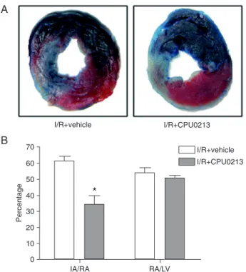
Infarct size was established by TTC/Evans blue double-staining method. Evans blue stains the myocardium blue, representing non-ischemic tissue, while the remaining area represents the risk area. TTC stains the myocardium red due to complete dehydrogenase activity, while the infarct area, lacking dehydrogenase activity, remains unstained (white; Figure 2A). CPU0213 pretreatment resulted in a significant
ETA and ETB receptor antagonist protects against myocardial I/R injury 1151
reduction of IA/RA compared to the I/R + vehicle group (I/R
+ vehicle: 61.3 ± 3.2 vs I/R + CPU0213: 34.0 ± 5.5%; P <
0.05), representing a 44.5% reduction in infarct size (Figure 2B). There was no significant difference in RA/LV, indicating a similar position of the coronary artery ligation.
Cardiac function
The left ventricular function and structure were assessed by echocardiography after 30-min ischemia and 6-h reperfusion. Rep-resentative M-mode echocardiographs of the three groups are shown in Figure 3A, B, and C. The echocardiograph revealed decreased EF and FS in the I/R + vehicle group com-pared to the Sham group. CPU0213 pretreat-ment increased EF and FS by 17.2% (I/R +
vehicle: 58.4 ± 2.8 vs I/R + CPU0213: 68.5
± 2.2%; P < 0.05) and 27.4% (I/R + vehicle:
26.6 ± 1.6 vs I/R + CPU0213: 33.9 ± 1.7%;
P < 0.01), respectively, compared to the I/R + vehicle group (Figure 3D), suggesting that CPU0213 improved myocardial contractile
function. There were no significant differ
-ences in other parameters between the I/R + vehicle group and the I/R + CPU0213 group (data not shown).
ET-1 levels in plasma, and CK and LDH activity in serum
Basal ET-1 levels in plasma of no I/R injury animals were low. After 30 min of ischemia and 6 h of reperfusion, ET-1 levels
were significantly elevated above basal lev
-els (Sham: 20.8 ± 2.1 vs I/R + vehicle: 34.1
± 4.4 pg/mL; P < 0.05). ET-1 levels were inhibited by CPU0213 (I/R + vehicle: 34.1 ±
4.4 vs I/R + CPU0213: 20.4 ± 2.8 pg/mL; P
< 0.05; Figure 4A).
Serum CK and LDH activities are ad-ditional markers of myocardial injury. Se-rum samples were obtained from the rats subjected to 30 min of ischemia and 6 h of reperfusion. CPU0213 pretreatment reduced CK and LDH activity compared to the I/R + vehicle group, indicating a protective role of CPU0213 (Figure 4B and C).
MDA levels in serum and MPO activity in the myocardium
MDA is a marker of lipid peroxidation, which is considered to be a major mecha-nism of tissue damage. To investigate whether CPU0213 had an effect on lipid peroxidation, we measured serum MDA levels. MDA levels were increased in the
I/R + vehicle group compared to the Sham group, but were reduced by 41.9% in the I/R + CPU0213 group (I/R
+ vehicle: 9.2 ± 1.3 vs I/R + CPU0213: 5.4 ± 0.5 nM; P <
0.05; Figure 5A), suggesting that CPU0213 can reduce lipid peroxidation.
We also determined MPO activity in ischemic myocardial tissues after 30 min of ischemia and 6 h of reperfusion. Rats
Figure 3. Cardiac function examined by echocardiography after ischemia-reper-fusion. Representative M-mode echocardiographs of Sham (A), I/R + vehicle (B), and I/R + CPU0213 groups (C). D, CPU0213 pretreatment increased ejection fraction (EF) and fractional shortening (FS) compared to the I/R + vehicle group. I/R = ischemia/reperfusion. Data are reported as means ± SEM for 7 rats/group.
#P < 0.001 vs Sham, *P < 0.05 vs I/R + vehicle (one-way ANOVA followed by the
Bonferroni post hoc test).
Figure 4. Endothelin-1 (ET-1) levels in plasma, and creatine kinase (CK) and lactate dehydrogenase (LDH) in serum after ischemia-reperfusion. A, ET-1 levels in plasma. B, Total CK activity in serum. C, Total LDH activity in serum. Data are reported as means ± SEM for 6 rats/group. #P < 0.05 vs Sham, *P < 0.05 vs I/R +
pretreated with CPU0213 had a 32.8% reduction in MPO activity compared to vehicle-treated rats (I/R + vehicle: 752.7
± 71.0 vs I/R + CPU0213: 505.9 ± 53.0
U/mg protein; P < 0.05; Figure 5B), suggesting that CPU0213 can inhibit neutrophil infiltration.
Akt and eNOS phosphorylation
To explore potential signal transduc-tion pathways activated by CPU0213, we examined the Akt/eNOS pathway since it has been reported that a
non-selective ETA/ETB antagonist increased
Akt and eNOS phosphorylation in the hearts of streptozotocin-induced dia-betic rats (17). We found a significant increase in the phosphorylation status of Akt in the ischemic myocardium in the I/R + CPU0213 group compared to the I/R + vehicle group (1.24-fold; Figure 6A). The phosphorylation status of Akt was suppressed by I/R, but was partly restored by CPU0123. A similar effect was observed with eNOS phosphoryla-tion (Figure 6B). CPU0213 pretreatment had no effect on total Akt or eNOS in the myocardium of all three groups. Thus, the data suggest a role for the Akt/eNOS pathway in mediating the effects of CPU0213.
Discussion
The major finding of the present study was that CPU0213 pretreatment immediately prior to coronary
occlu-sion significantly reduced myocardial infarct size, which is consistent with previous studies using other ET antagonists (7,10,11).
To further investigate the effect of CPU0213 on myo-cardial function, we assessed left ventricular function and structure by echocardiography. Interestingly, administration of CPU0123 improved left ventricular contractile function as indicated by a significant increase in EF and FS.
ET-1 is important for the modulation of cardiac contractil-ity. Both exogenous and endogenous ET-1 have been shown
to exert acute positive inotropic effects in vivo, which may be
mediated via ETA receptor activation (18,19). Furthermore,
the inotropic response to ET-1 was suggested to be biphasic
because of an overall positive inotropic effect of ETA
recep-tor stimulation and an ETB receptor-mediated decrease in
contractility at low ET-1 concentrations (20). In contrast,
we found that the ETA/ETB receptor antagonist CPU0213
increased contractile function compared to the I/R + vehicle
group. There are several possible reasons for this discrep-ancy. First, in our model, a large number of myocardial cells showed necrosis and apoptosis because of ischemia, and it is unclear whether ET-1 has positive inotropic effects under pathological conditions. Furthermore, even if ET-1 induces positive inotropy in surviving cardiomyocytes, this may be
insufficient to maintain left ventricular function. In the pres
-ent study, we found that CPU0213 pretreatm-ent decreased infarct size and attenuated cardiomyocyte injury following I/R, resulting in preservation of left ventricular contraction function. These data are consistent with a previous study
using a selective ETA antagonist (7). Administration of
bosentan, a non-selective ETA/ETB receptor antagonist,
after occlusion of the LAD, caused a significant decline in systolic and diastolic arterial pressure and heart rate dur-ing reperfusion (11). Thus, CPU0213 may improve cardiac function by reducing both the afterload and heart rate, which may also lead to decreased myocardial oxygen demand
Figure 5. Malondialdehyde (MDA) levels in serum and myeloperoxidase (MPO) activ-ity in myocardial tissues after ischemia-reperfusion. A, MDA levels in serum. B, MPO activity in myocardial tissues. Data are reported as means ± SEM for 6 rats/group. #P <
0.01 vs Sham, *P < 0.05 vs I/R + vehicle (one-way ANOVA followed by the Bonferroni post hoc test).
Figure 6. Representative Western blots showing (A) phospho-Akt (protein kinase B; Ser473) and total Akt and (B) phospho-eNOS (endothelial nitric oxide synthase; Ser1177) and total eNOS in ischemic myocardial tissues after ischemia-reperfusion. Quantitative densitometry data are reported as means ± SEM for 4 rats/group. #P < 0.05 vs Sham;
ETA and ETB receptor antagonist protects against myocardial I/R injury 1153
and attenuate myocardial injury during reperfusion. Although we demonstrated that CPU0213 resulted in an improvement of EF and FS, these improvements were not
as marked as the reduction in infarct size and the inflam
-matory parameters. There are two explanations: myocardial stunning is characterized by viable cardiomyocytes with depressed function after brief episodes of I/R (21) and early reperfusion during acute myocardial infarction results in a mixture of infarcted and stunned myocardium (22). This is confirmed by Figure 2 of our study, where there is evidence of live myocardium in the twilight zone between the infarct area and the non-ischemic area. The tissue salvaged by
reperfusion may require days or weeks to recover its con
-tractile function (23). As such, it may take more than 6 h to show clear improvement of EF and FS in the CPU0213-treated group. Furthermore, ET-1 is only one factor in the complex system of cardiac dysfunction after I/R, and there are also numerous alternative influences that can affect the recovery of cardiac function.
Numerous studies have demonstrated that plasma ET-1 levels increase after I/R (5,6). In the present study, plasma ET-1 levels also significantly increased after 30 min of ischemia and 6 h of reperfusion, while CPU0213 largely blocked this effect. This result is in contrast to previous
studies using other ETA/ETB receptor antagonists (24,25).
The ETB receptor is suggested to be responsible for
clear-ing circulatclear-ing ET-1, and therefore non-selective ETA/ETB
receptor antagonists block the ETB receptors and increase
plasma ET levels. By contrast, Feng et al. (15) showed that, in a model of L-thyroxin-induced cardiomyopathy, CPU0213 significantly inhibited elevated expression of ET-1 and dramatically decreased prepro-endothelin-1 (preproET-1) mRNA levels. Furthermore, in a model of septic shock, CPU0213 limited the up-regulation of endothelin convert-ing enzyme (ECE) and preproET-1 mRNA compared to the untreated septic shock group (26). The ET gene encodes preproET-1, which is cleaved into a precursor termed big ET. Big ET is secreted from endothelial cells and is con-verted to ET-1 by ECE (27,28). This may be explained by the improved hemodynamics and reduced ET-1 production following CPU0213. Verma et al. (29) also demonstrated that attenuating the release of ET-1 following I/R resulted
in enhanced cardiomyocyte tolerance to I/R in vitro.
There-fore, we measured total CK and LDH activity in serum and found that both CK and LDH activity increased after I/R, but decreased after pretreatment with CPU0213.
Oxidative stress is generally accepted to play an impor-tant role in myocardial I/R (30). In addition, oxidative stress can elevate ET-1 levels, and ET-1 has been demonstrated to increase the production of reactive oxygen species (ROS) (31). Although oxidative stress is an established
consequence of ETA receptor activation, a similar action of
the ETB receptor remains controversial (32,33). To
deter-mine the actions of the non-selective endothelin antagonist CPU0213 on oxidative stress, we measured serum levels
of MDA, the metabolite of lipid peroxidation (34), which reflects ROS production. Serum MDA levels increased in the I/R + vehicle group, but were markedly decreased in the I/R + CPU0213 group, indicating that CPU0213 can also reduce lipid peroxidation and oxidative stress, consistent with previous data (11).
Neutrophil accumulation and activation in cardiac tis-sues can damage the myocardium. During the period of reperfusion, neutrophils can rapidly plug microvessels
and capillaries, thus contributing to the “no-reflow” phe
-nomenon (35). Plasma ET-1 levels were found to predict angiographic no-reflow after successful primary or rescue PCI in patients with acute myocardial infarction (36). There-fore, we measured the peroxidase enzyme MPO found in neutrophils, reflecting neutrophil infiltration. MPO activity in the ischemic myocardium was decreased by CPU0213,
indicating that CPU0213 suppresses myocardial inflamma
-tion. Gonon et al.(37) reported a similar response with a
selective ETA receptor antagonist, LU 135252. Thus, the
anti-inflammation actions of CPU0213 may contribute to its cardioprotective action.
The relationship between ET-1 and the Akt/eNOS
path-way remains unclear. In an endothelial cell system, ETB
receptor activation led directly to Akt and eNOS activation (38). By contrast, administration of an endothelin receptor antagonist improved cardiac function by normalizing vascu-lar endothelial growth factor (VEGF) levels and increased the phosphorylation status of Akt and eNOS in hearts of streptozotocin-induced diabetic rats (11). We found that myocardial I/R blunted the phosphorylation status of Akt, which was partially restored by CPU0213. These data are inconsistent with a previous study (39), but may be ex-plained by the different durations of I/R. In that study, after 45 min of ischemia followed by 60 min of reperfusion, there were no differences in the phosphorylation status of Akt or eNOS between Sham, vehicle- and BQ123- (a selective
ETA receptor antagonist) treated groups. By contrast, we
showed that following 30-min ischemia and 6-h reperfusion, phospho-Akt and phospho-eNOS were impaired by I/R, but partly restored by CPU0213. Thus, it is possible that the short duration of reperfusion used by Hoshino et al. (39)
was insufficient to impair phosphorylation status. In agree
-ment with our data, non-selective ETA/ETB but not selective
ETA receptor antagonists improved endothelium-dependent
vasodilation in individuals with insulin resistance (40). These
data suggest that a non-selective ETA/ETB receptor
antago-nist may have additional benefits compared to a selective
ETA receptor antagonist. However, the exact mechanisms
the relationship between the Akt/eNOS pathway and non-selective endothelin antagonists in I/R.
We found that the non-selective ETA/ETB receptor
antagonist CPU0213 significantly attenuated myocardial I/R injury when administered immediately prior to coronary occlusion. CPU0213 treatment was associated with a de-crease in ET-1 levels, oxidative stress, and myocardial in-flammation. Moreover, CPU0213 improved the deterioration observed in the Akt/eNOS pathway following myocardial I/R
injury. Thus, CPU0213 may represent a promising strategy for the treatment of acute myocardial reperfusion.
Acknowledgments
We thank Professor D.Z. Dai for generously providing CPU0213 and Dr. Y.H. Yong for technical assistance with echocardiography. Research supported by grants from the National Science Foundation of China (#81070275).
References
1. Yanagisawa M, Kurihara H, Kimura S, Tomobe Y, Kobayashi M, Mitsui Y, et al. A novel potent vasoconstrictor peptide produced by vascular endothelial cells. Nature 1988; 332: 411-415.
2. Kedzierski RM, Yanagisawa M. Endothelin system: the double-edged sword in health and disease. Annu Rev Phar-macol Toxicol 2001; 41: 851-876.
3. Tirapelli CR, Casolari DA, Yogi A, Montezano AC, Tostes RC, Legros E, et al. Functional characterization and expression of endothelin receptors in rat carotid artery: involvement of nitric oxide, a vasodilator prostanoid and the opening of K+
channels in ETB-induced relaxation. Br J Pharmacol 2005; 146: 903-912.
4. Davie N, Haleen SJ, Upton PD, Polak JM, Yacoub MH, Morrell NW, et al. ET(A) and ET(B) receptors modulate the proliferation of human pulmonary artery smooth muscle cells. Am J Respir Crit Care Med 2002; 165: 398-405. 5. Han B, Ghanim D, Peleg A, Uretzky G, Hasin Y. Loss of
systemic endothelial function post-PCI. Acute Card Care 2008; 10: 79-87.
6. Vago H, Soos P, Zima E, Geller L, Keltai K, Roka A, et al. Changes of endothelin-1 and big endothelin-1 levels and action potential duration during myocardial ischemia-reperfusion in dogs with and without ventricular fibrillation. J Cardiovasc Pharmacol 2004; 44 (Suppl 1): S376-S379. 7. Goyal SN, Bharti S, Arora S, Golechha M, Arya DS.
En-dothelin receptor antagonist BQ-123 ameliorates myocar-dial ischemic-reperfusion injury in rats: a hemodynamic, biochemical, histopathological and electron microscopic evidence. Biomed Pharmacother 2010; 64: 639-646. 8. Eitel I, Nowak M, Stehl C, Adams V, Fuernau G, Hildebrand
L, et al. Endothelin-1 release in acute myocardial infarction as a predictor of long-term prognosis and no-reflow as -sessed by contrast-enhanced magnetic resonance imaging. Am Heart J 2010; 159: 882-890.
9. Climent B, Fernandez N, Garcia-Villalon AL, Monge L, San-chez A, Dieguez G. Effects of antagonists for endothelin ET(A) and ET(B) receptors on coronary endothelial and myocardial function after ischemia-reperfusion in anesthe-tized goats. Vascul Pharmacol 2006; 44: 384-390.
10. Ozdemir R, Parlakpinar H, Polat A, Colak C, Ermis N, Acet A. Selective endothelin a (ETA) receptor antagonist (BQ-123) reduces both myocardial infarct size and oxidant injury. Toxicology 2006; 219: 142-149.
11. Singh AD, Amit S, Kumar OS, Rajan M, Mukesh N. Cardio-protective effects of bosentan, a mixed endothelin type A and B receptor antagonist, during myocardial ischaemia and
reperfusion in rats. Basic Clin Pharmacol Toxicol 2006; 98: 604-610.
12. Richard V, Kaeffer N, Hogie M, Tron C, Blanc T, Thuillez C. Role of endogenous endothelin in myocardial and coronary endothelial injury after ischaemia and reperfusion in rats: studies with bosentan, a mixed ETA-ETB antagonist. Br J Pharmacol 1994; 113: 869-876.
13. Bohm F, Pernow J. The importance of endothelin-1 for vas-cular dysfunction in cardiovasvas-cular disease. Cardiovasc Res 2007; 76: 8-18.
14. Qi MY, Xia HJ, Dai DZ, Dai Y. A novel endothelin receptor antagonist CPU0213 improves diabetic cardiac insufficiency attributed to up-regulation of the expression of FKBP12.6, SERCA2a, and PLB in rats. J Cardiovasc Pharmacol 2006; 47: 729-735.
15. Feng Y, Dai DZ, Na T, Cui B, Wang T, Zhang Y, et al. Endothelin receptor antagonist CPU0213 suppresses ven-tricular fibrillation in L-thyroxin induced cardiomyopathy. Pharmacol Rep 2007; 59: 306-314.
16. Yip HK, Wu CJ, Chang HW, Yang CH, Yu TH, Chen YH, et al. Prognostic value of circulating levels of endothelin-1 in pa-tients after acute myocardial infarction undergoing primary coronary angioplasty. Chest 2005; 127: 1491-1497. 17. Jesmin S, Zaedi S, Shimojo N, Iemitsu M, Masuzawa K,
Yamaguchi N, et al. Endothelin antagonism normalizes VEGF signaling and cardiac function in STZ-induced dia-betic rat hearts. Am J Physiol Endocrinol Metab 2007; 292: E1030-E1040.
18. Konrad D, Oldner A, Wanecek M, Rudehill A, Weitzberg E, Biber B, et al. Positive inotropic and negative lusitropic ef-fects of endothelin receptor agonism in vivo. Am J Physiol Heart Circ Physiol 2005; 289: H1702-H1709.
19. Eszlari E, Czobel M, Molnar G, Bogats G, Kaszaki J, Nagy S, et al. Modulation of cardiac contractility through endothe-lin-1 release and myocardial mast cell degranulation. Acta Physiol Hung 2008; 95: 267-285.
20. Qi XL, Sia YT, Stewart DJ, Wei G, Nguyen QT, Cernacek P, et al. Myocardial contractile responsiveness to endothelin-1 in the post-infarction rat model of heart failure: effects of chronic quinapril. J Mol Cell Cardiol 2001; 33: 2023-2035. 21. Braunwald E, Kloner RA. The stunned myocardium:
pro-longed, postischemic ventricular dysfunction. Circulation 1982; 66: 1146-1149.
22. Jennings RB, Reimer KA. Factors involved in salvaging ischemic myocardium: effect of reperfusion of arterial blood. Circulation 1983; 68: I-25-I-36.
myo-ETA and ETB receptor antagonist protects against myocardial I/R injury 1155
cardial function by coronary artery reperfusion 1, 2, and 3 hours after occlusion in conscious dogs. Circ Res 1983; 53: 235-247.
24. Brunner F, Leonhard B, Kukovetz WR, Mayer B. Role of endothelin, nitric oxide and L-arginine release in ischaemia/ reperfusion injury of rat heart. Cardiovasc Res 1997; 36: 60-66.
25. Rossi P, Wanecek M, Konrad D, Oldner A. Tezosentan coun-teracts endotoxin-induced pulmonary edema and improves gas exchange. Shock 2004; 21: 543-548.
26. He HB, Dai DZ, Dai Y. CPU0213, a novel endothelin receptor antagonist, ameliorates septic renal lesion by suppressing ET system and NF-kappaB in rats. Acta Pharmacol Sin 2006; 27: 1213-1221.
27. Yanagisawa M, Inoue A, Ishikawa T, Kasuya Y, Kimura S, Kumagaye S, et al. Primary structure, synthesis, and biological activity of rat endothelin, an endothelium-derived vasoconstrictor peptide. Proc Natl Acad Sci U S A 1988; 85: 6964-6967.
28. Sawamura T, Kimura S, Shinmi O, Sugita Y, Kobayashi M, Mitsui Y, et al. Characterization of endothelin converting enzyme activities in soluble fraction of bovine cultured en-dothelial cells. Biochem Biophys Res Commun 1990; 169: 1138-1144.
29. Verma S, Maitland A, Weisel RD, Li SH, Fedak PW, Pomroy NC, et al. Hyperglycemia exaggerates ischemia-reperfu-sion-induced cardiomyocyte injury: reversal with endothelin antagonism. J Thorac Cardiovasc Surg 2002; 123: 1120-1124.
30. Maulik N, Yoshida T, Das DK. Oxidative stress developed during the reperfusion of ischemic myocardium induces apoptosis. Free Radic Biol Med 1998; 24: 869-875. 31. Loomis ED, Sullivan JC, Osmond DA, Pollock DM,
Pol-lock JS. Endothelin mediates superoxide production and vasoconstriction through activation of NADPH oxidase and uncoupled nitric-oxide synthase in the rat aorta. J Pharmacol Exp Ther 2005; 315: 1058-1064.
32. Cheng TH, Shih NL, Chen SY, Wang DL, Chen JJ. Reactive
oxygen species modulate endothelin-I-induced c-fos gene expression in cardiomyocytes. Cardiovasc Res 1999; 41: 654-662.
33. Dong F, Zhang X, Wold LE, Ren Q, Zhang Z, Ren J. En-dothelin-1 enhances oxidative stress, cell proliferation and reduces apoptosis in human umbilical vein endothelial cells: role of ETB receptor, NADPH oxidase and caveolin-1. Br J Pharmacol 2005; 145: 323-333.
34. Gao F, Yao CL, Gao E, Mo QZ, Yan WL, McLaughlin R, et al. Enhancement of glutathione cardioprotection by ascorbic acid in myocardial reperfusion injury. J Pharmacol Exp Ther 2002; 301: 543-550.
35. von Dobschuetz E, Hoffmann T, Messmer K. Inhibition of neutrophil proteinases by recombinant serpin Lex032 reduces capillary no-reflow in ischemia/reperfusion-induced acute pancreatitis. J Pharmacol Exp Ther 1999; 290: 782-788. 36. Niccoli G, Lanza GA, Shaw S, Romagnoli E, Gioia D,
Bur-zotta F, et al. Endothelin-1 and acute myocardial infarction: a no-reflow mediator after successful percutaneous myocar -dial revascularization. Eur Heart J 2006; 27: 1793-1798. 37. Gonon AT, Gourine AV, Middelveld RJ, Alving K, Pernow J.
Limitation of infarct size and attenuation of myeloperoxidase activity by an endothelin A receptor antagonist following ischaemia and reperfusion. Basic Res Cardiol 2001; 96: 454-462.
38. Liu S, Premont RT, Kontos CD, Huang J, Rockey DC. En-dothelin-1 activates endothelial cell nitric-oxide synthase via heterotrimeric G-protein betagamma subunit signaling to protein jinase B/Akt. J Biol Chem 2003; 278: 49929-49935. 39. Hoshino S, Kikuchi Y, Nakajima M, Kimura H, Tsuyama S,
Uemura K, et al. Endothelial NO synthase (eNOS) phos-phorylation regulates coronary diameter during ischemia-reperfusion in association with oxidative stress. Free Radic Res 2005; 39: 481-489.