Whole-Body Vibration Training Increases Myocardial Salvage Against
Acute Ischemia in Adult Male Rats
Shahnaz Shekarforoush
1and Mohammad Reza Naghii
2 Islamic Azad University,1 Arsanjan Branch, Fars – IranSport Physiology Research Center, Baqiyatallah University of Medical Sciences,2 Teerã – Iran
Mailing Address: Shahnaz •
Arsanjan - University Blv. Arsanjan Branch, Islamic Azad University, 7188855989, Shiraz, Fars – Iran
E-mail: sh.shekar@yahoo.com, shek@iaua.ac.ir
Manuscript received March 20, 2018, revised manuscript June 19, 2018, accepted July 23, 2018
DOI: 10.5935/abc.20180252
Abstract
Background: Whole body vibration training (WBV) is a new training program, which is safe and effective. It can be followed by the public. However, data on the safety and efficacy of vibration on myocardial ischemia reperfusion (IR) injury are lacking.
Objective: To examine the effect of WBV on the tolerance of the myocardium to acute IR injury in an experimental rat model.
Methods: Twenty-four male Wistar rats were divided into control and vibration groups. Vibration training consisted of vertical sinusoidal whole body vibration for 30 min per day, 6 days per week, for 1 or 3 weeks (WBV1 and WBV3 groups, respectively). All the rats were submitted to myocardial IR injury. Myocardial infarct size and ischemia-induced arrhythmias were assessed. Differences between variables were considered significant when p < 0.05.
Results:No differences were observed between the groups regarding the baseline hemodynamic parameters. Infarct
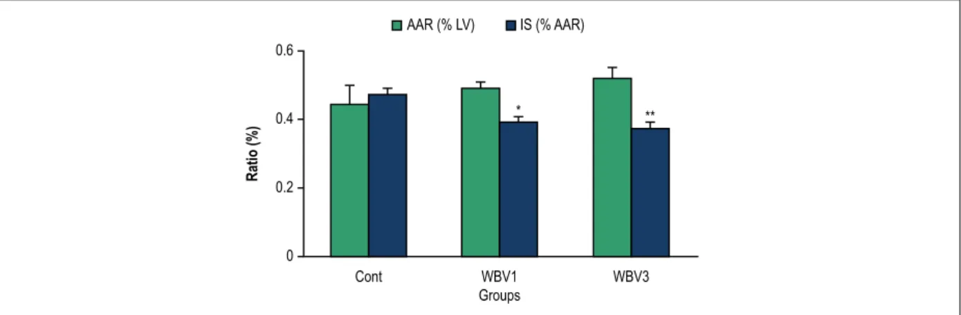
size was smaller in the experimental group (control, 47 ± 2%; WBV1, 39 ± 2%; WBV3, 37 ± 2%; p < 0.05, vs. control). Vibration produced a significant decrease in the number and duration of ventricular tachycardia (VT) episodes compared to the control value. All ventricular fibrillation (VF) episodes in the vibration groups were self-limited, while 33% of the rats in the control group died due to irreversible VF (p = 0.02).
Conclusion: The data showed that vibration training significantly increased cardiac tolerance to IR injury in rats, as evidenced by reduction in the infarct size and cardiac arrhythmias, and by facilitating spontaneous defibrillation. (Arq Bras Cardiol. 2018; [online].ahead print, PP.0-0)
Keywords: Rats Wistar; Body Composition; Vibration; Osteoporosis/prevention and control; Blood Viscosity; Ischemia; Cardiovascular Diseases; Ischemic Preconditioning.
Introduction
Whole body vibration training (WBV) has been recently proposed as an exercise training method with a potential for improving body composition and preventing osteoporosis and bone mass loss.1 In recent years, some studies have
shown that WBV may be a beneficial training mode in patients with multiple sclerosis,2 type 2 diabetes,3 chronic
obstructive pulmonary disease,4 and heart transplant
recipients.5 The effects of WVB on the cardiovascular
system were investigated in a number of published studies. Decreased arterial stiffness after WBV can reduce the risk of cardiovascular disease.6,7 An experiment conducted by
Robbins et al.8 showed a significant increase in blood flow
velocity with no significant changes in heart rate, blood pressure or peripheral skin temperature. Increased muscle
blood volume and blood flow velocity after vibration exercise were attributed mainly to the effect of vibrations in reducing blood viscosity and increasing its velocity through the arteries.9 These findings indicate that WBV may represent a
mild form of exercise for the cardiovascular system.10
Cardiovascular disease (CVD), which is induced by ischemia, is the leading cause of death worldwide. Restoration of blood flow, after a period of ischemia, can elicit pathological processes that exacerbate injury due to the ischemia itself.11
Preconditioning describes a pretreatment or premaneuver that is able to adapt the myocardium to ischemic stress. We have demonstrated some preconditioning interventions in previous experiments, reducing infarct size and arrhythmias.12,13
The cardioprotective effect of exercise preconditioning was reported as a reduction in infarct size in previous studies.14,15
Methods
Male Wistar rats weighing 250 to 300 g (10-12 weeks old) were obtained from the animal house of Shiraz University of Medical Sciences and housed under standard conditions, with free access to food and water. The investigation was approved by the University Ethics Committee in accordance with the Guide for the Care and Use of Laboratory Animals.
Experimental designs
A total of 24 rats were randomly assigned to 1 of 3 treatment groups (control vs. two experimental groups) by picking numbers out of a hat. The sample size (n) was established based on studies that evaluated the effects of exercise against myocardial IR injury.16,17 Animals in the vibration groups were
placed in a compartment attached to a vibration platform (Crazy Fit Massager/Model: YD 1002, Union Brilliant Group Co., LTD, Fujian, China). The vibration training consisted of a 5-min cycle on day 1, followed by an extra 5-min cycle each time for the next five sessions in the first week and then each rat was exposed to vertical sinusoidal vibration for 30 min per session (3 × 10 min cycles), 6 days a week for one week (WBV1 group) or 3 weeks (WBV3 group). The animals were given 1–2 min rest break between the cycles. The vibration was performed at mode 1 with amplitude of 1–10 mm and at a frequency of 10–50 Hz. The speed of mode 1 in each cycle increased gradually and then decreased with the same trend within each time period. The control animals remained in their cages and were placed over the vibration platform, without vibration treatment. Each training session was performed between 8.30–10.00 A.M.18
Surgical procedure
The protocol used has been thoroughly described in detail in our previous publication.12 Briefly, 24 hours after
the last training session, the animals were anesthetized and ventilated with room air enriched with oxygen at a rate of 70 breaths per min. A standard limb lead II electrocardiogram was monitored and recorded throughout the experiment. Catheters were inserted into the left carotid artery and tail vein for monitoring of blood pressure and infusion of Evans blue solution, respectively. After the thoracotomy, a 6-0 silk suture was passed around the left anterior descending coronary artery (LAD). Following a stabilization period of 20 min, the LAD was occluded for 30 min of ischemia and released for 120 min of reperfusion. Rectal temperature was continuously monitored and maintained at 37 ± 0.5°C.
Determination of infarct size and area at risk
At the end of reperfusion, the LAD was reoccluded and 1 mL of 2% solution of Evans Blue dye (Sigma, St. Louis, MO) was injected into the tail vein to identify the non-perfused area, also known as area at risk (AAR), from the perfused area. The rats were then killed with a pentobarbital overdose and their hearts were excised and frozen for one hour. The atria and right ventricle were removed, and the left ventricle was cut into transverse slices of 2 mm thickness from the apex to the
base. Tissue samples were then incubated with a 1% solution of 2,3,5 triphenyltetrazolium chloride (Sigma)] for 20 min at 37°C, and subsequently fixed in 10% phosphate-buffered formalin for one hour. Viable myocardium was stained red by triphenyltetrazolium chloride, whereas necrotic myocardium appeared as pale yellow. In each slice, areas at risk and infarcted areas were determined by computerized planimetry using an image analysis software (Image Tool, University of Texas, San Antonio, TX). Infarct size (IS) was expressed as percentage of the AAR (IS/AAR).12
Assessment of ventricular arrhythmias
Ischemia-induced ventricular arrhythmias were determined in accordance with the Lambeth conventions19 including
ventricular ectopic beat as premature ventricular complexes (PVC), ventricular tachycardia (VT) as a run of four or more consecutive ventricular premature beats at a rate faster than the resting sinus rate, and ventricular fibrillation (VF) as a signal for which individual QRS deflection can no longer be distinguished from one another. Complex forms (bigeminy and salvos) were added to PVC count and not analyzed separately. In order to determine the incidence of VT and VF, they were recorded as either occurring or not occurring during the first 30 min of ischemia in each group.
Statistical analyses
Unless stated otherwise, the results were expressed as Mean ± SD. All data were processed with the SPSS 16.0 statistical package for Windows version. The normality of distributions was verified by the Kolmogorov-Smirnov test. Fisher exact test (Chi-square) was used to analyze the incidence of VT and VF. Analysis of baseline, ischemia, and reperfusion HR and BP was done by repeated measures analysis of variance (ANOVA). The other data were analyzed using one-way ANOVA and then significant differences were examined by Tukey’s post-hoc test. Differences between the groups were considered significant at a level of p < 0.05.
Results
Hemodynamic parameters
Table 1 summarizes the hemodynamic data. There were no significant differences at baseline values for heart rate (HR) and mean arterial blood pressure (MBP) among the groups. Ischemia caused a marked reduction in blood pressure without any significant effect on the HR in the groups. MBP was nearly restored to the baseline level during the reperfusion period.
Infarct size
Figure 1 – Infarct size (IS) and area at risk (AAR) following 30-min of ischemia and 120-min of reperfusion in rats. LV: left ventricle; * p < 0.05 and ** p < 0.01 compared with the control group. Cont: control; WBV1: whole body vibration training for one week; WBV3: whole body vibration training for 3 weeks.
0.6
0.4
0.2
0
Cont WBV1
Groups
WBV3
Ratio (%)
AAR (% LV) IS (% AAR)
* **
Table1 – Hemodynamics parameters in the experimental groups
Group Baseline Ischemia Reperfusion
HR MBP HR MBP HR MBP
Cont 346 ± 48 113 ± 21 349 ± 51 100 ± 15* (0.04) 361 ± 44 107 ± 17
WBV1 373 ± 41 114 ± 7 385 ± 25 97 ± 8** (0.001) 383 ± 26 107 ± 13
WBV3 376 ± 26 107 ± 15 372 ± 18 91 ± 7* (0.01) 378 ± 27 108 ± 15
p-value 0.282 0.670 0.130 0.267 0.399 0.996
Note: Data presented as mean ± SD (P-value). HR: heart rate; MBP: mean arterial blood pressure. Cont: control, WBV1: whole body vibration training for one week, WBV3 = whole body vibration training for 3 weeks. *p < 0.05, **p < 0.01 compared to baseline value.
Ischemia-induced arrhythmias
Table 2 represents the number of PVC, VT and VF episodes and their duration during the 30-min ischemic period. The arrhythmias occurred after approximately 5–7 min of ischemia. The number of PVC decreased non-significantly in the experimental groups (p = 0.702). Vibration produced a significant decrease in the number and duration of VT episodes compared to the control value. The mean duration of reversible VF in the WBV3 group was reduced from 32.3 ± 19.4 s in the control group to 13.7 ± 10.3 s (as a non-significant trend). Although the longest VF episodes in the vibration groups lasted as much as 116 s, all VF episodes were self-limited. However, the longest observed non-fatal VF episode in the control group was 87 s. and 33% of the rats died due to irreversible VF (p = 0.02). The occurrence (% incidence per group) of VT during the 30-min ischemia was 100, 100 and 88% (p = 0.35) and the occurrence of VF was 75, 63 and 50% in the control, WBV1 and WBV3 groups, respectively (p = 0.58).
The numbers of premature ventricular complexes (PVC), the ventricular tachycardia (VT) and ventricular fibrillation (VF) episodes and duration are shown as means ± SEM. * p < 0.05 and ** p < 0.01 compared with the control group.
Cont: control; WBV1: whole body vibration training for one week; WBV3: whole body vibration training for 3 weeks.
Discussion
There are three main findings of the present study. First, WBV caused a significant decrease in IS following 30 min of ischemia and 120 min of reperfusion. Second, WBV had a protective effect on ischemia-induced arrhythmia. Third, all VF episodes were self-limited in the vibration groups, so the vibration improved arrhythmia-related mortality.
There are conflicting results regarding the effect of WBV on BP and HR. Performing dynamic exercise on a vertical vibration platform (30-35 Hz, 2 mm) for 12 weeks resulted in decreased systolic blood pressure in patients suffering from type 2 diabetes.20 Figueroa et al.’s study showed that 6 weeks
of WBV decreased systemic arterial stiffness and systolic blood pressure in young overweight/obese normotensive women.7
Unlike these results, it was demonstrated that one session of exercise with vibration increased systolic and diastolic blood pressure and stroke volume compared with exercise with no vibration in sedentary adults.21 In contrast, some
researchers have reported that WBV had no effect on the systolic and diastolic blood pressure, which is similar to our study results.6,8,22 These conflicting results may be explained
Table2 – Incidence and duration of ventricular arrhythmias during 30 min of ischemia
Groups PVC (n) VT VF
Episodes Duration Episodes Duration
Cont 283 ± 50 31 ± 4 70 ± 14 2.0 ± 0.9 32.3 ± 19.4
WBV1 271 ± 32 17 ± 1* 54 ± 19 2.3 ± 0.8 33.2 ± 17.9
WBV3 229 ± 55 13 ± 3** 12 ± 4* 1.0 ± 0.6 13.7 ± 10.3
p-value 0.702 0.002 0.018 0.559 0.475
PVC: premature ventricular complexes; VT: ventricular tachycardia; VF: ventricular fibrillation; WBV1: whole body vibration training for one week; WBV3: whole body vibration training for 3 weeks
The ischemia reperfusion model in the experimental animals provides an option to evaluate the occurrence of ischemia-induced arrhythmias and infarct size after an intervention. Posa et al. demonstrated that 6 weeks of voluntary exercise was protective against IR injury by reducing the myocardial infarct size.23 An important finding is that
one-to-several days of exercise can also reduce myocardial damage due to IR injury.24 Studies have demonstrated
that regular exercise increases antioxidant capacity in the heart, which can minimize oxidative stress following IR.25
During all sporting activities, externally-applied forces induce vibrations within the body tissues.10 WBV has been proposed as
an efficient alternative to moderate intensity exercise.26
Although recent studies have suggested that WBV leads to improvements in numerous health outcomes, including bone mineral density,27 muscle strength, or cardiovascular
fitness,28 no research has been performed so far to evaluate
the effects on IR injury.
The present study demonstrated that WBV is able to reduce myocardial infarct size and ischemia-induced arrhythmia during IR injury in rats. In the course of myocardial infarction, ventricular arrhythmias such as VT and VF are the most important cause of mortality.29 There was no difference in the ratio of AAR/LV between
the control and vibration animals, indicating thatall animals suffered a comparable degree of ischemic area. Therefore, the reductionof infarct size and arrhythmia in vibration-treatedanimals was due to the effect of the training. There are two types of VF: a sustained VF (SVF) that never terminates spontaneously and requires electrical defibrillation and a transient VF (TVF) that terminates by itself and spontaneously reverts into a sinus rhythm. Although it was believed for many years that TVF appears only in small mammals (rats, guinea pigs and rabbits), no differences were found in cardiac muscle mass, heart rate and action potential duration between animals with TVF and those with SVF. Intercellular uncoupling during ischemia most likely due to an increase in the intracellular Ca2+ and H+ ions or a decrease in the intracellular cAMP may
lead to SVF. Therefore, any defibrillating intervention should prevent intercellular uncoupling, most probably by increasing the intracellular concentration of cAMP, decreasing elevated [Ca2+]
i or preventing Ca
2+ overload.30 The results of the present
study suggested that all VF episodes were self-limited in the vibration groups. Thus, vibration training could reduce the risk of sudden death during ischemia, through both attenuation of the ischemia-induced arrhythmia and facilitation of spontaneous defibrillation. The exact mechanism of action by which vibration reduces the incidence of fatal VF episodes cannot be directly
fibrillation threshold in trained hearts during acute regional ischemia was shown in previous studies.31 Additionally, exercise
training has been reported to increase the levels of cAMP32 and
to improve cardiomyocyte function and diastolic Ca2+ control
in rats with post-infarction heart failure.33,34 Several studies have
also shown a positive correlation between infarct size and the occurrence of severe ventricular arrhythmias.35,36
Currently, exercise training has been introduced as the only practical method of providing cardioprotection against IR injury. If vibration-induced protection is nearly as effective as the exercise, it could be an alternative to exercise training, especially for those who are unable to perform traditional exercises. Delineating the mechanisms mediating vibration-induced protection against IR injury is important and could lead to the development of pharmacological or molecular approaches against cardiovascular diseases.
Limitations of the study
One of the limitations of the present study is that it was carried out on rats. Even though the large number of animal studies have conducted and contributed much to our understanding of disease mechanisms, their findings for predicting the effectiveness of strategies in humans has remained controversial.37,38 Therefore, the results need to be
confirmed by clinical trials in the future.
The frequency, amplitude, and the time of exposure of the subjects to vibration are important variations in clinical and experimental trials. However, due to lack of knowledge regarding optimum training protocols, the method was based on the methodology available in our laboratory. The proposed method has shown that is effective in improving health status by influencing cardiovascular disease (CVD) risk factors.18,39
We recommend evaluating the various vibration regimes on the IR injury in future studies.
Conclusions
1. Gilsanz V, Wren TA, Sanchez M, Dorey F, Judex S, Rubin C. Low-level, high-frequency mechanical signals enhance musculoskeletal development of young women with low BMD. J Bone Miner Res. 2006;21(9):1464-74.
2. Santos-Filho SD, Cameron MH, Bernardo-Filho M. Benefits of whole-body vibration with an oscillating platform for people with multiple sclerosis: a systematic review. Mult Scler Int. 2012;2012:274728.
3. Behboudi L, Azarbayjani MA, Aghaalinejad H, Salavati M. Effects of aerobic exercise and whole body vibration on glycaemia control in type 2 diabetic males. Asian J Sports Med. 2011;2(2):83-90.
4. Gloeckl R, Heinzelmann I, Kenn K. Whole body vibration training in patients with COPD: a systematic review. Chron Respir Dis. 2015;12(3):212-21.
5. Crevenna R, Fialka-Moser V, Rödler S, Keilani M, Zöch C, Nuhr M, et al. Safety of whole-body vibration exercise for heart transplant recipients. Phys Rehab Kur Med. 2003;13(5):286-90.
6. Lai CL, Chen HY, Tseng SY, Liao WC, Liu BT, Lee MC, et al. Effect of whole-body vibration for 3 months on arterial stiffness in the middle-aged and elderly. Clin Interv Aging. 2014 May 12;9:821-8.
7. Figueroa A, Gil R, Wong A, Hooshmand S, Park SV, Vicil F, et al. Whole-body vibration training reduces arterial stiffness, blood pressure and sympathovagal balance in young overweight/obese women. Hypertens Res. 2012;35(6):667-72.
8. Robbins D, Yoganathan P, Goss-Sampson M. The influence of whole body vibration on the central and peripheral cardiovascular system. Clin Physiol Funct Imaging. 2014;34(5):364-69.
9. Kerschan-Schindl K, Grampp S, Henk C, Resch H, Preisinger E, Fialka-Moser V, et al. Whole-body vibration exercise leads to alterations in muscle blood volume. Clin Physiol. 2001;21(3):377-82.
10. Cardinale M, Wakeling J. Whole body vibration exercise: are vibrations good for you? Br J Sports Med. 2005;39(9):585-9.
11. Kalogeris T, Baines CP, Krenz M, Korthuis RJ. Cell biology of ischemia/ reperfusion injury. Int Rev Cell Mol Biol. 2012;298:229-317.
12. Shekarforoush S, Foadoddini M. Cardiac effects of cupping: myocardial infarction, arrhythmias, heart rate and mean arterial blood pressure in the rat heart. Chin J Physiol. 2012;55(4):253-8.
13. Shekarforoush S, Safari F. Lactation protects against myocardial ischemia-reperfusion injury in rats. Acta Physiol Hung. 2015;102(4):372-9.
14. Kavazis AN. Exercise preconditioning of the myocardium. Sports Med. 2009;39(11):923-35.
15. Parra VM, Macho P, Sanchez G, Donoso P, Domenech RJ. Exercise preconditioning of myocardial infarct size in dogs is triggered by calcium. J Cardiovasc Pharmacol. 2015;65(3):276-81.
16. Brown DA, Lynch JM, Armstrong CJ, Caruso NM, Ehlers LB, Johnson MS, et al. Susceptibility of the heart to ischaemia-reperfusion injury and exercise-induced cardioprotection are sex-dependent in the rat. J Physiol. 2005;564(Pt 2):619-30.
17. Venditti P, Masullo P, Di Meo S, Agnisola C. Effects of prolonged aerobic exercise on myocardial responses to ischaemia-reperfusion in the rat. Exp Physiol. 2001;86(3):341-8.
18. Naghii MR, Hedayati M. Whole body vibration as a safe exercise training method induces no impaired alterations on rat plasma antioxidant biomarkers. Acta Physiol Hung. 2013;100(3):321-8.
19. Walker MJ, Curtis MJ, Hearse DJ, Campbell RW, Janse MJ, Yellon DM, et al. The Lambeth Conventions: guidelines for the study of arrhythmias in ischaemia infarction, and reperfusion. Cardiovasc Res. 1988;22(7):447-55.
20. Baum K, Votteler T, Schiab J. Efficiency of vibration exercise for glycemic control in type 2 diabetes patients. Int J Med Sci. 2007;4(3):159-63.
21. Dias T, Polito M. Acute cardiovascular response during resistance exercise with whole-body vibration in sedentary subjects: a randomized cross-over trial. Res Sports Med. 2015;23(3):253-64.
22. Salari NM, Movaseghi F. Effect of Whole Body Vibration Training on Blood Pressure and Heart Rate in Iranian Inactive Middle-aged Women. Int J Sport Stud. 2014;4(7):756-9.
23. Posa A, Szabo R, Kupai K, Barath Z, Szalai Z, Csonka A, et al. Cardioprotective effects of voluntary exercise in a rat model: role of matrix metalloproteinase-2. Oxid Med Cell Longev. 2015;2015:876805.
24. Quindry JC, Hamilton KL. Exercise and cardiac preconditioning against ischemia reperfusion injury. Curr Cardiol Rev. 2013;9(3):220-9.
25. Tao L, Bei Y, Zhang H, Xiao J, Li X. Exercise for the heart: signaling pathways. Oncotarget. 2015;6(25):20773-84.
26. Maddalozzo GF, Iwaniec UT, Turner RT, Rosen CJ, Widrick JJ. Whole-body vibration slows the acquisition of fat in mature female rats. Int J Obes (Lond). 2008;32(9):1348-54.
References
Acknowledgment
The present paper was financially supported by Arsanjan Branch, Islamic Azad University.
Author contributions
Conception and design of the research, writing of the manuscript and critical revision of the manuscript for intellectual content: Shekarforoush S, Naghii MR; acquisition of data, analysis and interpretation of the data, statistical analysis and obtaining funding: Shekarforoush S.
Potential Conflict of Interest
No potential conflict of interest relevant to this article was reported.
Sources of Funding
There were no external funding sources for this study.
Study Association
This study is not associated with any thesis or dissertation work.
Ethics approval and consent to participate
27. Gusi N, Raimundo A, Leal A. Low-frequency vibratory exercise reduces the risk of bone fracture more than walking: a randomized controlled trial. BMC Musculoskelet Disord. 2006; Nov 30;7:92.
28. Bogaerts AC, Delecluse C, Claessens AL, Troosters T, Boonen S, Verschueren SM. Effects of whole body vibration training on cardiorespiratory fitness and muscle strength in older individuals (a 1-year randomised controlled trial). Age Ageing. 2009;38(4):448-54.
29. Longo D, Fauci A, Kasper D, Hauser S. Harrison’s principles of internal medicine. 18th ed. New York: The McGraw- Hill; 2011.
30. Tribulova N, Manoach M. Factors determining spontaneous ventricular defibrillation. Exp Clin Cardiol. 2001;6(2):109-13.
31. Noakes TD, Higginson L, Opie LH. Physical training increases ventricular fibrillation thresholds of isolated rat hearts during normoxia, hypoxia and regional ischemia. Circulation. 1983;67(1):24-30.
32. Ray Hamidie RD, Yamada T, Ishizawa R, Saito Y, Masuda K. Curcumin treatment enhances the effect of exercise on mitochondrial biogenesis in skeletal muscle by increasing cAMP levels. Metabolism. 2015;64(10):1334-47.
33. Kemi OJ, MacQuaide N, Hoydal MA, Ellingsen O, Smith GL, Wisloff U. Exercise training corrects control of spontaneous calcium waves in hearts from myocardial infarction heart failure rats. J Cell Physiol. 2012;227(1):20-6.
34. Johnsen AB, Hoydal M, Rosbjorgen R, Stolen T, Wisloff U. Aerobic interval training partly reverse contractile dysfunction and impaired Ca2+ handling in atrial myocytes from rats with post infarction heart failure. PLoS One. 2013;8(6):e66288.
35. Grande P, Pedersen A. Myocardial infarct size: correlation with cardiac arrhythmias and sudden death. Eur Heart J. 1984;5(8):622-7.
36. Herlitz J, Hjalmarson A, Swedberg K, Waagstein F, Holmberg S, Waldenstrom J. Relationship between infarct size and incidence of severe ventricular arrhythmias in a double-blind trial with metoprolol in acute myocardial infarction. Int J Cardiol. 1984;6(1):47-60.
37. van der Worp HB, Howells DW, Sena ES, Porritt MJ, Rewell S, O’Collins V, et al. Can animal models of disease reliably inform human studies? PLoS Med. 2010;7(3):e1000245.
38. Leong XF, Ng CY, Jaarin K. Animal models in cardiovascular research: hypertension and atherosclerosis. Biomed Res Int. 2015;2015:528757.
39. Naghii MR, Darvishi P, Ebrahimpour Y, Ghanizadeh G, Mofid M, Hedayati M, et al. Effect of combination therapy of fatty acids, calcium, vitamin D and boron with regular physical activity on cardiovascular risk factors in rat. J Oleo Sci. 2012;61(2):103-11.