BIOMEDICAL SCIENCES
AND
CLINICAL INVESTIGATION
www.bjournal.com.br
www.bjournal.com.br
Braz J Med Biol Res, January 2010, Volume 43(1) 25-35
Expression of beta 2 integrin (CD18) in embryonic mouse and
chicken heart
L.A.M. Oliveira, R.K. Baker, S.E. Klewer and G.T. Kitten
Institutional Sponsors
Expression of beta 2 integrin (CD18) in
embryonic mouse and chicken heart
L.A.M. Oliveira
1, R.K. Baker
2, S.E. Klewer
3and G.T. Kitten
11Departamento de Morfologia, Instituto de Ciências Biológicas,
Universidade Federal de Minas Gerais, Belo Horizonte, MG, Brasil
2Departments of Cell Biology and Anatomy, 3Department of Pediatrics, Steele Children’s Research Center,
University of Arizona School of Medicine, Tucson, AZ, USA
Abstract
Integrins are heterodimeric receptors composed of α and β transmembrane subunits that mediate attachment of cells to the extracellular matrix and counter-ligands such as ICAM-1 on adjacent cells. β2 integrin (CD18) associates with four different α
(CD11) subunits to form an integrin subfamily, which has been reported to be expressed exclusively on leukocytes. However,
recent studies indicate that β2 integrin is also expressed by other types of cells. Since the gene for β2 integrin is located in
the region of human chromosome 21 associated with congenital heart defects, we postulated that it may be expressed in the developing heart. Here, we show the results from several different techniques used to test this hypothesis. PCR analyses
indi-cated that β2 integrin and the αL, αM, and αX subunits are expressed during heart development. Immunohistochemical studies in both embryonic mouse and chicken hearts, using antibodies directed against the N- or C-terminal of β2 integrin or against its α subunit partners, showed that β2 integrin, as well as the αL, αM, and αX subunits, are expressed by the endothelial and
mesenchymal cells of the atrioventricular canal and in the epicardium and myocardium during cardiogenesis. In situ hybridization
studies further confirmed the presence of β2 integrin in these various locations in the embryonic heart. These results indicate that the β2 integrin subfamily may have other activities in addition to leukocyte adhesion, such as modulating the migration and
differentiation of cells during the morphogenesis of the cardiac valves and myocardial walls of the heart.
Key words: Beta 2 integrin; CD18; Cardiac development; Epithelial-mesenchymal transformation; Down syndrome; Mouse and chicken cardiogenesis
Introduction
Integrins are transmembrane protein receptors that mediate cell-matrix and cell-cell adhesion. They consist of non-covalently linked heterodimers, which are composed of alpha (α) and beta (β) subunits (1). To date, 18 α and 8 β subunits have been characterized and shown to assemble into at least 24 distinct integrin heterodimers in humans (1,2). Integrins link their extracellular ligands to intracellular focal adhesion complexes and cytoskeletal elements and function as dynamic, bidirectional signaling receptors (3). Cells often express more than one type of integrin complex, which can exist in either an active or inactive conformational state (4). Changes in the expression of different types of integrins provide a cell with a mechanism by which it can interact with and respond to the unique extracellular matrix (ECM) in its immediate environment (5). Since integrin function is critical for a variety of cellular processes, the variables
mentioned above allow for a wide range of responses related to cell adhesion, motility, proliferation, differentia-tion, and morphogenesis (1).
The various α/β integrin heterodimers can be clustered into four families based on their ligand-binding charac-teristics (2). Members of the Leu-Asp-Val (LDV)-binding integrin family (α4β1, α4β7, α9β1, αEβ7, and the four members of the β2 subfamily) recognize a structurally related acid motif sequence in their ligands. The β2 integrin subfamily consists of the β2 subunit (also termed CD18) in combination with one of four different α (CD11) subunits: αL (CD11a), αM (CD11b), αX (CD11c), or αD (CD11d). β2 integrins and their ligands, consisting of various ECM pro-teins and immunoglobulin-type counter-receptors such as the intercellular adhesion molecules, have been intensely studied in circulating leukocytes and play an important role
Correspondence: G.T. Kitten, Departamento de Morfologia, ICB, UFMG, 30270-901 Belo Horizonte, MG, Brasil. USA efax: +815-377-0222. E-mail: [email protected]
in the immune-inflammatory response (6). β2 integrin is es-sential for leukocyte binding to and transmigration through the endothelial cell layer (7). Leukocytes from genetically engineered mice, which lack β2 integrin, do not adhere or transmigrate efficiently through the endothelium at sites of injury (8). Mutation or deletion of specific sites within either the extracellular or the cytoplasmic domains of β2 integrin also leads to reduced levels of β2 integrin expression and function (9). Similar deficiencies are observed in patients suffering from leukocyte adhesion deficiency-1 in which neutrophils displaying low levels of β2 integrin expression fail to arrive at sites of injury (7).
Although β2 integrin is often described as a “leukocyte-specific integrin”, or as being “expressed exclusively in hematopoietic cells” (10), and has been mainly studied in relation to its role during leukocyte adhesion and inflam-matory responses (6), a few studies have shown that it is also expressed by other cell types such as human umbili-cal vein endothelial cells (HUVEC) (11), cancer cells (12) and bone marrow stromal stem cells (13). However, little is known about the function of β2 integrin outside of its leukocyte-associated activities. While there have been numerous studies on integrins β1 and β3 and their associ-ated alpha subunits in various tissues during development (5,14), there have been very few studies addressing the expression or function of β2 integrin during embryonic development. Chamoux et al. (15) described the specific expression of the β2 integrin subunit in chromaffin cells during development of the fetal adrenal gland. These investigators postulated that β2 integrin may play a role in the migration of neural crest-derived pheochromoblasts through the fetal adrenal cortex to the center of the gland, where they colonize and are important in the formation of the chromaffin cells within the adrenal medulla. A study in
Xenopus embryos, in which the timing of appearance of various beta integrins was analyzed, noted that β2 integrin was expressed at high levels in late tailbud stages, but its distribution was not determined (16).
Integrin-mediated cellular interactions are essential during embryogenesis, including cardiac morphogenesis (17-19). During the initial stages of cardiac development, the embryonic heart tube is composed of three layers: the external myocardium, the internal endothelial layer and the cardiac jelly, an acellular layer containing various types of ECM proteins (20-22). As development proceeds, the cardiac jelly expands to form endocardial cushions in the atrioventricular (AV) canal and outflow tract. Within the endocardial cushions, signals originating from the myo-cardium activate a subset of endothelial cells to undergo an epithelial-mesenchymal transformation (EMT) and migrate into the cardiac jelly (23). These mesenchymal cells acquire fibroblast-like characteristics and are re -sponsible for synthesizing most of the connective tissue proteins in the valve leaflets and membranous portion of the interventricular septum (21).
Among the most common types of congenital heart defects are atrioventricular septal defects (AVSDs). Nearly 70% of all AVSDs are diagnosed in infants with trisomy 21 (Down syndrome) (24). This association has led to speculation that genes on chromosome 21 are important in AV valve development. Interestingly, the β2 integrin gene locus has been mapped to the region on human chromosome 21, which has been linked to congenital heart defects (24), raising the possibility that it may play a role in AV valve morphogenesis. Therefore, the objective of the present study was to define the temporal and spatial distribution of β2 integrin in the developing embryonic heart during cardiac morphogenesis. Results from PCR,
in situ hybridization and immunolocalization studies have demonstrated that β2 integrin and several of its α integrin partners are expressed by endothelial, mesenchymal, epi-cardial, and myocardial cells in both mouse and chicken embryonic hearts. These results indicate that β2 integrin can be expressed by non-hematopoietic embryonic cells where it may mediate various morphogenetic events such as those which occur during cardiogenesis.
Material and Methods
Embryos
Swiss mice were allowed to mate and pregnancy was detected by the presence of a copulation plug. The appear-ance of the plug was designated as day 0.5 of gestation. Embryos were dissected from the pregnant females after the required period of gestation. Fertilized White Leghorn eggs were incubated at 38°C and the chicken embryos were collected at stages HH10 to HH28 (25). All protocols involving animals were reviewed and approved by the Ethics Committee for Animal Experimentation (CETEA) of the Federal University of Minas Gerais.
Immunostaining and confocal microscopy
anti-goat, Cy3-conjugated goat anti-rabbit and Cy5-conjugated donkey anti-goat (Jackson Laboratory, USA) were used as secondary antibodies. Primary and secondary antibodies were diluted in 0.1% BSA/0.01% Tween 20/PBS. After label-ing with secondary antibodies for 1-2 h at room temperature, sections were washed in PBS and mounted in 10% 1.0 M Tris-HCl, pH 9.0/90% glycerol. In control experiments, primary antibodies were omitted from the staining protocol. Sections were analyzed using a laser scanning confocal microscope (Zeiss 510META, Germany).
We used fluorescent and hybrid routing protocols (HRP) to immunolocalize β2 integrin in embryonic chicken hearts. Immunofluorescent staining methods for β1 and β2 inte-grins were the same as used for mouse tissues. For HRP experiments, staged chicken embryos were dissected in PBS, fixed in 4% paraformaldehyde, paraffin embedded and sectioned. Sections were deparaffinized and hydrated and antigen retrieval was performed using 0.1 M citrate buf-fer, pH 5.5, for 15 min in a microwave oven. Endogenous peroxidase activity was blocked by immersing slides in 3% hydrogen peroxide in methanol. Slides were blocked with 2% BSA, 0.1% Tween 20 in PBS and incubated with polyclonal antibodies against β2 integrin (Santa Cruz Biotechnology, Inc., sc-6623 and sc-6625) as described above. Antibodies were diluted in 0.2% BSA, 0.01% Tween 20 in PBS. Sections were washed in PBS, incubated with HRP-conjugated secondary antibody at room temperature (Santa Cruz Biotechnology, Inc., sc-2020), and then washed again in PBS. Color reaction was carried out in the dark with 0.01% diaminobenzidine tetrahydrochloride and 0.03% H2O2 in PBS. In control experiments, primary antibodies were omitted from the staining protocol. Images were captured using a Leica microscope (Germany) equipped with Normarski Optics.
Reverse transcriptase PCR (RT-PCR) and quantitative real-time PCR
Total RNA was extracted from White Leghorn chicken embryonic tissues using Trizol (Gibco, USA). For PCR expression studies, whole hearts and AV canal regions
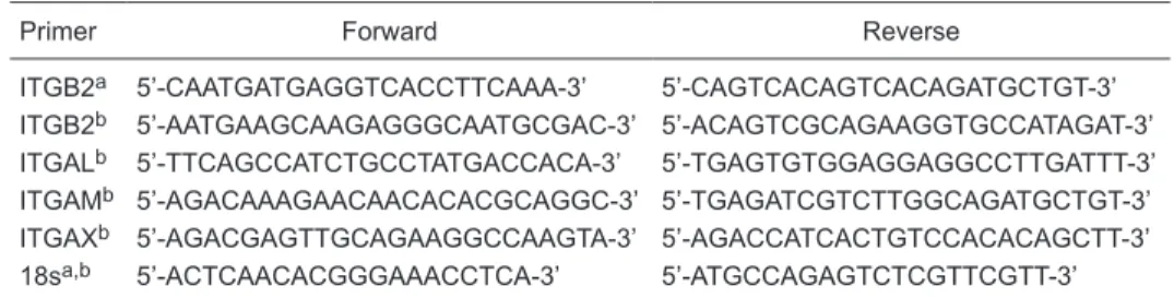
were microdissected from stage HH14-28 embryos. All samples were DNase treated (Ambion, USA) prior to reverse transcription using the iScript cDNA synthesis kit (Bio-Rad, USA). Samples were analyzed in a Nanodrop ND-100 ana-lyzer (Thermo Fisher Scientific Inc., USA) and 1 µg template was added per PCR assay following the manufacturer’s instruction. cDNA was synthesized using Hot Start PCR with AmpliTaq Gold DNA polymerase (Applied Biosystems, USA). cDNA concentration was established with the Quanti-iT Oligreen ssDNA reagent (Molecular Probes, USA) as described previously (26). Real-time PCR was performed with Platinum SYBR Green UDG (Invitrogen, USA) using 100 ng cDNA per reaction under the following conditions: 50°C for 2 min, 95°C for 2 min, and 35 cycles as follows: 94°C for 30 s, 58°C for 15 s, and 72°C for 45 s. A minimum of 3 different samples per stage were used and experiments were performed in triplicate for each sample. Primer pairs for each integrin subunit were designed using SciTools PrimerQuest (http://www.idtdna.com/Scitools/Applications/ Primerquest) and obtained commercially (Integrated DNA Technologies, USA). In order to avoid false results due to genomic DNA contamination, primers were designed to cross intron/exon boundaries. Separate forward and reverse primers for β2 integrin were used in RT-PCR and real-time PCR experiments. RT-PCR conditions were 95°C for 10 min and 35 cycles as follows: 94°C for 30 s, 60°C for 30 s, and 72°C for 45 s. Control reactions for RT-PCR and real-time PCR were performed using primers for the 18s ribosomal subunit. No-template control reactions were performed in every experiment. Additional controls, no enzyme and no primer, were also run in initial experiments (Table 1).
In situ hybridization
Sense and antisense avian β2 integrin riboprobes corresponding to 387-1120 bp (GenBank NM_205251) were produced by RT-PCR from stage HH20 AV canal and subsequently cloned into pBlueScript II KS+ (Stratagene, USA). Sense (control) probe reactions were included in each experiment to determine the level of non-specific binding.
Whole-mount embryo in situ hybridization protocols
Table 1. List of real-time PCR and RT-PCR primers.
Primer Forward Reverse
ITGB2a 5’-CAATGATGAGGTCACCTTCAAA-3’ 5’-CAGTCACAGTCACAGATGCTGT-3’
ITGB2b 5’-AATGAAGCAAGAGGGCAATGCGAC-3’ 5’-ACAGTCGCAGAAGGTGCCATAGAT-3’
ITGALb 5’-TTCAGCCATCTGCCTATGACCACA-3’ 5’-TGAGTGTGGAGGAGGCCTTGATTT-3’
ITGAMb 5’-AGACAAAGAACAACACACGCAGGC-3’ 5’-TGAGATCGTCTTGGCAGATGCTGT-3’
ITGAXb 5’-AGACGAGTTGCAGAAGGCCAAGTA-3’ 5’-AGACCATCACTGTCCACACAGCTT-3’
18sa,b 5’-ACTCAACACGGGAAACCTCA-3’ 5’-ATGCCAGAGTCTCGTTCGTT-3’ aReal-time PCR; bRT-PCR. ITGB2 = beta 2 integrin; ITGAL = alpha L integrin; ITGAM = alpha M
and digoxigenin-labeled riboprobe synthesis for chicken β2 integrin probes were as described (27). Images of whole-mount embryos were captured on a dissecting microscope (Leica model MZ125).
For in situ hybridization analysis on tissue sections, staged chicken embryos were collected in PBS, fixed overnight in a 6:3:1 mixture of ethanol/formalin/acetic acid, embedded in paraffin and sectioned. Sections were de-waxed, hydrated, proteinase K treated (20 µg/ml) and then post-fixed in 4% paraformaldehyde in PBS. Sections were incubated in pre-hybridization solution at 65°C prior to overnight incubation with hybridization solution contain-ing a riboprobe at 65°C. Slides were washed in SSC/50% formamide/0.1% Tween 20 at 65°C, blocked in 20% sheep serum/MABT/2% blocking powder at room temperature and then incubated overnight with anti-digoxigenin antibody diluted in the same blocking solution at 4°C. Slides were washed in MABT at room temperature and once in NTMT buffer prior to color reaction with NBT (Roche, USA) and BCIP (Roche) in NTMT at 37°C. Images were captured using a Leica microscope equipped with Normarski Optics.
Results
β2 integrin expression in the developing mouse
heart
mRNA transcripts of β2 integrin, as well as its α partners - αL, αM, and αX integrins - were successfully amplified by RT-PCR using RNA extracted from embryonic day 13.5
(ED13.5) whole hearts (Figure 1). These results demon-strate that β2 integrin mRNA is present in the embryonic heart, but do not rule out the possibility that it was derived from a small number of hematopoietic cells, which remain in the lumen of the hearts after the collection and washing procedures.
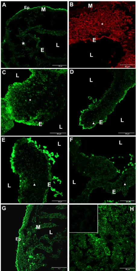
In order to determine which cells actually express β2 integrin, we performed immunohistochemical localization experiments with two antibodies directed against different domains in β2 integrin (i.e., N- or C-terminal epitopes). Positive control sections of 1-month-old mouse thymus incubated with either of these antibodies showed strong immunolabeling of thymocytes (Figure 2H) as compared to secondary antibody control sections in which primary antibodies had been omitted from the incubation proce-dure (Figure 2H, inset). β2 integrin immunolabeling was performed in mouse embryos obtained from stages ED10.5 to 19.5. At ED10.5 and ED11.5, β2 integrin immunostaining was observed in the endothelial layer of the cushions, on the surface of mesenchymal cells within the cardiac jelly, and in the epicardium (Figure 2A,B). At ED10, the epicardial layer had not yet completed its migration and covered the external surface of the myocardium. A low level of staining was also observed in some areas of the myocardium.
At ED13.5, when active mesenchymal cell migration is still occurring in the cushions, a strong signal was observed in the endothelium and mesenchymal cells (Figure 2C). At ED15.5, β2 integrin signal decreased, but was still present in the valve endothelial cells and in mesenchymal cells, which were differentiating into the fibroblast-like, interstitial cells of the valve leaflets (Figure 2D). A similar pattern of β2 integrin expression continued to be seen during AV valve maturation at ED17.5 (Figure 2E) but became weaker in valve endothelial cells and fibroblasts of the prenatal mouse AV valve at ED19.5 (Figure 2F). At ED15.5, the epicardium was strongly labeled and positive myocardial cells were observed in both the mural and trabecular por-tions of the myocardium (Figure 2G). These observapor-tions show that β2 integrin is expressed by non-hematopoietic cells during the early stages of mouse heart development, including in cells involved in the EMT, which occurs in the forming valves. Occasionally, intensely immunolabeled cells were seen within the lumen of the heart (Figure 2D-F), with specific staining well above background autofluorescence, indicating high levels of β2 integrin expression. Although we have not characterized these cells, it is possible that they are hematopoietic cells, hemangioblasts, and/or en -dothelial progenitor cells, all of which are known to express β2 integrin (28).
Immunolocalization of the α integrin partners of β2
integrin in embryonic mouse heart
As described above, mRNA transcripts for αL, αM, and αX were shown to be present in the embryonic heart (see Figure 1). Immunohistochemical procedures using
Figure 1. Detection of β2 integrin and its α subunit partners in
ED13.5 embryonic mouse hearts by RT-PCR. Integrin subunit expression was analyzed in ED13.5 embryonic mouse hearts by
RT-PCR with specific primers: Lane 1, 1-kb DNA ladder; Lanes 2 and 3, the presence of β2 integrin was confirmed by using two
different sets of primers, which generated the expected bands of 269 or 702 bp; Lane 4, αL (CD11a) 282 bp; Lane 5, αM (CD11b) 264 bp; Lane 6, αX (CD11c) 508 bp. No bands were detected
Figure 2. Immunofluorescent localiza
-tion of β2 integrin in mouse tissue. A-G, Sections of embryonic mouse hearts: A, ED10.5; B, ED11.5; C, ED13.5; D, ED15.5;
E, ED17.5; F, ED19.5; G, ED15.5.
Identi-cal patterns of β2 integrin immunostaining
were obtained using the N- or C-terminal-
specific anti-β2 integrin antibodies (data
not shown). Secondary antibody control sections of embryonic hearts, which were included in every experiment, displayed
no signal. β2 was present in endothelium
(E), epicardium (Ep), myocardium (M), mesenchymal cells in the cardiac jelly (*),
and fibroblast-like cells in valve leaflets
(arrowheads). Immunolabeled myocardial cells were observed in both the mural and trabecular portions of the myocardium (G). Intensely positive cells were occa-sionally seen within the lumen (L) of the heart in some sections (D-F). These cells were not characterized, but may represent hematopoietic cells, hemangioblasts, or endothelial progenitor cells (see text). H, Sections of 1-month-old mouse thymus.
β2 integrin was localized on the cell sur -face of thymocytes. H (inset), Control sec-tions in which the primary antibody was omitted displayed only a low level of
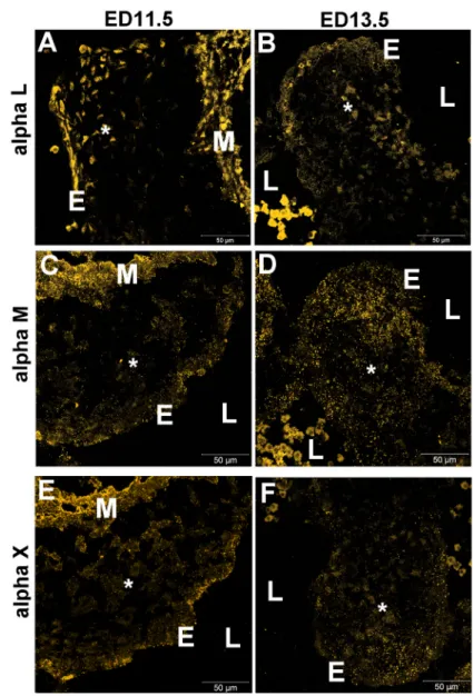
antibodies specific for these α integrin subunits allowed us to determine more precisely their distribution pattern. Endothelial cells of the AV canal cushion, mesenchymal cells inside the cardiac jelly and myocardial cells expressed αL integrin at ED11.5, during the initial stages of EMT (Figure 3A) and at ED13.5 (Figure 3B). Immunolabeling for αM integrin displayed the same pattern as observed for αL integrin in the AV canal at ED11.5 and ED13.5 (Figure 3C,D). An antibody specific for αX integrin showed that it was also expressed in the AV canal at ED11.5 and ED13.5
(Figure 3E,F), but with slightly less reactivity in the myo-cardial layer. The positive results obtained with antibodies against the αL, αM and αX partners of β2 integrin indicate that various α/β2 integrin heterodimers are present in the embryonic heart.
β2 integrin expression in developing avian embryos
The distribution of β2 integrin was not unique to mouse heart since we also confirmed expression in embryonic chicken heart. RNA from whole hearts and AV canal
seg-Figure 3. Immunofluorescent localization of the α integrin subunit partners of β2 integrin in embryonic mouse hearts. Sections of
ED11.5 (A,C,E) and ED13.5 (B,D,F) mouse hearts were immunolabeled with antibodies against specific α integrin subunits: A,B, αL
(CD11a); C,D, αM (CD11b); E,F, αX (CD11c). No immunostaining was observed in control sections in which the primary antibody was
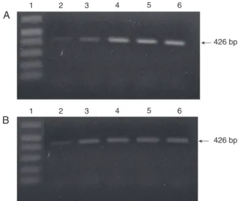
ments isolated a) at the onset of the EMT (HH14), b) during active mesenchymal cell invasion (HH17-HH20), and c) during endocardial cushion remodeling (HH25 and HH28) were used as templates for RT-PCR and real-time PCR. β2 integrin transcripts were expressed in all embryonic chick hearts and AV canal samples analyzed with RT-PCR (Figure 4A,B). Results of RT-PCR experiments using the 18s ribo-somal subunit as a control displayed similar band densities in all of the stages tested (data not shown). Real-time PCR showed that cardiac β2 integrin mRNA levels increase in
the AV canal during the remodeling of endocardial cushion EMT and subsequent remodeling of the valves (Figure 5). Real-time PCR controls with 18s showed equal levels in all stages (data not shown).
In situ hybridization protocols were performed on whole mounts and sections ofchicken embryos to define the spatial distribution of β2 integrin mRNA transcripts during endo -cardial cushion EMT and AV valve remodeling. At HH14, β2 integrin was expressed in the cardiac myocardium and endocardium (Figure 6A,C). At HH20, β2 integrin mRNA was detected in a more pronounced manner in the myo-cardium, epicardium and endomyo-cardium, and was expressed in mesenchymal cells invading the endocardial cushions (Figure 6B,D). At HH25 and HH28, when the cushions are heavily populated by mesenchymal cells, β2 integrin continued to be detected clearly in the endothelial layer,
Figure 4. RT-PCR analysis of β2 integrin in embryonic chicken
hearts. RT-PCR analysis of β2 integrin subunit expression was
performed using total RNA isolated from whole embryonic chick-en hearts (A) or dissected atriovchick-entricular canal regions (B). Lane 1, 1-kb DNA ladder; lane 2, HH14; lane 3, HH17; lane 4, HH20;
lane 5, HH25, and lane 6, HH28.
Figure 5. Relative quantitation of β2 integrin expression in the
chicken atrioventricular (AV) canal. mRNA was isolated from dis-sected AV canals at different stages of development and
ana-lyzed using real-time PCR. β2 integrin mRNA was detectable at stage HH14, increased significantly at HH17, during the period
when numerous mesenchymal cells are actively migrating in the endocardial cushions, and progressively increased in subsequent stages. mRNA fold changes were calculated relative to the values obtained for HH14. Data are reported as means ± SEM for three experiments.
Figure 6. Localization of β2 integrin mRNA in chicken embryos
using in situ hybridization. Positive labeling of β2 integrin mRNA
was detected in whole mounts (A,B) and sections (C-F) of em-bryonic chicken hearts using an antisense riboprobe. A,C, HH14;
B,D, HH20; E, HH25, and F, HH28. E (inset), Control in which
HH25 showed no labeling with a sense probe for β2 integrin. At =
atrium; M = myocardium; * = mesenchymal cells; arrows = heart;
mesenchymal cells, epicardium, and myocardium (Figure 6E,F). Control sections hybridized with β2 integrin sense probe were negative (Figure 6E, inset).
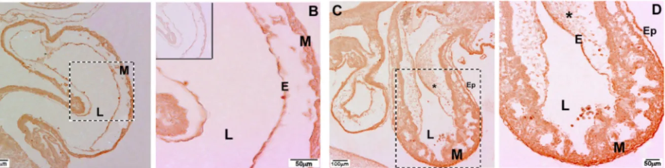
Immunohistochemical studies using HRP-conjugated secondary antibodies were also performed in the develop-ing chick. At HH14 β2 integrin staindevelop-ing was localized in the endothelium and the myocardium (Figure 7A,B). At HH20,
β2 integrin staining was more pronounced and was detected in endothelial layer, mesenchymal cells, epicardium, and myocardium (Figure 7C,D), matching the distribution pattern seen with in situ hybridization. As described by others (18), β1 integrin was also present in the cardiac endothelium, myocardium and mesenchymal cells of the embryonic chicken heart (Figure 8).
Figure 7. Immunohistochemical localization of β2 integrin in embryonic chicken hearts. A, Positive immunolabeling for β2 integrin was
detected in the endothelial and myocardial layers of stage HH14 chicken heart. B, Higher magnification of the boxed area in A. C, β2 in -tegrin was present in the endothelium, mesenchymal cells, epicardium, and myocardium at HH20. D, Higher magnification of the boxed
area in C. B (inset), Control section adjacent to that seen in A showing no reaction when the primary antibody was omitted. E = endo-E = endo-thelium; Ep = epicardium; L = lumen; M = myocardium; * = mesenchymal cells. Magnification bars: A, C = 100 µm; B, D = 50 µm.
Figure 8. Immunofluorescent localization of
β2 and β1 integrins in embryonic chicken
heart. Immunostaining using Cy3-conjugat-ed secondary antibodies. Sections of stag-es HH17 (A) and HH20 (B) chicken hearts
showing β2 integrin localization. C, β1 in -tegrin in stage HH20 heart. D, No staining was detected when the primary antibody was omitted from the staining protocol. E = endothelium; L = lumen; M = myocardium;
Discussion
The spatial and temporal distribution and multiple bind-ing functions of integrins have been intensely studied since the earliest reports characterizing these matrix receptors appeared in the literature (1). The fact that integrins play important roles in numerous cellular processes such as migration, adhesion, and signal transduction, as well as in morphogenesis and pathological process, continues to make them a focal point in research. A number of integrins have been characterized in the developing heart (18,19). The β2 integrin subfamily has been extensively studied, but most of this research centers on its role in the adhesion, migration and diapedesis of lymphocytes and leukocytes (6). Although the β2 integrin family is frequently referred to as being “leukocyte specific” (10), in the present study, using PCR, in situ hybridization, and immunohistochemical methods, we demonstrate that β2 integrin is also expressed by endothelial, mesenchymal, epicardial, and myocardial cells during normal heart development in both mammalian and avian embryos. It is noteworthy that three different alpha integrin partners for β2 integrin (i.e., αL, αM, and αX) were also detected during heart formation. The functional impor-tance of each of these integrin α/β2 combinations during the process of cardiogenesis remains to be determined.
The functional role of β2 integrin has been studied in β2 integrin null mice (29). These mice are viable and do not have any grossly obvious abnormalities in the heart or any other organs, but do display a phenotype closely resembling that of leukocyte adhesion deficiency type 1 patients, including leukocytosis, impaired leukocyte adhe-sion and migration, and the occurrence of mucocutaneous infections. However, it is important to note that in this study, the number of homozygous mice was lower than predicted by Mendelian genetics, suggesting that some β2 null off-spring die prenatally. In addition, 10-40% of newborn β2 null animals were reported to die perinatally due to unre-ported causes. Previous studies have shown that β1, and possibly other β integrins, are able to compensate, at least partially, for the loss of β2 integrin (30). Such compensatory mechanisms may allow some of the β2 null embryos to develop into adults. Other studies have shown that β2 null mice often have additional defects. For example, Miura et al. (13) reported that β2 null mice display certain features of osteoporosis, including decreased bone mineral density and increased trabecular bone space. Thus, based on the results presented here, a detailed morphometric analysis of the hearts of β2 null mice is warranted.
To our knowledge, there have been relatively few other studies demonstrating the presence of β2 integrin in devel-oping tissues. The presence of high levels of β2 integrin mRNA was noted in the late tailbud stage of Xenopus em-bryos, but specific tissue distribution was not determined (16). A study of ECM components and their integrin recep-tors in the human fetal adrenal gland demonstrated that β2
integrin is expressed by neural crest-derived chromaffin cells, which migrate through the fetal cortex on their way to the center of the gland, where they colonize and help form the medulla (15). Whether β2 integrin is expressed by neural crest cells in other locations during development has not been reported. Several other studies have shown that β2 integrin is also expressed in cell types other than leukocytes, such as HUVEC and cancer cells (11,12). Al-though it is clear that a number of non-hematopoietic cells can express β2 integrin, little is known about the function of β2 integrin outside its leukocyte-associated activities.
The ligands, which bind to β2 integrin and its α partners in the embryonic heart, have not yet been determined. The β2/α heterodimers may interact with counter-receptors (eg., VCAM, ICAM) on adjacent cells and thus modulate cell-cell adhesion, or bind to ECM proteins. β2 integrins have been shown to bind to various ECM proteins, including collagens, fibronectin, fibrinogen, plasminogen, and heparin (2). Col-lagens, fibronectin and numerous other matrix proteins are present in most embryonic tissues, including the cardiac jelly (20,21,31-33). Interestingly, β2 integrins also bind to CCN1 (CYR61), a secreted, cysteine-rich protein associ-ated with the ECM that promotes cell adhesion, migration, proliferation, differentiation, and survival or death in a cell type-dependent manner (34). CCN1 is expressed in the endothelium, cushion mesenchymal cells and myocardium, and is thought to be essential for tissue remodeling during cardiac development. Ccn1 null mice suffer embryonic lethality and display impaired cardiac valvuloseptal mor-phogenesis, which results in AVSDs (35). Ccn1 deficiency
does not affect endocardial cushion tissue formation or EMT, but results in accelerated apoptosis in cushion tissue and reduced gelatinous activity in the ventricular septum and AV valves.
References
1. Hynes RO. Integrins: bidirectional, allosteric signaling ma-chines. Cell 2002; 110: 673-687.
2. Humphries JD, Byron A, Humphries MJ. Integrin ligands at a glance. J Cell Sci 2006; 119: 3901-3903.
3. Harburger DS, Calderwood DA. Integrin signalling at a glance. J Cell Sci 2009; 122: 159-163.
4. Arnaout MA, Goodman SL, Xiong JP. Structure and mechan-ics of integrin-based cell adhesion. Curr Opin Cell Biol 2007; 19: 495-507.
5. Meighan CM, Schwarzbauer JE. Temporal and spatial regu-lation of integrins during development. Curr Opin Cell Biol
2008; 20: 520-524.
6. Springer TA. Traffic signals for lymphocyte recirculation and
leukocyte emigration: the multistep paradigm. Cell 1994; 76: 301-314.
7. Hogg N, Bates PA. Genetic analysis of integrin function in man: LAD-1 and other syndromes. Matrix Biol 2000; 19: 211-222.
8. Palazzo AJ, Jones SP, Girod WG, Anderson DC, Granger DN, Lefer DJ. Myocardial ischemia-reperfusion injury in
CD18- and ICAM-1-deficient mice. Am J Physiol 1998; 275: H2300-H2307.
9. Tan SM, Hyland RH, Al-Shamkhani A, Douglass WA, Shaw JM, Law SK. Effect of integrin beta 2 subunit truncations on
LFA-1 (CD11a/CD18) and Mac-1 (CD11b/CD18) assembly,
surface expression, and function. J Immunol 2000; 165: 2574-2581.
10. Hogg N, Henderson R, Leitinger B, McDowall A, Porter J, Stanley P. Mechanisms contributing to the activity of integ-rins on leukocytes. Immunol Rev 2002; 186: 164-171. 11. Langeggen H, Berge KE, Johnson E, Hetland G. Human
umbilical vein endothelial cells express complement
recep-tor 1 (CD35) and complement receprecep-tor 4 (CD11c/CD18) in vitro. Inflammation 2002; 26: 103-110.
12. Karmakar S, Mukherjee R. Integrin receptors and ECM pro-teins involved in preferential adhesion of colon carcinoma cells to lung cells. Cancer Lett 2003; 196: 217-227. 13. Miura Y, Miura M, Gronthos S, Allen MR, Cao C, Uveges
TE, et al. Defective osteogenesis of the stromal stem cells predisposes CD18null mice to osteoporosis. Proc Natl Acad Sci U S A 2005; 102: 14022-14027.
14. Bokel C, Brown NH. Integrins in development: moving on, responding to, and sticking to the extracellular matrix. Dev Cell 2002; 3: 311-321.
15. Chamoux E, Bolduc L, Lehoux JG, Gallo-Payet N. Identifi -cation of extracellular matrix components and their integrin receptors in the human fetal adrenal gland. J Clin Endocrinol Metab 2001; 86: 2090-2098.
16. Ransom DG, Hens MD, DeSimone DW. Integrin expression in early amphibian embryos: cDNA cloning and characteriza-tion of Xenopus beta 1, beta 2, beta 3, and beta 6 subunits.
Dev Biol 1993; 160: 265-275.
17. Clark EB, Markwald RR, Takao A, Hamilton RM. Develop-mental mechanisms of heart disease. New York: Futura; 1995.
18. Simpson DG, Reaves TA, Shih D, Burgess W, Borg TK, Terracio L. Cardiac integrins: The ties that bind. Cardiovasc Pathol 1998; 7: 135-143.
19. Danen EH, Sonnenberg A. Integrins in regulation of tissue development and function. J Pathol 2003; 200: 471-480. 20. Kitten GT, Markwald RR, Bolender DL. Distribution of
base-ment membrane antigens in cryopreserved early embryonic hearts. Anat Rec 1987; 217: 379-390.
21. Little CD, Rongish BJ. The extracellular matrix during heart development. Experientia 1995; 51: 873-882.
22. Peacock JD, Lu Y, Koch M, Kadler KE, Lincoln J. Temporal and spatial expression of collagens during murine atrioven-tricular heart valve development and maintenance. Dev Dyn
2008; 237: 3051-3058.
23. Person AD, Klewer SE, Runyan RB. Cell biology of cardiac cushion development. Int Rev Cytol 2005; 243: 287-335. 24. Korenberg JR, Bradley C, Disteche CM. Down syndrome:
molecular mapping of the congenital heart disease and duodenal stenosis. Am J Hum Genet 1992; 50: 294-302. 25. Hamburger V, Hamilton HL. A series of normal stages in the
development of the chick embryo. J Morphol 1951; 88: 49-92.
26. Tavares AL, Mercado-Pimentel ME, Runyan RB, Kitten GT. TGF beta-mediated RhoA expression is necessary for epithelial-mesenchymal transition in the embryonic chick heart. Dev Dyn 2006; 235: 1589-1598.
27. Nieto MA, Patel K, Wilkinson DG. In situ hybridization analy-sis of chick embryos in whole mount and tissue sections.
Methods Cell Biol 1996; 51: 219-235.
28. Chavakis E, Aicher A, Heeschen C, Sasaki K, Kaiser R,
El Makhfi N, et al. Role of beta2-integrins for homing and
neovascularization capacity of endothelial progenitor cells.
J Exp Med 2005; 201: 63-72.
during the migration of the cushion tissue mesenchyme, but it is important to remember that β2 integrin is also ex -pressed in the epicardium and myocardium during heart development. Additional studies will be needed in order to clarify the functional importance of β2 integrin in each of these locations during cardiac morphogenesis.
Acknowledgments
The authors thank Lorenza Carvalhaes, Othon Gervá-sio and André Tavares for their comments and help with
29. Scharffetter-Kochanek K, Lu H, Norman K, van Nood N, Munoz F, Grabbe S, et al. Spontaneous skin ulceration and defective T cell function in CD18 null mice. J Exp Med 1998; 188: 119-131.
30. Henderson RB, Lim LH, Tessier PA, Gavins FN, Mathies M, Perretti M, et al. The use of lymphocyte function-associated
antigen (LFA)-1-deficient mice to determine the role of LFA-1, Mac-LFA-1, and alpha4 integrin in the inflammatory response
of neutrophils. J Exp Med 2001; 194: 219-226.
31. Klewer SE, Krob SL, Kolker SJ, Kitten GT. Expression of type VI collagen in the developing mouse heart. Dev Dyn
1998; 211: 248-255.
32. Gittenberger-de Groot AC, Bartram U, Oosthoek PW, Bar-telings MM, Hogers B, Poelmann RE, et al. Collagen type VI expression during cardiac development and in human fetuses with trisomy 21. Anat Rec A Discov Mol Cell Evol Biol 2003; 275: 1109-1116.
33. Carvalhaes LS, Gervasio OL, Guatimosim C, Heljasvaara R,
Sormunen R, Pihlajaniemi T, et al. Collagen XVIII/endostatin
is associated with the epithelial-mesenchymal transforma-tion in the atrioventricular valves during cardiac develop-ment. Dev Dyn 2006; 235: 132-142.
34. Chen CC, Lau LF. Functions and mechanisms of action of CCN matricellular proteins. Int J Biochem Cell Biol 2009; 41: 771-783.
35. Mo FE, Lau LF. The matricellular protein CCN1 is essential for cardiac development. Circ Res 2006; 99: 961-969. 36. Murphy M, Insoft RM, Pike-Nobile L, Derbin KS, Epstein LB.
Overexpression of LFA-1 and ICAM-1 in Down syndrome thymus. Implications for abnormal thymocyte maturation. J Immunol 1993; 150: 5696-5703.
37. Yakubenko VP, Belevych N, Mishchuk D, Schurin A, Lam
SC, Ugarova TP. The role of integrin alpha D beta2 (CD11d/ CD18) in monocyte/macrophage migration. Exp Cell Res