Ecto nucle o tidase activitie s in
Se rto li ce lls fro m im m ature rats
Departamento de Bioquímica, Instituto de Ciências Básicas da Saúde, Universidade Federal do Rio Grande do Sul, Porto Alegre, RS, Brasil E.A. Casali, T.R. da Silva,
D.P. Gelain, G.R.R.F. Kaiser, A.M.O . Battastini, J.J.F. Sarkis and E.A. Bernard
Abstract
Sertoli cells have been shown to be targets for extracellular purines such as ATP and adenosine. These purines evoke responses in Sertoli cells through two subtypes of purinoreceptors, P2Y2 and PA1. The signals to purinoreceptors are usually terminated by the action of ectonucleotidases. To demonstrate these enzymatic activities, we cultured rat Sertoli cells for four days and then used them for different assays. ATP, ADP and AMP hydrolysis was estimated by measuring the Pi released using a colorimetric method. Adenosine deaminase activity (EC 3.5.4.4) was determined by HPLC. The cells were not disrupted after 40 min of incubation and the enzymatic activities were considered to be ectocellularly localized. ATP and ADP hydrolysis was markedly increased by the addition of divalent cations to the reaction medium. A competition plot demonstrated that only one enzymatic site is responsible for the hydrolysis of ATP and ADP. This result indicates that the enzyme that acts on the degradation of tri- and diphosphate nucleosides on the surface of Sertoli cells is a true ATP diphosphohydrolase (EC 3.6.1.5) (specific activities of 113 ± 6 and 21 ± 2 nmol Pi mg-1 min-1 for ATP and ADP, respectively). The ecto-5-nucleotidase (EC 3.1.3.5) and ectoadenosine deaminase activities (specific activities of 32 ± 2 nmol Pi mg-1 min-1 for AMP and 1.52 ± 0.13 nmol adenosine mg-1 min-1, respectively) were shown to be able to terminate the effects of purines and may be relevant for the physiological control of extracellular levels of nucleotides and nucleo-sides inside the seminiferous tubules.
Co rre spo nde nce E.A. Bernard
Departamento de Bioquímica Rua Ramiro Barcellos, 2600, Anexo 90035-003 Porto Alegre, RS Brasil
Fax: + 55-51-316-5540 E-mail: elenbern@ vortex.ufrgs.br
Received September 6, 2000 Accepted July 2, 2001
Ke y wo rds
·Sertoli cells
·Purine nucleotides
·ATP diphosphohydrolase
·Ecto-5’-nucleotidase
·Ectoadenosine deaminase
Intro ductio n
Extracellular purines interact with spe-cific receptors (purinoreceptors) on the sur-face of cells activating several biological processes (for reviews, see 1-3). Previous studies have demonstrated that adenosine nucleotides can modulate Sertoli cell re-sponses through the purinoreceptors present on these cells (4-10).
The hormonal regulation of Sertoli cell
the same study, Filippini et al. (5) demon-strated that ATP can activate inositol phos-pholipid turnover and Ca2+
signaling. Regarding adenosine, a product of ATP degradation by ectonucleotidases, Rivkees (11) localized and characterized receptors for this structure in rat testis tissue and dem-onstrated that Sertoli cells have the PA1 re-ceptor subtype on the plasma membrane. In testis tissue, the PA1 receptors inhibit adenylyl cyclase activity (4,10). Adenosine inhibition of the hormonal effects of FSH in Sertoli cells is reversed by pertussis toxin (8). This fact suggests that the regulation of a cyclic nucleotide-dependent pathway is one of the transduction mechanisms by which adeno-sine regulates the functions of these cells.
The extracellular hydrolysis of ATP to adenosine by ectonucleotidases has been re-ported for several cell types (12-20). These enzymatic activities can regulate the extra-cellular concentration of adenine nucleo-tides and nucleosides modulating their local effects. Degradation of ATP and other nucleo-tides occurs through a cascade of cell sur-face-bound enzymes such as ecto-ATPase (EC 3.6.1.3), ectoapyrase/ATP diphospho-hydrolase/NTPDase (EC 3.6.1.5), and ecto-5-nucleotidase (EC 3.1.3.5), resulting in the formation of ADP, AMP and adenosine (21). The presence of apyrase activity has been well demonstrated in a large number of mam-malian sources (12,13,15). Apyrase is the enzyme that hydrolyzes ATP and ADP (and other tri- and diphosphate nucleosides) to the monophosphate esters plus inorganic phosphate (Pi), releasing 2 mol Pi/mol ATP and 1 mol Pi/mol ADP.
Barbacci et al. (22) identified and char-acterized a possible ecto-ATPase activity in rat Sertoli cells. In the present study, we demonstrate that Sertoli cells in culture are able to promote the hydrolysis of ATP, ADP and AMP and we present evidence for the first time that the enzymes responsible for nucleotide hydrolysis are a true ectoapyrase (ATP and ADP hydrolysis) and an
ecto-5-nucleotidase (AMP hydrolysis). The kinetic parameters for nucleotide hydrolysis and the effect of divalent cations, calcium and mag-nesium, on enzymatic activities were deter-mined. Moreover, we show that Sertoli cells can control extracellular adenosine levels through ectoadenosine deaminase activity (EC 3.5.4.4).
Mate rial and Me tho ds
Mate rial
Culture medium (DMEM/F-12) and soy-bean trypsin were purchased from Gibco (Grand Island, NY, USA). Lactate dehydro-genase (LDH) kit, soybean trypsin inhibitor S, DNase I, collagenase I, hyaluronidase I-S, nucleotides, nucleosides and HEPES were obtained from Sigma (St. Louis, MO, USA), FBS from Cultilab Ltda. (São Paulo, SP, Brazil), and 24-well plates from Costar Co. (Cambridge, MA, USA). Tetrabutyl ammo-nium chloride was purchased from Fluka Chemika (Neu Ulm, Switzerland). All other chemical reagents were of the highest avail-able grade.
Se rto li ce ll culture s
Primary cultures of Sertoli cells from 17-to 19-day-old Wistar rats were prepared as previously described (23). Briefly, the testis was sequentially digested with 0.25% tryp-sin and DNase (10 µg/ml) for 30 min at 37o
C to remove the interstitial tissue. The seminif-erous tubules obtained were dissociated with collagenase (1 mg/ml) and hyaluronidase (1 mg/ml) to separate Sertoli cells from myoid and germ cells for centrifugation at 40 g for 10 min. The cultures were grown to conflu-ence on 24-multiwell plates (approximately 0.6 x 10⁶
cells/well or 100 µg protein/well) at 34o
Hanks-buffered saline solution and main-tained for three days more in serum-free DMEM/F-12 (1:1). On the fourth day of culture, the Sertoli cell monolayers were used for the ectonucleotidase assays. The Sertoli cell cultures were estimated to be more than 95% pure, as determined by bright light and phase contrast microscopy and al-kaline phosphatase cytochemistry (24). Cel-lular integrity was determined on the basis of LDH activity. The Sertoli monolayers were incubated with reaction medium for 40 min and then samples were taken for the determi-nation of LDH with a Sigma kit (LD-L 10, catalog No. 228-10) at 37o
C in a Cobas Mira automatic incubator. LDH activity is reported as unit of enzyme per mg protein (U/mg).
Ecto nucle o tidase assays
Sertoli cell monolayers were washed three times with the reaction medium containing 135 mM NaCl, 5 mM KCl, 10 mM glucose and 10 mM HEPES, pH 7.4. The reaction was started by adding the substrate (ATP, ADP or AMP) to the reaction medium con-taining 135 mM NaCl, 5 mM KCl, 10 mM glucose and 10 mM HEPES, pH 7.4, plus CaCl2 and/or MgCl2 (1, 2 and 5 mM, as indicated) and EDTA (2 mM, as indicated). The final volume was 0.2 ml and incubation was carried out at 34o
C. After incubation, a supernatant sample was taken and mixed with cold trichloroacetic acid to a final con-centration of 5%. This mixture was centri-fuged for 10 min at 16,000 g at 4o
C and aliquots were taken for the assay of released Pi according to the procedure of Chan et al. (25). Incubation time and protein concentra-tion were chosen in order to ensure the lin-earity of the reaction. Controls to correct for nonenzymatic hydrolysis of nucleotides were prepared by measuring the Pi released into the same reaction medium incubated with-out cells. The possible contamination with Pi released from cultures was avoided by incubating the monolayers in reaction
medi-um without nucleotides. The cultures did not present a measurable release of Pi at any time of incubation. All assays were done in quadruplicate. Km and Vmax were calculated by linear regression and presented graphi-cally by the Eadie-Hofstee plot. The ATP and ADP concentrations for the same hy-drolysis rate were obtained from the sub-strate curve and used to construct the Chevillard competition plot (26).
Ade no sine de am inase assay
On the fourth day of culture, the mono-layers were washed three times with adeno-sine deaminase assay (ADA) buffer consist-ing of 100 mM NaCl, 20 mM KCl, 4 mM MgCl2, 2 mM CaCl2, 10 mM NaHCO3, 5 mM glucose and 15 mM Tris, pH 7.4 (27). The reaction was started by adding adeno-sine (0.15 mM) to the same ADA buffer as described above. The final volume was 0.2 ml and the incubation was carried out at 34o
C. After incubation (0, 30 and 60 min), the supernatant was taken and maintained on ice. The samples were boiled for 3 min and centrifuged at 4o
deamina-tion of adenosine was not detected at any time.
Ce llular pro te in de te rm inatio n
After the assays, the Sertoli cell mono-layers were digested with 0.5 N NaOH and total protein was measured by the method of Lowry et al. (29).
Statistical analysis
The mean ± SEM data for groups of three or four experiments were analyzed by
ANOVA and the post hoc
Student-New-man-Keuls test using the statistical program SPSS 6.0 for Windows.
Re sults and D iscussio n
ATP, AD P and AMP hydro lysis
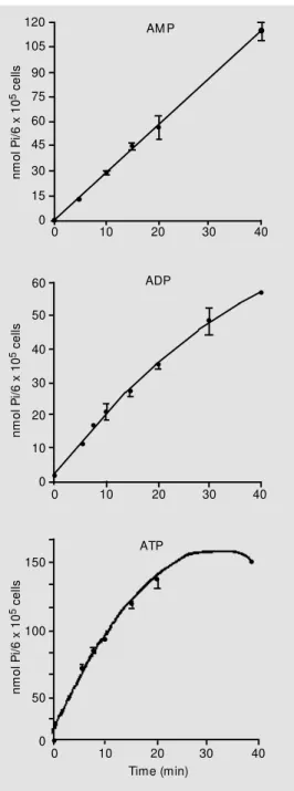
Sertoli cell cultures promoted ATP, ADP and AMP hydrolysis that was linear for at least 20 min (Figure 1). One possible prob-lem in the detection of apyrase activity (ATP and ADP hydrolysis) in most sources is the interference of 5-nucleotidase, which may cause an overestimation of ATPase and ADPase activity (30). The procedure used in the present study to avoid this problem was the limitation of both ATP and ADP hy-drolysis to less than 10%. In addition, all mammalian ecto-5-nucleotidase activities described are strongly inhibited by ATP and ADP in the low micromolar range (31). ATP diphosphohydrolase (apyrase) is an enzyme able to promote the removal of two phate groups of ATP but of only one phos-phate group of ADP. This enzyme presents divalent cation dependence and can be stim-ulated by Ca2+
and Mg2+
(12,15,30,32). The ecto-5-nucleotidase can also be stimulated by Mg2+
(14,31) but this activation was lower than the apyrase activation (26.3 ± 5% for the ecto-5-nucleotidase activation and 1490 ± 84% for the apyrase activation in relation to the control). In the presence of 2 mM EDTA, ATP and ADP hydrolysis was prac-tically negligible and AMP hydrolysis was lower than control (without the addition of divalent cations) (Figure 2). Thus, the en-zymes responsible for the hydrolysis of ATP, ADP and AMP in Sertoli cells could be cation activated. No significant differences were observed in enzymatic activation by different cations at the different
concentra-n
m
o
l
P
i/
6
x
1
0
5 c
e
lls
120
105
90
75
60
30
15
0 45
0 10 20 30 40
n
m
o
l
P
i/
6
x
1
0
5 c
e
lls
60
50
40
30
10
0 20
0 10 20 30 40
150
100
50
0
0 10 20 30 40
Time (min)
n
m
o
l
P
i/
6
x
1
0
5 c
e
lls
Figure 1. Time course of ATP, ADP and AM P hydrolysis. Ca2+
-ATP, Ca2+-ADP and M g2+-AM P
w ere incubated w ith Sertoli cell monolayers as described in M a-terial and M ethods. The data show n w ere from an experiment carried out in quadruplicate. Data are reported as means ± SEM .
AM P
ADP
n m o l P i m g p ro te in
-1 m
in -1 125 100 75 0 AM P ADP ATP + + * *+ *+ * + *+ *+ *+ *+ C o n tr o l 2 m M E D T A 1 m M C a 2 + 2 m M C a 2 + 1 m M M g 2 + 2 m M M g 2 + C o n tr o l 2 m M E D T A 1 m M C a 2 + 5 m M C a 2 + 2 m M C a 2 + 1 m M M g 2 + 2 m M M g 2 + 5 m M M g 2 + 2 m M C a 2 +/M g 2 + C o n tr o l 2 m M E D T A 1 m M C a 2 + 2 m M C a 2 + 5 m M C a 2 + 1 m M M g 2 + 2 m M M g 2 + 5 m M M g 2 + 2 m M C a 2 +/M g 2 +
Figure 2. Cation dependence on ectonucleotidase activities. Ser-toli cells w ere incubated w ith the reaction medium plus 1 mM AM P (gray bars), 1 mM ADP (w hite bars), and 1 mM ATP (black bars) plus cat ions or EDTA, as indicated. The data are representative of three different experiments: mean ± SEM (N = 4) of one typical experiment. * P<0.05 compared to the con-trol group; +P<0.05 compared to
the 2 mM EDTA group (Student-New man-Keuls test).
n m o l P i m g p ro te in
-1 m
in -1 8.00 6.40 4.80 3.20 1.60 0.00
0.0 0.2 0.4 0.6 0.8 1.0
P
Figure 3. The competition plot. The concentration at w hich the velocities w ere the same for ATP and ADP w as chosen for the Chevillard plot. The assay conditions are described in M a-terial and M ethods. The incuba-tion time w as 10 min; substrate A (ADP) at P = 0 w as 0.1 mM and substrate B (ATP) at P = 1 w as 0.04 mM . Data represent an experiment carried out in quintuplicate. The values are the mean ± SEM . No significant ference w as found betw een dif-ferent points.
tions tested for ATP, ADP and AMP hy-drolysis (Figure 2). This result indicates that maximal activation of the apyrase and ecto-5-nucleotidase activities is possible with 2 mM or less of both cations tested. No addi-tive effects were observed when the two divalent cations tested were added at the same time to the reaction medium, suggest-ing that both Ca2+
and Mg2+
are competing for the same activation site. It is important to note that there was a parallel profile of acti-vation for all substrates (ATP, ADP and AMP) with each cation added. Based on these results, we established as optimal con-ditions for measuring the ectonucleotidase activities the ratio of 1 mM/2 mM for the nucleotides/divalent cation. In this way the ectonucleotidase activities were measured in the physiological extracellular range of divalent cations and nucleotides.
A single active site
ATP and ADP hydrolysis could be cata-lyzed by an ATP diphosphohydrolase (apy-rase) or by enzyme combinations able to mimic apyrase activity. To show that ATP and ADP hydrolysis occurs due to an
ranging from 15 to 1000 µM for each sub-strate. Mg2+
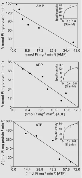
-AMP hydrolysis was determined at AMP concentrations ranging from 15 to 2000 µM. The results (Figure 4, inset) indi-cated that all the enzymatic activities in-creased with increasing nucleotide concen-trations until saturation with 1 mM sub-strate. The Eadie-Hofstee plot for the hy-drolysis of ATP, ADP and AMP is shown in Figure 4. The Michaelis constant (Km, app) (Table 1) calculated by linear regression from the results in Figure 4 was closely similar for ATP and ADP hydrolysis (131 ± 17.4 and 110 ± 29 µM, respectively). It is important to note that a similar Km value for both sub-strates is also a characteristic of other apy-rases described in the literature (15,30). The Sertoli cell cultures were able to hydrolyze other di- and triphosphate nucleosides such as GTP, GDP, ITP and IDP (data not shown). The hydrolysis of different di- and triphos-phate nucleotides is another important char-acteristic of apyrases from various sources (12,15,16,30). AMP hydrolysis has a calcu-lated Km of 410 ± 73 µM (Table 1). All ecto-5-nucleotidases described have Km values in the micromolar range (31). Variations in kinetic data could be the result of species-and/or tissue-specific forms of the enzyme, analysis of impure preparations or of varia-tions in the assay condivaria-tions (31). The IMP and GMP hydrolysis was only 33 and 25% of AMP hydrolysis (12 and 8 nmol Pi mg pro-tein-1
min-1
, respectively) under the same conditions. The AMP substrate preference is another characteristic of ecto-5-nucleotidase of several tissues (31). These results indicate that the extracellular AMP hydrolysis occur-ring in Sertoli cells is performed by an ecto-5-nucleotidase.
Ce llular inte grity
The lack of intracellular LDH release during the assays indicated the cellular in-tegrity of cultures. The Sertoli cells remained intact during 40 min of incubation in the
Table 1. Kinetic parameters for ATP, ADP and AM P hydrolysis.
Enzyme activity Km (µM ) Vmax (nmol Pi mg protein-1 min-1)
ATPase 131 ± 17.4 59.5 ± 4.6
ADPase 110 ± 29.0 15.4 ± 0.5
AM Pase 410 ± 73.4 43.3 ± 7.7
The kinetic constants w ere determined using linear regression analysis applied to the data in Figure 4. The values of Km and Vmax are the mean ± SEM of four experiments
for ATPase and of three experiments for ADPase and AM Pase activities. The mean values of the kinetic parameters did not differ significantly (P<0.01).
V ( n m o l P i m g p ro te in
-1 m
in
-1) 150
120 90 60 30 0 85 68 51 34 17 0
0.0 8.6 17.2 25.8 34.4 43.0
40 30 20 10 0 S p e ci fi c ac ti vi ty
0 0.8 1.6 [S] (mM )
AM P
(nmol Pi mg-1 min-1) [AM P]
16 12 8 4 S p e ci fi c ac ti vi ty
0 0.4 0.8 [S] (mM ) 0
0.0 3.4 6.8 10.2 13.6 17.0
(nmol Pi mg-1 min-1) [ADP]
600 480 360 240 120 0 80 40 20 0 S p e ci fi c ac ti vi ty
0 0.4 0.8 [S] (mM )
0.0 14.4 28.8 43.2 57.6 72.0
(nmol Pi mg-1 min-1) [ATP]
ADP ATP V ( n m o l P i m g p ro te in
-1 m
in -1) V ( n m o l P i m g p ro te in
-1 m
in
-1)
Figure 4. Eadie-Hofstee plots of ATP, ADP and AM P hydrolysis. Reaction rate w as measured by released Pi as described in M a-t erial and M ea-t hods. Resula-t s w ere obtained w ith a nucleotide concentration ranging from 15 to 2000 µM for each substrate. Data w ere plotted using Eadie-Hofstee plots and w ith the inset of nonlinear regression for three substrates. Best-fit analysis indi-cated a linear relationship. Plots are for representative experi-ments carried out in quadrupli-cate. The data for the nonlinear regression plot are reported as mean ± SEM . V is nmol Pi mg protein-1 min-1 and [S] is the
sub-strate concentration in mM .
Kine tic param e te rs o f ATPase , AD Pase and
AMPase activitie s
Ca2+
-ATP and Ca2+
reaction medium. Only 5% of LDH activity of lysed cells (1.33 ± 0.19 U/mg protein) was measured during 40 min of incubation. In this way the participation of cytosolic en-zymes in extracellular nucleotide hydrolysis was excluded.
Ade no sine de aminase activity
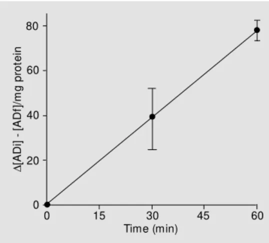
It has been previously demonstrated that testis tissue contains adenosine deaminase mRNA (33) and we have investigated whether this enzyme is present on the plasma membrane surface of Sertoli cells and its capacity to degrade extracellular adenosine. Sertoli cells are able to degrade the extracel-lular adenosine by an ectoadenosine deami-nase activity (Figure 5). The ectoadenosine deaminase is a key enzyme in purine me-tabolism that catalyzes the irreversible trans-formation of adenosine and 2-deoxyadeno-sine to ino2-deoxyadeno-sine and 2-deoxyino2-deoxyadeno-sine, respec-tively. Its enzymatic activity is homoge-neously distributed along the cell surface in some cells (34). The Sertoli cell ectoadeno-sine deaminase activity was linear up to 60 min of incubation (Figure 5) and had a spe-cific activity of 1.52 ± 0.13 nmol adenosine mg protein-1
min-1
. The decrease in adeno-sine levels in the extracellular medium was of the same order of magnitude as the in-crease in inosine and hypoxanthine (data not shown) and this demonstrates that the aden-osine decrease is a result of adenaden-osine deami-nase activity rather than of cellular adeno-sine uptake. The presence of dipyridamole (10 µM), a classical inhibitor of adenosine uptake (35), in the ADA buffer did not alter the extracellular adenosine concentration but caused 30% inhibition of ectoadenosine deaminase activity. A possible explanation for this result is the participation of ecto-adenosine deaminase in a protein complex that can have multiple functions such as catalytic deamination activity, coupled with adenosine receptors and with selective chan-nels by uptake of extracellular adenosine
(34,36). The presence of dipyridamole can alter the conformation of the protein com-plex with a modification in the enzymatic activity.
The physio lo gical ro le
The physiological role of ectonucleoti-dases is unknown but it has been speculated that their function could be the control of extracellular concentration of purines. The modulation of the levels of different nucleo-tides may involve cross-talking between the diverse pathways activated in purinergic receptor subtypes. The general way to con-trol the concentration of adenine nucleotides is the sequential activity of ecto-ATPases, ecto-ATP diphosphohydrolases (apyrase) and ecto-5-nucleotidases (12-17,19,20,30). The adenosine released from cells or resulting from extracellular ATP hydrolysis can be deaminated by the ectoadenosine deaminase activity, producing inosine (18,34,36).
Adenosine and its antagonists, such as caffeine, have been long postulated to influ-ence the male reproductive system. Several reports have demonstrated the presence of adenosine receptors in Sertoli cells (10,11) and their modulation by adenosine and dif-ferent adenosine analogues (4,8,9). The purinoreceptor subtype P2Y2 is present in these cells (5) and its modulation by ATP has been well demonstrated (5-7). In Sertoli
D
[A
D
i]
[
A
D
f]
/m
g
p
ro
te
in
80
60
40
20
0
0 15 30 45 60
Time (min)
cells, extracellular ATP and adenosine can modulate the hormonal response to FSH, decreasing cAMP levels (4,5,8-10), and can regulate anionic (6) and transferrin secretion (7) and the intracellular levels of Ca2+
by inositol phospholipid turnover (5). Thus, the control of extracellular levels of adenosine nucleotides is of high physiological rel-evance. However, the mechanism control-ling purine concentrations in testicular tis-sue is poorly known. We show in this study that Sertoli cells were able to hydrolyze ATP and ADP (Figure 1) and that only one active site is responsible for the hydrolysis of the two nucleotides (Figure 3). Barbacci et al. (22) identified and characterized one pos-sible ecto-ATPase in rat Sertoli cells. The Km calculated for the possible ecto-ATPase is the same we found for ATP and ADP hy-drolysis (Table 1), indicating that one en-zyme is responsible for the hydrolysis of the two nucleotides. The results obtained in the competition plot confirm this hypothesis. The cation requirements demonstrated by Barbacci et al. (22) for ATP hydrolysis are equal to those for ADP hydrolysis demon-strated by us (Figure 2). On the other hand, our results cannot exclude the co-expression of two enzymes (a fact demonstrated in the rat brain) (37) because the ATP hydrolysis demonstrated by Barbacci et al. (22) was slightly inhibited by ADP. In other tissues it has been postulated that the control of extra-cellular nucleotide concentration is due to the action of enzymatic complexes with the possible participation of two or more ecto-enzymes (21). The most obvious physiologi-cal role for apyrase in Sertoli cells, in anal-ogy to other tissues, is to participate in an enzyme chain together with a 5-nucleoti-dase for the complete hydrolysis of ATP to adenosine.
AMP hydrolysis occurs through the ac-tion of an ecto-5-nucleotidase releasing adenosine that could create a secondary sig-nal for PA1 receptors. The Sertoli cell
ecto-5-nucleotidase has proved to be cation acti-vated and in the presence of normal extracel-lular concentrations of Ca2+
and Mg2+ (1-2 mM) its activity is increased compared to the control activity (without cation addition) and the 2 mM EDTA group (Figure 2). The Km/
Vmax values (Table 1) and the substrate
pref-erence are in accordance with the other char-acteristics of ecto-5-nucleotidase from sev-eral tissues (31).
In order to terminate the purine extracel-lular cascade of nucleotides and nucleosides, the ectoadenosine deaminase produces ino-sine that can be taken up and/or degraded by the cells. We have demonstrated that Sertoli cells have an ectoadenosine deaminase ac-tivity with the same buffer requirement as for other cells (Figure 5) (28). In analogy, this enzyme can participate together with an ATP diphosphohydrolase and the ecto-5-nucleotidase in the control of the extracellu-lar adenosine levels and eliminate the purine cascade.
The physiological control of ectonucle-otidase activities is unknown. Moreover, Franco et al. (34,36) have demonstrated that some ectoenzymes can play the role of one enzyme and that of a receptor which can be internalized when the substrate is in its ac-tive site. Another possibility is the co-local-ization of ectoenzyme and receptor at the same site on the plasma membrane. Recep-tor desensitization can occur by endocytosis of membrane fragments where the ectoen-zyme and the receptor in question are pres-ent. Some authors have shown that ecto-nucleotidases can lose their activities in re-sponse to a cell signal (38,39) and that ectonucleotidase activities initially localized on the membrane surface are internalized into endoplasmic vesicles (40).
Re fe re nce s
1. El-M oatassim C, Dornand J & M ani J (1992). Extracellular ATP and cell signal-ling. Biochimica et Biophysica Acta, 1134: 31-45.
2. Dubyak GR & El-M oatassim C (1993). Sig-nal transduction via P2-purinergic recep-t ors f or exrecep-t racellular ATP and orecep-t her nucleotides. American Journal of Physiol-ogy, 265: C577-C606.
3. Ralevic V & Burnstock G (1998). Recep-tors for purines and pyrimidines. Pharma-cological Review s, 50: 413-492. 4. Cont i M , Boit ani C, DeM anno DA,
M igliaccio S, M onaco L & Szymeczek C (1989). Characterization and function of adenosine receptors in the testis. Annals of the New York Academy of Sciences, 564: 39-47.
5. Filippini A, Riccioli A, Cesaris P de, Paniccia R, Teti A, Stefanini M , Conti M & Ziparo E (1994). Activation of inositol phospholipids and calcium signaling in rat Sertoli cells by P2 purinergic receptors: m odulation of follicle-stim ulating hor-mone responses. Endocrinology, 134: 1537-1545.
6. Ko WH, Chan HC, Chew SB & Wong PYD (1998). Regulated anion secretion in the epithelia from Sertoli cells of immature rats. Journal of Physiology,512: 471-480. 7. M eroni SB, Cánepa DF, Pellizari EH & Schteingart HF (1998). Effects of puriner-gic agonists on aromatase and gamma-glutamyl transpeptidase activities and on transferrin secretion in cultured Sertoli cells. Journal of Endocrinology, 157: 275-283.
8. M onaco L, DeM anno DA, M artin M W & Conti M (1988). Adenosine inhibition of the hormonal response in the Sertoli cell is reversed by pertussis toxin. Endocrinol-ogy, 122: 2692-2698.
9. M onaco L, Toscano M V & Conti M (1984). Purine modulation of the hormonal re-sponse of the rat Sertoli cell in culture.
Endocrinology, 115: 1616-1624. 10. Stiles GL, Pierson G, Sunay S & Parsons
WJL (1986). The rat testicular A1 adeno-sine receptor-adenylate cyclase system.
Endocrinology, 119: 1845-1851. 11. Rivkees SA (1994). Localization and
char-acterization of adenosine receptor expres-sion in rat testis. Endocrinology, 135: 2307-2313.
12. Battastini AM O, Oliveira EM , M oreira CM , Bonan CD, Sarkis JJF & Dias RD (1995). Solubilization and characterization of an ATP diphosphohydrolase (EC 3.6.1.5) from rat brain synaptic plasma
mem-branes. Biochemistry and M olecular Biol-ogy International, 37: 209-219.
13. Candinas D, Koyamada N, M iyatake T, Siegel J, Hancock W W , Bach FH & Robson SC (1996). Loss of rat glomerular ATP diphosphohydrolase activity during reperfusion injury is associated w ith oxi-dative stress reactions. Thrombosis and Haemostasis, 76: 807-812.
14. Darvish A, Pomerantz WR, Zografides P & M etting P (1996). Contribution of cytoso-lic and membrane-bound 5' -nucleotidase to cardiac adenosine production. Ameri-can Journal of Physiology, 271: H2162-H2167.
15. Frasseto SS, Dias RD & Sarkis JJF (1993). Characterization of an ATP diphosphohy-drolase activity (apyrase, EC 3.6.1.5) in rat blood platelets. M olecular and Cellular Biochemistry,129: 47-55.
16. Ishikaw a H, Tamiya T, Tsuchiya T & M at-sumoto J (1984). A novel ATP-ADPase from M imosa pulvinus. Comparative Bio-chemistry and Physiology,78B: 59-61. 17. Kohring K & Zimmermann H (1998).
Up-regulation of ecto-5’nucleotidase in hu-man neuroblastoma SH-SYY cells on dif-ferentiation by retinoic acid or phorbol-ester. Neuroscience Letters, 258: 127-130.
18. Lloyd HGE & Fredholm BB (1995). In-volvement of adenosine deaminase and adenosine kinase in regulating extracellu-lar adenosine concentration in rat hippo-campal slices. Neurochemistry Interna-tional, 26: 387-395.
19. M inelli A, M oroni M , Trinari D & M ezza-soma I (1997). Hydrolysis of extracellular adenine nucleotides by equine epididy-mal spermatozoa. Comparative Biochem-istry and Physiology,117B: 531-534. 20. Node K, Kitakaze M , M inamini T, M
ichi-hiko T, Inoue M , Hori M & Kamada T (1997). Action of ecto-5' -nucleotidase by protein kinase C and its role in ischaemic tolerance in canine heart. British Journal of Pharmacology, 120: 273-281. 21. Grobben B, Anciaux K, Roymans D, Stefan
C, Bolen M , Esmans EL & Slegers H (1999). An ecto-nucleotide pyrophos-phatase is one of the main enzymes in-volved in the extracellular metabolism of ATP in rat C6 glioma. Journal of Neuro-chemistry, 72: 826-834.
22. Barbacci E, Filippini A, Cesaris P De & Ziparo E (1996). Identification and charac-terization of an ecto-ATPase in rat Sertoli cells. Biochemical and Biophysical Re-search Communications,222: 273-279.
23. Rocha AB, Guma FCR, Casali EA, Scherer GS, Achaval M E & Bernard EA (1997). Influence of the biomatrix on the re-sponse of Sertoli cells to FSH. Archives of Physiology and Biochemistry, 105: 1-5. 24. Palombi F & Di Carlo C (1988). Alkaline
phosphatase is a marker for myoid cells in cultures of peritubular and tubular tissue.
Biology of Reproduction, 39: 1101-1109. 25. Chan K, Delfert D & Junger KD (1986). A direct colorimetric assay for the Ca2+
-ATPase activity. Analytical Biochemistry, 157: 375-380.
26. Chevillard C, Cárdenas M L & Cornish-Bow den A (1993). The competition plot: a simple test of w hether tw o reactions oc-cur at the same active site. Biochemical Journal, 289: 599-604.
27. Trams GE & Lauter CJ (1974). On the sidedness of plasm a m em brane en-zymes. Biochimica et Biophysica Acta, 345: 180-197.
28. Voelter W, Zech K, Arnold P & Ludw ig G (1980). Determination of selected pyrim-idines, purines and their metabolites in serum and urine by reversed-phase ion-pair chromatography. Journal of Chroma-tography, 199: 345-354.
29. Low ry OH, Rosebrough NJ, Farr AL & Randall RJ (1951). Protein measurement w ith the Folin phenol reagent. Journal of Biological Chemistry, 193: 265-275. 30. Sarkis JJF & Salto C (1991).
Characteriza-tion of a synaptosomal ATP diphosphohy-drolase from the electric organ of Tor-pedo marmorata. Brain Research Bulletin, 26: 871-876.
31. Zimmermann H (1992). 5' -Nucleotidase: molecular structure and functional as-pects. Biochemical Journal, 285: 345-365. 32. Kettlun AM , Alvarez A, Quintar R, Valen-zuela M A, Collados L, Aranda E, Banda A, Chayet L, Chiong M , M ancilla M & Traverso-Cori A (1994). Human placental ATP-diphosphohydrolase biochem ical characterization, regulation and function.
International Journal of Biochemistry, 26: 437-488.
33. M eng J, Zhang F, Huhtaniemi I & Pakari-nen P (1997). Characterization and devel-opmental expression of a testis-specific adenosine deam inase m RNA in t he mouse. Journal of Andrology, 18: 88-95. 34. Franco R, M allol J, Casadó V, Lluis C,
261-268.
35. M eghji P & New by AC (1990). Sites of adenosine formation, action and inactiva-tion in the brain. Neurochemistry Interna-tional,16: 227-232.
36. Saura CA, M allol J, Canela EI, Lluis C & Franco R (1998). Adenosine deaminase and A1 adenosine receptors internalize together follow ing agonist-induced recep-tor desensitization. Journal of Biological Chemistry, 273: 17610-17617.
37. Kegel B, Braun N, Heine P, M aliszew ski CR & Zimmermann H (1997). An ecto-ATPase and an ecto-ATP diphosphohydro-lase are expressed in rat brain. Neuro-pharmacology, 36: 1189-1200.
38. Robson SC, Kaczmarek E, Siegel JB, Candinas D, Kosiak K, M illan M , Hancock WW & Bach FH (1997). Loss of ATP di-phosphohydrolase activity w ith endotheli-al activation. Journal of Experim ental M edicine, 185: 153-163.
39. Kobayashi T, Okada T, Saz EG del & Seguchi H (1997). Internalization of ecto-ATPase activity in human neutrophils upon stimulation w ith phorbol ester or formyl peptide. Histochemistry and Cell Biology, 107: 353-363.