Susana Trivinho-Strixino
1Giovanni Strixino
1ABSTRACT. The pupae and larvae of Caladomyia ortoni Säwedall, 1981 and Caladomyia riotarumensis Reiff, 2000, from São Paulo State, are described and illustrated. Caladomyia ortoni Säwedall, 1981 = Nimbocera paulensis Trivinho-Strixino & Strixino, 1991, syn. nov.
KEYWORDS.Caladomyia; Chironomidae; Diptera; immature stages; São Paulo, Brazil.
INTRODUCTION
The genus Caladomyia was described by SÄWEDALL (1981)
with material collected in Amazonas by E. J. Fittkau and F. Reiss in the decades of ‘60 and ‘70. In his work this author describes
eight new species. In the revision of the genus by REIFF (2000)
new species were described and recognized 18 species for the genus, of which one in Central America, 16 in the Amazonian region and one in São Paulo State. Although the species of this genus are apparently common in the chironomid fauna of South America, little is known about their immatures. Besides the
description of the pupal exuviae of C. spixi by SÄWEDALL (1981),
the only species whose larvae and pupa stages are known is
Caladomyia friederi Trivinho-Strixino & Strixino, 2000. It lives associated to aquatic macrophytes of the littoral areas of ponds and reservoirs at São Paulo State. The studies that have been carried on by the group of the Laboratório de Entomologia Aquática da Universidade Federal de São Carlos tries to incorporate information about the Chironomidae species of São Paulo State, and to maintain larval rearing in laboratory conditions to establish the complete identity of the adults and immature of this family.
The immature stages of the two species here described (C.
ortoni e C. riotarumensis) were reared in laboratory and
specialists of the München Museum confirmed the adult
identification. The larvae of C. ortoni were originally identified
as belonging to the genus Nimbocera and described as N.
paulensis Trivinho-Strixino & Strixino, 1991. Only recently, after the complete establishment of the relation between immature and adult, a correct taxonomic identification was possible.
The morphological terminology follows SÆTHER (1980). The
term “taeniae” was used for any broad flattened seta on the
pupa (LANGTON 1994).
Caladomyia ortoni Säwedall (Figs 1 - 6)
Caladomyia ortoni Säwedall, 1981: 132 (adult male).
Nimbocera paulensis Trivinho-Strixino & Strixino, 1991: 175 (4th instar
larvae). Syn. nov.
Material examined: 2 larvae, São Paulo, São Carlos, Universidade Federal de São Carlos (UFSCar) campus, Fazzari reservoir, 5.IV.1992, leg. S. Trivinho-Strixino; 1 larva with pupa, São Paulo, São Carlos, Represa da Mata, Fazenda Canchim, 10.IX.1996, leg. S. Trivinho-Strixino; 1 pupal exuvia + pharate male on same slide, São Paulo, São Carlos, Represa da Colônia, Fazenda Canchim, 11.IX.2000, leg. S. Trivinho-Strixino; 2 male pupal exuviae; 1 female pupal exuvia, São Paulo, São Carlos, Represa da Mata, Fazenda Canchim, 30.X.2000, leg. S. Trivinho-Strixino; 1 male pupal exuvia + adult male on the same slide, Lagoa Cabiúnas, Rio de Janeiro, Macaé, 15.XII.1999, leg. A. Sanseverino.
Pupa (exuviae) (n=5). Abdominal length 3.0 mm.
Pupal exuvia pale yellowish. Cephalothorax: frontal setae elongate, slender, near 60 µm long, mounted apically on well-developed conical cephalic tubercles (Fig. 1). Thoracic horn slender and smooth, about 370 µm long. Thorax smooth with
1. Laboratório de Entomologia Aquática, Departamento de Hidrobiologia, Universidade Federal de São Carlos. Caixa Postal 676, 13565-905 São Carlos-SP, Brazil. E-mail: strixino@power.ufscar.br
The immature stages of two
Caladomyia
Säwedall, 1981
species, from São Paulo State, Brazil (Chironomidae,
with homogenous central field of fine shagreen sparse medially; III with a pair of anterior fields of fine shagreen and a pair of posteromedian curved long bands of spines; IV with a pair of median s-shaped long bands of spines; V with a median pair of s-shaped longitudinal band of short bifid or trifid spinules; VI with an anterior pair of oval patches of bifid and trifid spinules; VIII with fine shagreenation around the anterior pair of dorsal taeniate setae. Hook row occuping 1/2 width of segment. Pedes spurii A absent; B present on segment II. Segment VIII with broad posterolateral anal comb consisting of 12-13 marginal teeth and several overlapping ventral teeth (Fig. 4). Anal lobe well developed with complete fringe of about 27-35 taeniae in a single row and 2 pair of dorsal taeniae. Abdominal setation as Table I.
4th instar larva (complementary description).
The larva (originally described as Nimbocera paulensis)
presents as main characteristic the antennae mounted on proeminent pedestal with short distal spur and large Lauterborn organs arising from tip of segment 2 on long pedicels, spirally for about 2/3 of their length. Claws of posterior parapods simple (TRIVINHO-STRIXINO & STRIXINO 1991).
Dorsal surface of head. Clypeal (S3) simple with base plumose (Fig. 6). Labrum. SI comb-like, bases fused. SII distally plumose, situated on short pedestal; SIII simple, seta-like; SIV present(Fig. 5). Labral lamella well developed. Pecten epipharyngis consisting of 3 distally serrated scales. Premandible with 3 teeth; brush well developed.
Differential diagnosis. The presence and the pattern of
spine distribution on tergites III and IV observed in C. ortoni
in C. friedri and C. riotarumensis. The partially annulated
pedicels of Lauterborn organs and the absence of serrate claws
on posterior parapods differentiate the C. ortoni larvae from
those of C.friederi and C. riotarumensis larvae.
Caladomyia riotarumensis Reiff (Figs 7 – 18)
Caladomyia riotarumensis Reiff, 2000: 190 (adult male)
Material examined. 3 male pupal exuviae, 1 female pupal exuvia, 1 pupal exuvia with pharate male on the same slide and 3 larvae, São Paulo, São Carlos, Universidade Federal de São Carlos (UFSCar) campus, Lagoa Mayaca, 5.V.1998, leg. S. Trivinho-Strixino.
Pupa (exuvia) (n=5). Abdominal length 3.67 mm.
Pupal exuvia pale yellowish. Frontal apotome rugulose. Frontal setae elongate, slender, near 75 µm long. Cephalic tubercles absent (Fig. 7). Thoracic horn slender and smooth, about 300 µm long (Fig. 8). Thorax smooth with weak granulation close to median suture (Fig. 9). Wing sheath with short nose; pearl row absent. Thoracic setation: on both sides
3 precorneals (PC1-3)situated in front of the thoracic horn basal
ring; PC1 longer than PC2-3. Two lateral antepronotals
(LAps1-2). Four dorsocentrals (DC1-4) present and situated in
two widely separated pairs (Fig. 9).
Abdomen (Fig. 10). Pedes spurii A absent; B present on segment II. Hook row about 1/2 width of segment II. Tergite I without shagreen. II with anterior band of small points. III to V with anterior pair of rounded patches of spines enclosed by bands of fine points; VI with anterior pair of rounded patches
* T = taeniae
I II III IV V VI VII VIII 0-setae 1 1 1 1 1 1 1 dorsal 2 3 5 5 5 5 5 1 + 1T
ventral 1 3 4 4 4 4 4 lateral 3 3 3 3T* 3T 4T 5T
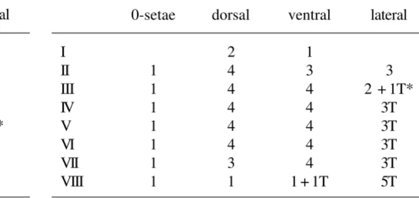
Table I. Pupal abdominal setation of Caladomyia ortoni (the numbers refers to pairs of setae).
* T = taeniae
I II III IV V VI VII VIII 0-setae 1 1 1 1 1 1 1 dorsal 2 4 4 4 4 4 3 1 ventral 1 3 4 4 4 4 4 1 + 1T
lateral
3 2 + 1T*
3T 3T 3T 3T 5T
Figs. 1-6. Caladomyia ortoni. Larva and pupa: 1, cephalic tubercle; 2, thorax, lateral; 3, abdomen, dorsal; 4, anal comb; 5, larval labro-epipharyngeal region; 6, larval antennal tubercles and clypeal setae S3.
1
0.1 mm2
0.5 mm4
0.05 mm
3
0.5 mm
5
0.1 mm
6
Figs. 7 – 11. Caladomyia riotarumensis. Pupa: 7, frontal apotome; 8, thoracic horn; 9, thorax, lateral; 10, abdomen, dorsal; 11, anal comb.
7
0.1 mm9
0.5 mm
0.5 mm
10
8
0.1 mm
11
0.1 mm
0.025 mm
Figs. 12 – 18.Caladomyia riotarumensis. Larva: 12, labro-epipharyngeal region; 13, antennal tubercles and clypeal setae S3; 14, premandible; 15, mandible; 16, mentum; 17, antenna; 18, posterior parapod claws.
12
13
17
0.1 mm
15
0.05 mm
14
0.05 mm
16
Received 28.X.2002; accepted 30.IX.2003
Head: Width 293 µm, length 375 µm. Clypeal (S3) simple
(Fig. 13). SI comb-like, bases fused; SII distally plumose, situated on pedestal about 1/2 as long as SII; SIII simple, seta-like; SIV present. Pecten epipharyngis with 3 distally serrated scales
(Fig. 12). Premandible 80-92 µm long with three teeth (Fig. 14);
brush well developed. Antennae 5-segmented on pedestal
60-70 µm long bearing a distinct apical tooth (Fig. 17); basal
segment 153 µm (148-157) longer than flagellum, with basal
ring organ and small seta in distal ½; segment 2 ½ as long as
segment 1; Lauterborn organs large, pedicels near 92 µm long,
longer than segment 3, their proximal half sclerotized. Mandible
(Fig. 15) about 110 µm with pale dorsal tooth; apical and 2 inner
teeth brown. Mentum (Fig. 16) 88 µm (84-95) large with pale
median tooth slightly notched laterally, 5 pairs of lateral teeth brown. Ventromental plates touching medially.
Abdomen with anal tubules curved down. Posterior parapods, in addition to simple hooks, with some serrate claws (Fig. 18).
Differential diagnosis. C. riotarumensis larva is similar to
C. friederi. The main difference is the large dimensions of the
first and the proportion of the antennal segments, and the
Caladomyia.
REFERENCES
LANGTON, P. H. 1994. If not “filaments”, then what? Chironomus6: 9. REIFF, N. 2000. Review of the mainly Neotropical genus Caladomyia
Säwedall, 1981, with description of seven new species (Insecta, Diptera, Chironomidae). In: M. BAEHR & M. SPIES (eds). Contributions to chironomid research in memory of Dr. Friedrich Reiss. Spixiana 23: 175-198.
SÆTHER, O. A. 1980. Glossary of chironomid morphology terminology (Diptera: Chironomidae). Entomologica Scandinavica Supplement 14: 1-51.
SÄWEDALL, L. 1981. Amazonian Tanytarsini II. Description of Caladomyia
n. gen. and eight new species (Diptera: Chironomidae).
Entomologica Scandinavica Supplement 12: 123-143. TRIVINHO-STRIXINO, S. & G. STRIXINO. 1991. Duas novas espécies de Nimbocera
Reiss (Diptera, Chironomidae) do Estado de São Paulo. Revista Brasileira de Entomologia 35(1): 173-178.
TRIVINHO-STRIXINO, S. & G. STRIXINO. 2000. A new species of Caladomyia