Scanning MscL Channels with Targeted
Post-Translational Modifications for Functional
Alterations
Irene Iscla☯, Robin Wray☯, Christina Eaton, Paul Blount *
Department of Physiology, University of Texas Southwestern Medical Center at Dallas, Dallas, Texas, United States of America
☯These authors contributed equally to this work. *[email protected]
Abstract
Mechanosensitive channels are present in all living organisms and are thought to underlie the senses of touch and hearing as well as various important physiological functions like osmoregulation and vasoregulation. The mechanosensitive channel of large conductance (MscL) fromEscherichia coliwas the first protein shown to encode mechanosensitive chan-nel activity and serves as a paradigm for how a chanchan-nel senses and responds to mechani-cal stimuli. MscL plays a role in osmoprotection inE.coli, acting as an emergency release valve that is activated by membrane tension due to cell swelling after an osmotic down-shock. Using an osmotically fragile strain in an osmotic down-shock assay, channel func-tionality can be directly determinedin vivo. In addition, using thiol reagents and expressed MscL proteins with a single cysteine substitution, we have shown that targeted post-transla-tional modifications can be performed, and that any alterations that lead to dysfuncpost-transla-tional proteins can be identified by thisin vivoassay. Here, we present the results of such a scan performed on 113 MscL cysteine mutants using five different sulfhydryl-reacting probes to confer different charges or hydrophobicity to each site. We assessed which of these tar-geted modifications affected channel function and the top candidates were further studied using patch clamp to directly determine how channel activity was affected. This comprehen-sive screen has identified many residues that are critical for channel function as well as highlighted MscL domains and residues that undergo the most drastic environmental changes upon gating.
Introduction
Mechano-transduction, the ability to sense and respond to mechanical stimuli, is thought to underlie the senses of touch, hearing, balance, proprioception as well as fundamental physio-logical processes, such as osmoregulation, vascular regulation, bladder control, and ischemia. Mechanosensitive channels are proposed to be the molecular entities underlying this sense at the cellular level, and are present in essentially all living organism from archaebacteria to plants
OPEN ACCESS
Citation:Iscla I, Wray R, Eaton C, Blount P (2015) Scanning MscL Channels with Targeted Post-Translational Modifications for Functional Alterations. PLoS ONE 10(9): e0137994. doi:10.1371/journal. pone.0137994
Editor:Zhe Zhang, Xuzhou Medical College, CHINA
Received:June 25, 2015
Accepted:August 25, 2015
Published:September 14, 2015
Copyright:© 2015 Iscla et al. This is an open access article distributed under the terms of the Creative Commons Attribution License, which permits unrestricted use, distribution, and reproduction in any medium, provided the original author and source are credited.
Data Availability Statement:All relevant data are within the paper and its Supporting Information files.
to mammals. The first cloned mechanosensitive channel is the bacterial mechanosensitive channel of large conductance, MscL, and it is to date one of the best studied, thus serving as a paradigm of how mechanosensors sense and respond to membrane stretch [1]. Although there are no obvious homologies between bacterial mechanosensitive (MS) channels and candidates for eukaryotic MS channels, there are structural similarities, including an amphipathic helix adjacent to the pore running parallel to the cytoplasmic side of the membrane, and a series of charges at the cytoplasmic region of a non-pore-forming transmembrane domain. In addition, channels from both kingdoms have shown modulation by some amphipathic compounds such as lysophosphatidylcholine (LPC) and arachidonic acid (AA), which alter the membrane lipid tension profile.
Mechanosensitive channels in bacteria serve a role in osmoprotection. MscL and the mechanosensitive channel of small conductance MscS have a concerted role in helping bacteria cope with sudden decreases in the osmolarity of their environment or osmotic down-shock. These channel’s large conductance of approximately 3 and 1 nS respectively allow for the rapid flow of solutes to reach a new osmotic balance and save the cells from lysis. They have a redun-dant role with only the double null strain being osmotically fragile. This phenotype can be res-cued with the expression, in trans, of either functional MscL or MscS and gives a sensitive screen to detect changes in channel functionin vivo.
The crystal structure of a closed state ofM.tuberculosisMscL was solved by the Rees group
revealing a homopentameric protein, with both N- and C-terminal predicted to be on the cyto-plasmic side [2,3]. The N-terminal domain appears as a helix running parallel to the cyto-plasmic membrane, the first transmembrane domain (TM1) forming the pore, the second transmembrane domain (TM2) in close contact with the lipids, the C-terminal domain form-ing a cytoplasmic helical bundle (Fig 1A). This bundle remains in place during channel gating [4]. Unfortunately, an open-state crystal structure of the channel has proved elusive to date, but a structural model of an open MscL based on electro paramagnetic resonance (EPR) studies has been generated, predicting a tilting of the two transmembrane domains during channel gat-ing [5].
The large conductance of 3nS and molecular sieving experiments predicting a 30 Å pore [6] suggest that the MscL channel undergoes a large conformational change upon channel gating with many residues and domains changing their local environment. Presumably, encouraging or discouraging these movements by making specific individual residues more hydrophilic or hydrophobic would lead to changes in the functional properties of the channel.
One of the great advantages of studying microbial channels is the ability to use screens to directly assess the functional properties of expressed proteins. Here we utilize such a screen to determine the activity of MscLin vivoby assessing phenotypic changes, and thus determine
which post-translational changes in MscL modified its function. Using a library of MscL cyste-ine mutants spanning 113 of the 136 residues, we studied the effects of five different post-trans-lational modificationsin vivo, thus totaling 678 potential changes, including the original
cysteine substitutions. Modifications that yielded interesting phenotypic changes have been further studied by patch clamp to determine their effects on channel activities. The data not only identify critical residues, but also make predictions of how the channel changes conforma-tion upon channel gating.
Materials and Methods
Strains and cell growth
All mutants were generated using the Mega Primer method as described [7] and were inserted in the pB10b or pB10d expression constructs [8–10].E.colistrain PB104 (ΔmscL::Cm) [9] was
used for electrophysiological and for stationary growth experiments. TheE.coliFRAG-1 [11]
derivative strain, MJF455ΔmscL::Cm,ΔmscSand MJF367ΔmscL::Cm[12] were used
experi-ments after osmotic down-shock and electrophysiology. Cultures were routinely grown in Len-nox Broth media (LB) (Fisher Scientific, Pittsburgh, PA), or citrate-phosphate defined medium (CphM) consisting of (per liter: 8.57 g of Na2HPO4, 0.87 g of K2HPO4, 1.34 g of citric acid, 1.0 g of NH4SO4, 0.001 g of thiamine, 0.1 g of Mg2SO4.7H2O, 0.002 g of (NH4)2SO4.FeS-O4.6H2O) plus 100μg/mL ampicillin, in a shaker-incubator at 37°C and rotated at 250 cycles per minute. Protein expression was induced by addition of 1 mM isopropyl-β -D-thiogalacto-pyranoside (IPTG) (Anatrace Inc, Maumee, OH).
Sulfhydrylreagents 4-hydroxybenzyl methanethiosulfonate (4-HB-MTS), benzyl metha-nethiosulfonate (MTSBn), decyl methametha-nethiosulfonate (decyl-MTS), 2-(Trimethylammonium) ethyl methanethiosulfonate Bromide (MTSET+) and 2-sulfonatoethyl methanethiosulfonate sodium salt (MTSES-) were obtained from Toronto Research Chemicals (North York, ON, Canada). MTS reagents were dissolved in methanol or ethanol, according to manufacturer’s
recommendation, to a 100mM concentration and stored at -20°C in argon; they were diluted to their final concentration right before using.
In Vivo
Functional Assays
Viability experiments were done using theE.coliMJF455 and MJF367 strains. Colonies were
grown overnight at 37°C in citrate phosphate medium plus 1 mM ampicillin. The overnight culture was diluted 1:20 in this defined medium, grown for 1h and then diluted to an OD600of 0.05 in the same medium supplemented with 0.5M NaCl. Cultures were induced at an OD600 0.2, for 30 minutes with 1 mM IPTG. The induced cultures were diluted 1:20 into (i) citrate-phosphate medium containing 0.5 M NaCl (mock shock); (ii) water (osmotic down-shock). Cells were then incubated at 37°C for 20 min, and then six consecutive 1:10 serial dilutions were made in medium containing either no salt (for the osmotic down-shock conditions) or 0.5 M NaCl (for the mock-shock conditions). These diluted cultures were plated and grown overnight and the colony-forming units were counted and averaged per experiment. To assess the effect of the different MTS reagents in cell viability they were added to the shock solution at the following concentrations: 50μM 4-HB-MTS, 5–10μM MTSBn, 10μM Decyl-MTS, 1mM MTSET+and 2mM MTSES-.
Electrophysiology
E.coligiant spheroplasts were generated and used in patch-clamp experiments as described
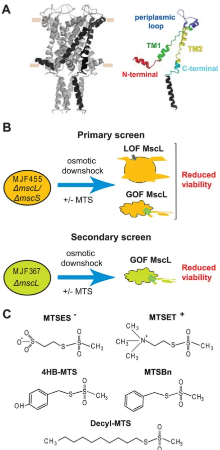
previously [13]. Excised, inside-out patches were examined at room temperature under sym-metrical conditions using a buffer comprised of 200 mM KCl, 90 mM MgCl2, 10 mM CaCl2, marked by horizontal tan lines. The domains, in which the study has been divided, are highlighted in an isolated subunit using different colors. (B) A schematic description of thein vivoscreens used to study the activity of the MscL cysteine substituted channels before and after post-translational modification with different MTS reagents. In a primary screen, the osmotically fragile strain MJF 455 is osmotically shocked with or without the MTS reagents present during the osmotic down-shock. Channels with reduced sensitivity (Loss of function LOF) or increased sensitive to tension (gain of function GOF), lead to cells with reduce viability in the primaryin vivoscreens. A secondary screen is done to distinguish between these two phenotypes. The secondary screen consists in MJF367 strain (not osmotically fragile) osmotically shocked with the MTS reagents present in during the osmotic down-shock. In the secondary screens a reduced viability indicates more sensitive channel or GOF. (C) The structure of the sulfhydryl reagents 2-sulfonatoethyl methanethiosulfonate sodium salt (MTSES-), ethyl methanethiosulfonate Bromide (MTSET+), 4-hydroxybenzyl methanethiosulfonate (4-HB-MTS), benzyl methanethiosulfonate (MTSBn), and decyl methanethiosulfonate (decyl-MTS), 2-(Trimethylammonium) are shown.
and 5 mM HEPES pH 6 (Sigma, St. Louis, MO). MTS reagents were added to the bath chamber or backfilled in the patch pipette at the following final concentrations: 50μM 4-HB-MTS, 100μM MTSBn, 10μM Decyl-MTS, 1mM MTSET+and 2mM MTSES-.
Recordings were performed at–20 mV (positive pipette). Data were acquired at a sampling rate of 20 kHz with a 5 kHz filter using an AxoPatch 200B amplifier in conjunction with Axo-scope software (Axon Instruments, Union City, CA). A piezoelectric pressure transducer (World Precision Instruments, Sarasota, FL) was used to monitor the pressure throughout the experiments. MscL pressure threshold was defined as the pressure at which openings were observed at least every 0.5 seconds. Measurements before and after treatment with MTS reagents were compared in the same patch as previously described [14]. Analysis was per-formed using Clampfit10 from Pclamp10 (Axon Instruments, Union City, CA).
Results
In vivo
screens
As the MscL channel gates, it changes conformation and several individual residues undoubt-edly change their local environment with some going into more hydrophilic environments, other going into more hydrophobic. If these changes are encouraged or inhibited, the function of the channel should be altered. Under the hypothesis that such changes in MscL channel function can be detected usingin vivoassays, we used a cysteine library (residues S2-L114,
excluding only the C-terminal helical bundle, which has little if any functional role [4,10,15], and post-translationally modified the channel with five different substitutions by using sulfhy-dryl probes. Each probe can potentially react with the cysteine, and since they have different charges or different affinities to the membrane, they conferred these properties to the cysteine substituted site. Therefore, by determining how these changes affect channel activityin vivo
andin vitro, we can infer what environmental changes take place during channel gating at that
site (Fig 1).
A combination of twoin vivoassays was used to determine the phenotype of the modified
channels. For the primaryin vivoscreen, we used theE.coliMJF 455 strain, null for two major
mechanosensitive channels. This is an osmotically fragile strain, whose viability is severely compromised upon a sudden reduction of the osmolarity of the media (osmotic down-shock) (S1A Fig). This phenotype can be rescued by expressionin transof WT MscL and to a different
extent by mutated MscL channels depending on the severity of the mutation on channel activ-ity. Although this is a very sensitive assay, two opposite channel phenotypes can lead to an observed reduced viability: channels that require very high pressures to gate, higher than the lytic pressure of the membrane, thus phenotypically loss of function (LOF), or channels that gate at lower than normal pressures and leak the contents of the cells, or gain of function (GOF) (Fig 1B). To distinguish between these two possible phenotypes, a secondin vivoscreen
can be used by evaluating the viability of a non-osmotically fragile strain MJF 367 (MscL null but still containing MscS), with or without the MTS reagents present during the osmotic down-shock. Only the misgating of MscL at lower than normal tensions, i.e. a GOF phenotype, causes a reduced viability; LOF phenotypes will not cause a reduced viability in these secondary screens because the cell contains MscS and thus is not osmotically fragile.
The ability of each mutated channel to rescue the osmotically fragileE.colistrain MJF
455 was assayed in anin vivoassay. Briefly, bacterial cells expressing each channel were grown
in a high osmolarity media and subsequently diluted in media of the same osmolarity (non-shocked) or into water to produce an osmotic down-shock; because the cysteine mutation itself could effect a LOF or GOF phenotype, some of the mutants demonstrated a loss-of-viability phenotype upon this osmotic down-shock (S1A–S1E Fig). In the same set of experiments, sam-ples were also shocked in the presence of the five different MTS reagents described above. For every mutant the plots represent the change in viability between shocked and shocked in the presence of the MTS reagents. Negative values indicate that the expressed channel malfunc-tions after modification with a probe, while a positive value means that the MTS treatment improves the functionality of that cysteine mutant, and thus its ability to better rescue the osmotically fragile strain. To distinguish modifications that yielded the greatest changes in via-bility, we used a benchmark of a 50% change in viability. For the clarity of the paper we present the data divided into protein domains.
The N-terminal region of
E.
coli
MscL: S1 and S1-TM1 linker
As described above, the effect of the cysteine substitutions in MscL channel activityin vivowas
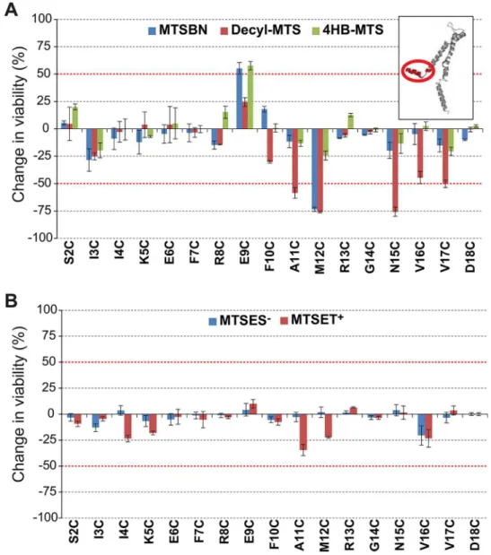
initially evaluated with no MTS reagents present during the osmotic down-shock. Of the 17 residues included in the N-terminal region (residues S2 to D18), 9 showed a lower viability due to a decrease in sensitivity or LOF phenotype and 3 appear to be due to a GOF phenotype (S1A Fig) [7,8]. Interestingly, this is the region in which the cysteine substitutions had the greatest effect on channel activity, thus highlighting its important role in channel gating [8].
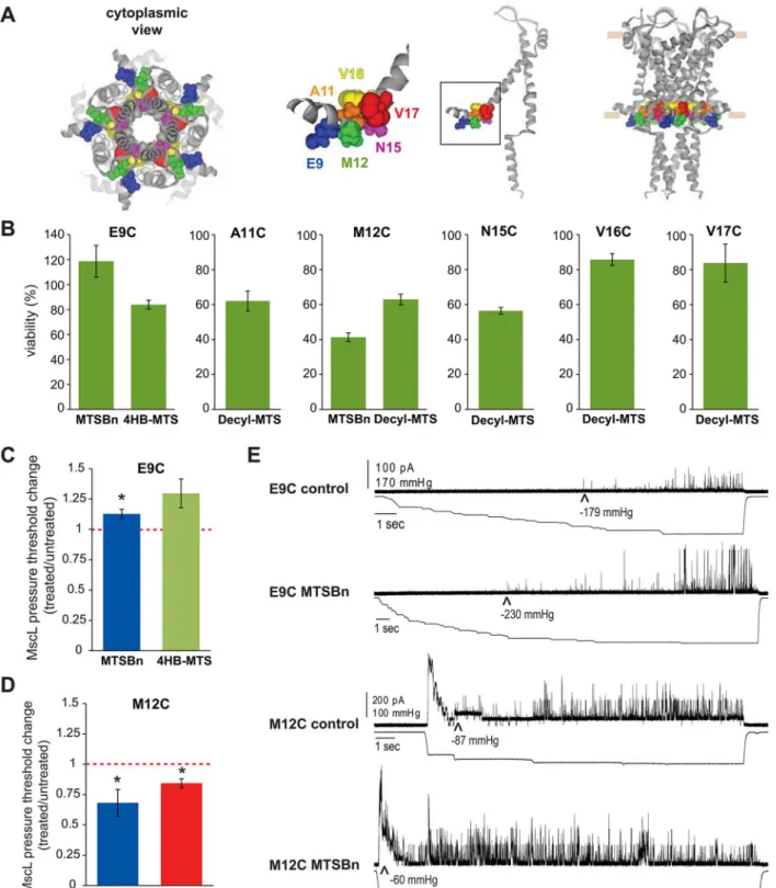
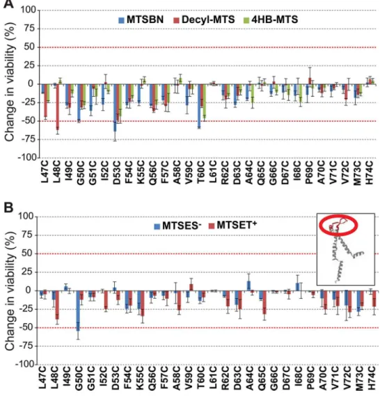
Post-translational modification with the hydrophobic MTS reagents MTSBn, decyl-MTS, and 4HB-MTS, produced greater than 50% changes in viability after an osmotic down-shock for channels modified at residues E9, A11, M12, N15, V16 and V17 (Figs2Aand3A). Interest-ingly, E9 showed an increase in viability as opposed to a decrease. These data would be consis-tent with the GOF phenotype observed for the untreated E9C mutated protein, as assayed by osmotic down-shock (seeS1 Figand [8]) being rectified by the modifications. No changes in the viability were observed when shocked in the presence of MTSES-and MTSET+(Fig 2B).
Secondary screens were performed in order to determine which channel phenotype caused the observed changes in viability. As explained inFig 1B, areduced viability in the secondary screens are the result of more sensitive channels, a GOF phenotype, while an unchanged viability indicates a LOF phenotype. Using the strain MJF367, which is only null for MscL and thus not osmotically fragile, hydrophobic substitutions at residues A11, M12 and N15 led to reduced via-bility, demonstrating that these modifications lead to a GOF phenotype (Fig 3A and 3B). In con-trast with these results, modification of V16 and V17 residues gave no indication of leading to a GOF phenotype after modification in the secondary screens, suggesting they led to a LOF phenotype. As discussed above, E9C is a GOF independent of any modification and it appears this phenotype is remediated by MTSBn and 4HB-MTS; these data could indicate that these modifications lead to a more wild type functional channel, or a LOF channel.
The first transmembrane domain: TM1
The effect of cysteine substitutions on MscL channel activityin vivowas studied for the pore
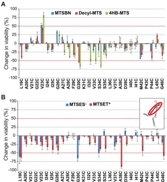
forming domain TM1 (residues L19-G46). Of the 25 residues included in this region 3 showed a lower viability due to a decrease in sensitivity or LOF phenotype and 6 due to a GOF pheno-type (S1B Fig)[7]; the latter are presumably because of the disruption of the hydrophobic lock that stabilizes the closed state of the channel [10].
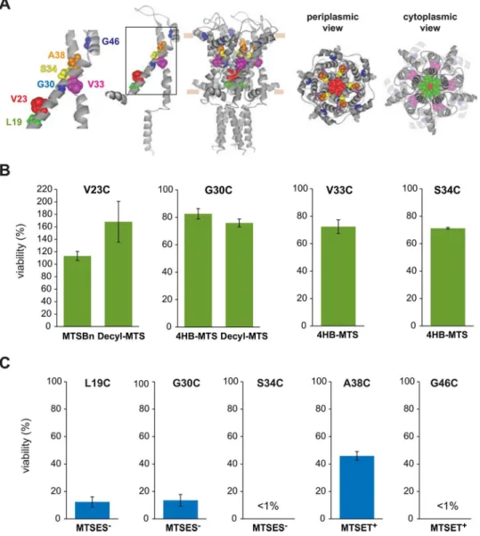
Post-translational modification in this region with the hydrophobic MTS reagents MTSBn, decyl-MTS, and 4HB-MTS, produced changes greater than 50% in their viability after an osmotic down-shock for mutants V23C, G30C, V33C, and S34C (Fig 4A). Again one of the
Fig 2. Effects of post translational modifications on the N-terminal domain of MscL determined byin vivoscreens.The viability of the osmotically fragile strain MJF455 expressing MscL cysteine substituted mutants from residues S2 to D18 (insert), was measured after an osmotic down-shock. The graphs show the differences in viability between non-treated and post-transnationally modified channels with (A) the hydrophobic MTS reagents MTSBn (blue), decyl-MTS (red) or 4HB-MTS (green) or (B) the negatively charged MTSES-(blue) and positively charged MTSET+(red). The red grid line indicates a±50% change that was used as a threshold for further studies.
Fig 3. Functional changes by substitutions in the N-terminal domain of MscL, determined byin vivoand patch clamp experiments.(A) The location of the residues showing changes in viability upon post-translational modifications is highlighted in the closed structure ofE.coliMscL. From right to left: a pentameric MscL is shown in a side view followed by a single subunit and a close up of the region and a cytoplasmic view of the pentameric channel. (B) Viability of MJF 367 (mscL-) shocked in the presence of the indicated MTS reagent is shown for each individual MscL mutant. (C) The changes in the pressure threshold required to gate E9C MscL caused by treatment with MTS reagents is graphed as the ratio between before and after modification of the same patch. The red line indicated no change. (D) The changes in the pressure threshold required to gate M12C MscL caused by MTS reagents is graphed as the ratio between before and after modification of the same patch. The red line indicates no change. (E) Representative traces of E9C MscL and M12C before (control) and after treatment with MTSBn. The upper traces correspond to the current and the lower traces the negative pressure applied to the patch.
mutated channels, V23C, showed an increased viability upon modification, suggesting some sort of remediation of the GOF phenotype induced by the cysteine mutation (seediscussionof E9C above). Changes in the viability were observed for mutants L19C, G30C, A38C and G46C, when shocked in the presence of MTSES-and MTSET+(Fig 4B).
To determine which channel phenotype caused the observed changes in viability, secondary screens were performed. As discussed above, the interpretation of data for V23C is an apparent remediation of the GOF phenotype by the hydrophobic probes. For mutants G30C, V33C and S34C, the hydrophobic substitutions didn’t affect the viability in the assay using the MJF367
MscL-null strain; on the other hand, charged substitutions yielded dramatic decreases in viabil-ity for L19C, G30C, S34C, A38C and G46C with this assay (Fig 5A and 5B). In essence, thein Fig 4. Effects of post translational modifications on the TM1 domain of MscL determined byin vivo
channel activity.The viability of the osmotically fragile strain MJF455 expressing MscL cysteine substituted mutants from residues L19 to G46 (insert), was measured after an osmotic down-shock. The graphs show the differences in viability between non-treated and post-transnationally modified channels with (A) the
hydrophobic MTS reagents MTSBn (blue), decyl-MTS (red) or 4HB-MTS (green) or (B) the negatively charged MTSES-(blue) and positively charged MTSET+(red). The red grid line indicates a±50% change that was used as a threshold for further studies.
vivodata for TM1 is consistent with a“hydrophobic lock”hypothesis which states that it is
largely the transient exposure of hydrophobic residues in TM1 to an aqueous environment that is the primary energy barrier for channel opening [10,19–22].
The TM1 domain has been previously well studied and many MscL channels with modifica-tions in this domain, including many of the sites identified in these screens, have previously been patched with results supporting the“hydrophobic lock”. One potential complication of working with channels with cysteine mutations in this domain is that while they may give GOF phenotypesin vivo, where the environment is highly reduced, they may form inter-subunit
disulfide bridges that lock the channel closed in patch clamp at ambient redox. This has been
Fig 5. Functional changes by substitutions in the TM1 domain of MscL, determined byin vivoand patch clamp experiments.(A) The location of the residues showing changes in viability upon post-translational modifications is highlighted in the closed structure ofE.coliMscL. From right to left: a pentameric MscL is shown in cytoplasmic view, a periplasmic view, and a side view where the approximate location of the membrane is shown. A single subunit and a close up of the region with the labeled residues is on the left. (B) Viability of MJF 367 (mscL-) cells, shock in the presence of the indicated hydrophobic MTS reagent is shown for each individual MscL mutant. (C) Viability of MJF 367 osmotically shocked with charged MTS reagents is shown for the indicated cysteine mutants.
previously reported [7], and an example is shown for V23C treated with MTSBn and decyl-MTS inS2 Fig.
The periplasmic loop
The effect of cysteine substitutions on MscL channel activityin vivowas studied for the
peripla-mic loop (residues L47-H74). Of the 28 residues included in this region, 3 showed a lower via-bility due to a decrease in sensitivity or LOF phenotype and 2 due to a GOF phenotype (S1C Figand [7]).
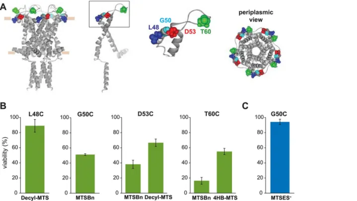
Post-translational modification in this region with the hydrophobic MTS reagents MTSBn, decyl-MTS, and 4HB-MTS, caused changes in viability after an osmotic shock for mutants L48C, G50C, D53C, and T60C (Fig 6A). A similar effect was observed for G50C when shocked in the presence of MTSES-(Fig 6B).
To determine which channel phenotype caused the observed changes in viability, secondary screens were performed. In these screens, only small if any decreases in viability were observed, suggesting that none of the modifications lead to a severe GOF phenotype, with the exception of T60C treated with MTSBn (Fig 7A and 7B).
The second transmembrane domain: TM2
The effect of cysteine substitutions on MscL’s channel activityin vivowas studied for the
sec-ond transmembrane domain TM2, (residues Y75-N100). I92C MscL was excluded from this study because expression of this mutant caused a severe reduction on bacterial growth [7]. Of the 25 residues included in these region 7 showed a lower viability due to a decrease in sensi-tivity or LOF phenotype and 2 due to a GOF phenotype (S1D Figand (Levin and Blount, 2004)).
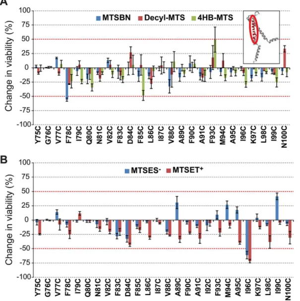
Post-translational modifications in this region with the hydrophobic MTS reagent MTSBn, changed the viability of F78C more than 50% when compared to only osmotic down-shock (Fig 8A). Changes in the viability of cells expressing I96C MscL were observed when shocked in the presence of MTSES-and MTSET+(Fig 8B).
To determine which channel phenotype caused the observed changes in the viability, sec-ondary screens were performed. MTSBn substitution at residue F78 and charged substitutions at residue I96, effected GOF phenotypes (Fig 9A and 9B). Interestingly, a patch clamp analysis of the effects of the MTSBn modification at site F78, showed opposite results relative to those observedin vivo;treatment with MTSBn produced a statistically significant increase in the
pressure threshold needed to gate the channels (treated/untreated 1.19 ± 0.01, n = 5, p<0.005
paired Student t-test).
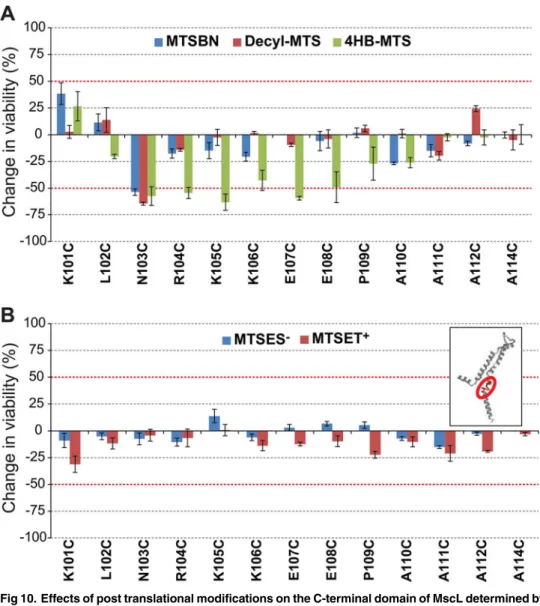
The C-terminal domain
The effect of cysteine substitutions on MscL’s channel activityin vivowas studied for the
sec-ond transmembrane domain TM2, (residues K101-A114). Of the 13 residues included in this region 2 showed a lower viability due to a GOF phenotype (S1F Figand [23]).
Post-translational modification in this region with the tyrosine like MTS reagent 4HB-MTS changed the viability of N103C to E108C more than 50% when compared to osmotic down-shock only. N103C viability was also affected by the presence of MTSBn and decyl-MTS (Fig 10A). No changes were observed in this region when shocked in the presence of the charged probes MTSES-or MTSET+(Fig 10B).
reported, N103 showed a dichotic behavior with MTSBn and decyl-MTS effecting a GOF phe-notype, but not 4HB-MTS apparently because of its movement into the membrane upon chan-nel gating [14].
The patch clamp results shown inFig 11Ccorrelate with thein vivoresults, with 4HB-MTS
increasing the pressure threshold needed to gate R104C, K105C and K106C. Interestingly MTSBn and decyl-MTS have opposite effect on residue R104, a pattern similar to the one observed for its neighbor residue N103 [14].
Discussion
The molecular mechanisms underlying the gating of mechanosensitive channels is a field of increasing interest since these channels are involved in essential physiological processes such
Fig 6. Effects of post translational modifications on the periplasmic loop of MscL determined byin vivochannel activity.The viability of the osmotically fragile strain MJF455 expressing MscL cysteine substituted mutants from residues L47 to H74 (insert), was measured after an osmotic down-shock. The graphs show the differences in viability between non-treated and post-transnationally modified channels with (A) the hydrophobic MTS reagents MTSBn (blue), decyl-MTS (red) or 4HB-MTS (green) or (B) the negatively charged MTSES-(blue) and positively charged MTSET+(red). The red grid line indicates a±50% change that was used as a threshold for further studies.
as vasoregulation, touch, hearing, pain, depression, and responses to ischemia. Interestingly, mechanosensitive channels from eukaryotic and prokaryotic organisms are found in struc-turally unrelated protein families. For example, the few eukaryotic mechanosensitive chan-nels known to date belong to very different families like the two pore potassium chanchan-nels (TRAAK, TREK-1, and TREK-2), the transient receptor potential family (TRPV4) and the Piezo channels [24–26]. The prokaryotic mechanosensitive channels MscL and MscS are also structurally unrelated. However, some of these channels have common mechanisms of acti-vation including modulation by amphipaths such as LPC and arachidonic acid, and there are also some analogous and common structural domains in otherwise very different families. For example anα- helix running along the cytoplasmic membrane and connected to the pore domain plays a crucial role in the gating of such different channels as TRPY1, TRAAK, TREK, MscL and MscS [2,8,27–30]. This highlights the relevance of a detailed understand-ing of the gatunderstand-ing mechanisms of mechanosensitive channels.
The choice of thein vivoscreens as the main approach to determine MscL channel activity
has the double advantage of giving relevant information about channel function in its native environment and comparing channel activity before and after post-translational modifications. These post-translational modifications at the site of the cysteine substituted residue confer it with different properties and their effects on channel activity can be directly evaluated. Further-more, the possibility to combine bacterial screens using different bacterial strains allows one to distinguish between different channel phenotypes and better understand the transitions of MscL domains during gating. It should be pointed out that negative results are difficult to interpret; a lack of effect could mean that the site is tolerant of changes, or it could simply be
Fig 7. Functional changes by substitutions in the periplasmic loop of MscL, determined byin vivoand patch clamp experiments.(A) The location of the residues showing changes in viability upon post-translational modifications is highlighted in the closed structure ofE.coliMscL. From right to left: a pentameric MscL is shown in cytoplasmic view, a periplasmic view, and a side view where the approximate location of the membrane is shown. A single subunit and a close up of the region with the labeled residues is on the left. (B) Viability of MJF 367 (mscL-) shock in the presence of the indicated MTS reagent is shown for each individual MscL mutant, for hydrophobic or (C) charged MTS reagents.
that the residue is buried and inaccessible to the reagents. This could, for instance, explain why there are only 2 hits within TM2, which is predicted to have ample lipid and protein-protein interactions, and both hits were toward the cusp of the domain. However, the predic-tions of large conformational changes occurring upon gating combined with a pore size large enough to easily pass the reagents to the cytoplasmic side would suggest that accessibility should be better than typical membrane proteins. Indeed, the assay identified several residues predicted to be inaccessible. One of the more extreme examples is L96, toward the cytoplasmic end of TM2, which was influenced by the charged MTSET+and MTSES-reagents; presumably this could only occur if the reagents crossed the open pore to gain cytoplasmic accessibility.
Early in the study of MscL, the N-terminal domain was shown to play a crucial role in chan-nel gating since deletion of just 11 amino acids lead to a non-functional chanchan-nel [10]. From the
Fig 8. Effects of post translational modifications on the TM2 domain of MscL determined byin vivochannel activity.The viability of the osmotically fragile strain MJF455 expressing MscL cysteine substituted mutants from residues Y75 to N100 (insert), was measured after an osmotic down-shock. The graphs show the differences in viability between non-treated and post-transnationally modified channels with (A) the hydrophobic MTS reagents MTSBn (blue), decyl-MTS (red) or 4HB-MTS (green) or (B) the negatively charged MTSES-(blue) and positively charged MTSET+(red). The red grid line indicates a±50% change that was used as a threshold for further studies.
first analysis of the crystal structure of theM.tuberculosisMscL, this region was not resolved,
but later in a reanalysis of the crystallographic data it was solved as aα- helix running along the cytoplasmic membrane [3,31]. Molecular dynamic simulation using this newly defined structure embedded in lipids, first proposed that the S1 region remains parallel to the mem-brane during MscL gating [32]. A systematic study of the disulfide bridging pattern,in vivo
and patch clamp data agreed with the proposed location of the N-terminal domain. From these data, we proposed a model in which this region would act as a sensor and stabilizer of the TM1 domain during channel opening [8]. Further studies using a combination of patch clamp, fluo-rescence resonance energy transfer (FRET) spectroscopy, electron paramagnetic resonance experiments, and molecular and Brownian dynamics simulations, suggest a similar role for this region [16]. Interestingly, an amphipathicα-helix connected to the pore domain by a flexible glycine, is found in various mechanosensitive channels as diverse as TRAAK, 1, TREK-2 [29,30,33], the yeast transient receptor potential channel, TRPY1 [27], and the bacterial mechanosensitive channel MscS [2,3].
Although several cysteine substitutions in the N-terminal region lead to altered channel phenotypes (S1 FigA), very few of the post-translational modifications produced changes in viability (Fig 2A and 2B). This lack of observed effect may be due to the fact that the N-terminal domain is densely packed with the cytoplasmic region of TM2 at the lipid interface [34]. This conformation could either interfere with the accessibility of MTS probes to the sites, or make the region more sensitive to the size of the probes. One such example is residue E9, which leads to a GOF phenotype when substituted to cysteine. Interestingly E9 is in close proximity to resi-due K101 as determined by disulfide trapping experiments, and favoring this interaction leads to a locked closed channel [34]. Although it has also been shown that electrostatic interactions between E9 and K101 are crucial for channel gating [23], we failed to see a remediation of E9C GOF phenotype when treated with MTSES-. As stated earlier, this lack of an effect could be due to inaccessibility, or to the bulkier MTS substitution affecting channel function in this tightly packed region. Hydrophobic substitutions at A11 and M12 conferred upon the channels a GOF phenotype (Fig 3B). As confirmed by direct study of the channels activity by patch clamp experiments, hydrophobic post-translational modifications in M12C with decyl and MTSBn, significantly lowered the channels pressure threshold (Fig 3D). This decrease in pressure threshold after a hydrophobic substitution suggests that while the channel opens these residues move to a more hydrophobic environment, which could be the lipid membrane since these res-idues are located at the cytoplasmic membrane interphase of TM1.
The R13 to D18 region, sometimes called the S1-TM1 linker of MscL, is highly conserved and has been proposed to contain a very conserved motive (NhhD), part of a common ances-tral sensor for both voltage and membrane tension [35,36]. Interestingly, residues N15 and D18 in Eco MscL have been proposed essential for the formation of a water chain across the channel that leads to and stabilizes the opening of MscL [32]. Cysteine substitutions of the most conserved residues in this domain, including R13, G14 and D18, produced very strong phenotypes when mutated to cysteines, causing a reduction in viability of about 90% (S1A Fig).
Fig 9. Functional changes by substitutions in the TM2 domain of MscL, determined byin vivoand patch clamp experiments.(A) The location of the residues showing changes in viability upon post-translational modifications is highlighted in the closed structure ofE.coliMscL. A pentameric MscL is shown side view where the approximate location of the membrane marked, followed by single subunit with the labeled residues. Cytoplasmic and periplasmic views are also shown below. (B) Viability of MJF 367 (mscL-) cells, shock in the presence of the indicated MTS reagent is shown for each individual MscL, for hydrophobic or C. charged MTS reagents.
R13C is a strong GOF mutant reducing growth by 70% and G14C and D18C are strong LOF mutants [7]. Surprisingly, modification of the cysteines with the MTS reagents used in this study did not significantly modify these phenotypes, as determined by ourin vivoscreen. In
one model, the R13 residues are proposed to approach their counterparts in neighboring sub-units during gating with the electrostatic repulsion acting as a resistive force to gating (Levin and Blount, 2004); the finding that neither MTSET+nor MTSES-sustitutions appear to reme-diate the GOF phenotype in our screens, suggests that either the residue is inaccessible, or that the model may be an over simplification. In another detailed model for the gating ofE.coli
MscL which proposed the tilting of the transmembrane domains during opening, G14 was sug-gested to act as a hinge in the transmission of tension between S1 and TM1[37,38], so it may not be surprising that few if any substitutions at this location are tolerated. Interestingly, G14 is
Fig 10. Effects of post translational modifications on the C-terminal domain of MscL determined byin vivochannel activity.The viability of the osmotically fragile strain MJF455 expressing MscL cysteine substituted mutants from residues K101 to A114 (insert), was measured after an osmotic down-shock. The graphs show the differences in viability between non-treated and post-transnationally modified channels with (A) the hydrophobic MTS reagents MTSBn (blue), decyl-MTS (red) or 4HB-MTS (green) or (B) the negatively charged MTSES-(blue) and positively charged MTSET+(red). The red grid line indicates a±50% change that was used as a threshold for further studies.
the hinge between the cytoplasmic amphipathicα-helix and the pore region, part of the com-mon structural feature shared by many otherwise structurally unrelated mechanosensitive channels.
The pore forming first transmembrane domain of MscL was found to be a hot mutational spot in a random mutagenesis study where many mutations, almost all to more hydrophilic residues, led to more sensitive or GOF channels [22]. A subsequent study mutated a single resi-due, G22, to all other amino acids and confirmed that at least for this site, G22, the GOF phe-notype correlated with the hydrophilicity of the residue [39]. There are several lines of data that suggest this domain twists in a clockwise (as seen from the periplasm) corkscrew fashion upon channel opening [5,19,40,41]. Furthermore, it has been proposed that a“hydrophobic lock”at the constriction point of the pore stabilizes the closed state of MscL and it is the tran-sient exposure of these residues to an aqueous environment upon channel gating that is the pri-mary energy barrier [13]. Cysteine substitutions in the cytoplasmic part of TM1 effected only GOF phenotypes (S1B Fig), [7]; for residue V23C, the proposed constriction point, post-trans-lational modification with hydrophobic probes reverted this phenotype (Fig 4A). A similar effect has been described for residue G26C [19,20,42], for which a longer time of treatment with decyl-MTS leads to a non-functional channel, probably due to an unbreakable hydropho-bic lock when more subunits are substituted. As shown for the V23C MscL (S2 Fig), the behav-ior in patch of this mutated channel is complicated, presumably due to disulfide bridging in the
in vitroredox environment. This is true for other TM1 mutants including R13C and G26C,
presumably due to their close proximity in the pentameric structure. It is interesting to point out that, in the case V23C, treatment with MTS reagents to cross-linked channels is indeed effective as seen for the reversion of the channel phenotypes, implying that the probes are able to break these cysteine bonds (S2 Fig).
The periplasmic loop of MscL is the least conserved part of the channel and probably the least studied. Early work showed that enzymatic digestion of the periplasmic loop, periplasmic and cytoplasmic extra membrane domains of MscL, or reconstitution of separately synthesized TM1 and TM2 domains, all lead to more sensitive channels that retained their mechanosensi-tive. These studies suggested that these domains tuned the mechanosensitivity level of the channel [43,44]. Consistent with this work, it was also found that select mutations at Q65 could lead to either GOF or LOF phenotypes [45]. In addition, a molecular dynamic simulation predicts that the loop region is highly mobile, and attributes this region a role in the stability of the open state as well as structural stability during the tilting of the membranes during gating [32]. Cysteine substitution in this region caused few but strong altered phenotypes. One inter-esting residue in the periplasmic loop region, Q56, is also known to modulate the kinetics of MscL, with aromatic substitutions drastically increasing the channel mean open time [10]. Our results agree with this previous observation, with the aromatic substitutions decreasing the via-bility of the cells (Fig 6A), although the change did not reach the 50% cut of established for this work. Interestingly, recent work using chimeras between MscL orthologues, found that residue I49 plays a critical role in channel kinetics, acting as a“clasp knife spring”controlling the
Fig 11. Functional changes by substitutions in the C-terminal domain of MscL, determined byin vivo
and patch clamp experiments.(A) The location of the residues showing changes in viability upon post-translational modifications is highlighted in the closed structure ofE.coliMscL. From left to right: a pentameric MscL is shown in a side view where the approximate location of the membrane marked, a single subunit and a close up of the region with the labeled residues followed by a cytoplasmic view. (B) Viability MJF 367 (mscL-) cells shock in the presence of the indicated MTS reagent is shown for each individual MscL mutant. C. The changes in the pressure threshold required to gate R104C, K105C and K106C MscL caused by MTS reagents is graphed as the ratio between before and after modification of the same patch. The red line indicates no change.
stability of the open and closed channel states of the channel [46]. At the neighboring residue L48, post-translational modification with decyl-MTS, effected a strong LOF phenotype, proba-bly because the anchoring of the protein at the membrane at this site impairs the mobility required to gate MscL.
The second transmembrane domain of MscL surrounds TM1, and is in close contact with the lipids. One of the hits in this region was I96C treated with either MTSET+or MTSES-. This residue has been previously shown to yield a GOF phenotype when mutated to a positive resi-due, and is predicted to interact with V23 upon channel gating [41]. But perhaps the most interesting residue within this domain is F78, which is predicted to face the membrane, and has been proposed as a membrane tension sensor in an asparagine scan of the periplasmic interface of TM2 [47]. Among the most conserved residues in this region are residues G76, F78, F85 and F93 [48], but interestingly cysteine substitutions have little (F78, F85) or the opposite effect on channel function (G76C), than the asparagine substitutions. While the F78C mutated protein had little if any phenotypein vivo, treatment with MTSBn, which should
reconstitute the aromatic ring to the site, led to a GOF phenotype. The fact that the disulfide linkage adds bulk to the residue and is not a strict reconstitution of the original structure can also account for this effect. To further complicate the interpretation, the channel phenotype, as assayed by patch clamp, appeared to be a LOF when treated with MTSBn. These data might be explained by subtle differences between thein vivoassay and patch clamp including the
mem-brane potential and a potential asymmetry between the two leaflets of the lipid bilayersin vivo
that may be lost upon spheroplasts preparation, which could contribute to the discrepancies. Given the differences between thein vivoand patch clamp experiments it is surprising that in
most instances the data from these two experimental approaches correlate well.
The cytoplasmic region of MscL at the lipid-cytoplasmic interphase has been proposed to enter a more hydrophobic environment to accommodate for the tilting of TM2 while channel gating [14,23]. We have proposed that a conserved charge cluster acts as a“knot in a rope”to stop the tilting and incorporation of the helix in the membrane Although deletion studies have shown that charges in the conserved RKKEE region (residues 104–108) are essential for chan-nel function, our data suggest that no single residue plays a critical function and instead it appears to be the property of the entire cluster that is important (S1E Fig). In our previous study, we concentrated on N103 and found a correlation between hydrophobicity of the substi-tution and channel sensitivity. Furthermore, fluorescence quenching measurements in the presence of brominated lipids suggested that the residue approaches the lipid membrane dur-ing channel gatdur-ing. Here we further find that the residue adjacent to N103, R104, displays a similar behavior in patch clamp experiments, with the very hydrophobic substitutions by MTSBn and decyl-MTS leading to more sensitive channels and the tyrosine like substitution to less sensitive channels (Fig 11C). Interestingly, restoration of, by post-translational modifica-tion with the charged MTS probes, did not have an effect in channel activityin vivo(Fig 10B).
Again the tight packing of this region might either impair probe accessibility or be too sensitive to the size of the probe.
Although MscL is probably the best studied MS channel, the lack of an open crystal struc-ture makes predicting the changes that occur upon gating difficult. However, because of the conformational changes are large, leading to what is the largest gated pore known, many resi-dues and sub-domains drastically change their local environment upon gating. Hence, studies in which the fundamental properties of specific residues are changed can give valuable clues for transition and open states. In the present work, usingin vivoscreens we identify important
channel function, as well as highlighting MscL domains that undergo the more drastic environ-mental changes upon gating.
Supporting Information
S1 Fig. Effect of single cysteine substitutions on MscL channel functionin vivo.The viability
after an osmotic down-shock of MJF 455 cells expressing single cysteine mutants is shown for substitutions in (A) the N-terminal region, (B) the first transmembrane domain (TM1), (C) the periplasmic loop, (D) the second transmembrane domain (TM2) and (E) the C-terminal domain ofE.coliMscL. The red dotted line is for reference that indicates the viability after an
osmotic down-shock of MJF 455 cells expressing wild type MscL. Red asterisks highlight mutants whose decrease in viability is due to a gain of function phenotype. Each bar represents the mean of at least three independent experiments. Error bars represent standard error. (EPS)
S2 Fig. Effect of post translational modifications on V23C MscL channel activity in patch clamp.A. The changes in the pressure threshold required to gate V23C MscL after MTS
treat-ment, are presented as the ratio between the pressure threshold before and after modification of the same patch. The red line indicates no change. B. Representative traces of V23C MscL and before (control) and after treatment with MTSBn and decyl-MTS. The negative pressure applied to the patch is under each trace on the right.p<0.007 paired Student t-test n
7.
(EPS)
Acknowledgments
The authors thank Drs. Hannah Malcolm and Limin Yang, for useful discussions and critical reading of the manuscript and Jonathan Padro Arroyo for technical support.
Author Contributions
Conceived and designed the experiments: PB II. Performed the experiments: II RW CE. Ana-lyzed the data: II RW CE. Wrote the paper: II RW PB.
References
1. Iscla I, Blount P. Sensing and responding to membrane tension: the bacterial MscL channel as a model system. Biophys J. 2012; 103(2):169–174. doi:10.1016/j.bpj.2012.06.021PMID:22853893
2. Bass RB, Strop P, Barclay M, Rees DC. Crystal structure ofEscherichia coliMscS, a voltage-modu-lated and mechanosensitive channel. Science. 2002; 298(5598):1582–1587. PMID:12446901
3. Steinbacher S, Bass R, Strop P, Rees DC. Structures of the prokaryotic mechanosensitive channels MscL and MscS. In: Hamill OP, editor. Mechanosensitive Ion Channels (a volume in the Current Topics in Membranes series). 58. St. Louis, MO: Elsievier Press; 2007. p. 1–20.
4. Yang LM, Wray R, Parker J, Wilson D, Duran RS, Blount P. Three routes to modulate the pore size of the MscL channel/nanovalve. ACS Nano. 2012; 6(2):1134–1141. doi:10.1021/nn203703jPMID:
22206349
5. Perozo E, Cortes DM, Sompornpisut P, Kloda A, Martinac B. Open channel structure of MscL and the gating mechanism of mechanosensitive channels. Nature. 2002; 418(6901):942–948. PMID:
12198539
6. Cruickshank CC, Minchin RF, Le Dain AC, Martinac B. Estimation of the pore size of the large-conduc-tance mechanosensitive ion channel ofEscherichia coli. Biophys J. 1997; 73(4):1925–1931. PMID:
9336188
8. Iscla I, Wray R, Blount P. On the structure of the N-terminal domain of the MscL channel: helical bundle or membrane interface. Biophys J. 2008; 95(5):2283–2291. doi:10.1529/biophysj.107.127423PMID:
18515388
9. Blount P, Sukharev SI, Moe PC, Schroeder MJ, Guy HR, Kung C. Membrane topology and multimeric structure of a mechanosensitive channel protein ofEscherichia coli. EMBO J. 1996a; 15(18):4798– 4805.
10. Blount P, Sukharev SI, Schroeder MJ, Nagle SK, Kung C. Single residue substitutions that change the gating properties of a mechanosensitive channel inEscherichia coli. Proc Natl Acad Sci USA. 1996; 93 (21):11652–11657. PMID:8876191
11. Epstein W, Davies M. Potassium-dependent mutants ofEscherichia coliK-12. J Bacteriol. 1970; 101 (3):836–843. PMID:4908783
12. Levina N, Totemeyer S, Stokes NR, Louis P, Jones MA, Booth IR. Protection ofEscherichia colicells against extreme turgor by activation of MscS and MscL mechanosensitive channels: identification of genes required for MscS activity. EMBO J. 1999; 18(7):1730–1737. PMID:10202137
13. Blount P, Moe PC. Bacterial mechanosensitive channels: integrating physiology, structure and func-tion. Trends Microbiol. 1999; 7(10):420–424. S0966-842X(99)01594-2 [pii] PMID:10498951
14. Iscla I, Wray R, Blount P. Anin vivoscreen reveals protein-lipid interactions crucial for gating a mechan-osensitive channel. FASEB J. 2011; 25(2):694–702. doi:10.1096/fj.10-170878PMID:21068398
15. Häse CC, Ledain AC, Martinac B. Molecular dissection of the large mechanosensitive ion channel (Mscl) ofE.coli—mutants with altered channel gating and pressure sensitivity. J Membr Biol. 1997; 157 (1):17–25. PMID:9141355
16. Corry B, Hurst AC, Pal P, Nomura T, Rigby P, Martinac B. An improved open-channel structure of MscL determined from FRET confocal microscopy and simulation. J Gen Physiol. 2010; 136(4):483–494. doi:
10.1085/jgp.200910376PMID:20876362
17. White SH, Wimley WC. Membrane protein folding and stability: physical principles. Annu Rev Biophys Biomol Struct. 1999; 28:319–365. doi:10.1146/annurev.biophys.28.1.319PMID:10410805
18. Enkvetchakul D, Jeliazkova I, Bhattacharyya J, Nichols CG. Control of inward rectifier K channel activity by lipid tethering of cytoplasmic domains. J Gen Physiol. 2007; 130(3):329–334. PMID:17698595 19. Bartlett JL, Levin G, Blount P. Anin vivoassay identifies changes in residue accessibility on
mechano-sensitive channel gating. Proc Natl Acad Sci USA. 2004; 101:10161–10165. PMID:15226501 20. Bartlett JL, Li Y, Blount P. Mechanosensitive channel gating transitions resolved by functional changes
upon pore modification. Biophys J. 2006; 91(10):3684–3691. PMID:16935962
21. Blount P, Schroeder MJ, Kung C. Mutations in a bacterial mechanosensitive channel change the cellu-lar response to osmotic stress. J Biol Chem. 1997; 272(51):32150–32157. PMID:9405414
22. Ou X, Blount P, Hoffman RJ, Kung C. One face of a transmembrane helix is crucial in mechanosensi-tive channel gating. Proc Natl Acad Sci USA. 1998; 95(19):11471–11475. PMID:9736761
23. Zhong D, Blount P. Electrostatics at the membrane define MscL channel mechanosensitivity and kinet-ics. FASEB J. 2014; 28(12):5234–5241. doi:10.1096/fj.14-259309PMID:25223610
24. Anishkin A, Loukin SH, Teng J, Kung C. Feeling the hidden mechanical forces in lipid bilayer is an origi-nal sense. Proc Natl Acad Sci U S A. 2014; 111(22):7898–7905. doi:10.1073/pnas.1313364111PMID:
24850861
25. Coste B, Mathur J, Schmidt M, Earley TJ, Ranade S, Petrus MJ, et al. Piezo1 and Piezo2 are essential components of distinct mechanically activated cation channels. Science. 2010; 330(6000):55–60. doi:
10.1126/science.1193270PMID:20813920
26. Coste B, Xiao B, Santos JS, Syeda R, Grandl J, Spencer KS, et al. Piezo proteins are pore-forming sub-units of mechanically activated channels. Nature. 2012; 483(7388):176–181. doi:10.1038/nature10812 PMID:22343900
27. Zhou X, Su Z, Anishkin A, Haynes WJ, Friske EM, Loukin SH, et al. Yeast screens show aromatic resi-dues at the end of the sixth helix anchor transient receptor potential channel gate. Proc Natl Acad Sci USA. 2007; 104(39):15555–15559. PMID:17878311
28. Brohawn SG, Campbell EB, MacKinnon R. Domain-swapped chain connectivity and gated membrane access in a Fab-mediated crystal of the human TRAAK K+ channel. Proc Natl Acad Sci U S A. 2013; 110(6):2129–2134. doi:10.1073/pnas.1218950110PMID:23341632
29. Brohawn SG, del Marmol J, MacKinnon R. Crystal structure of the human K2P TRAAK, a lipid- and mechano-sensitive K+ ion channel. Science. 2012; 335(6067):436–441. doi:10.1126/science.
30. Dong YY, Pike AC, Mackenzie A, McClenaghan C, Aryal P, Dong L, et al. K2P channel gating mecha-nisms revealed by structures of TREK-2 and a complex with Prozac. Science. 2015; 347(6227):1256– 1259. doi:10.1126/science.1261512PMID:25766236
31. Chang G, Spencer RH, Lee AT, Barclay MT, Rees DC. Structure of the MscL homolog from Mycobac-terium tuberculosis: A gated mechanosensitive ion channel. Science. 1998; 282:2220–2226. PMID:
9856938
32. Jeon J, Voth GA. Gating of the mechanosensitive channel protein MscL: the interplay of membrane and protein. Biophys J. 2008; 94(9):3497–3511. doi:10.1529/biophysj.107.109850PMID:18212020 33. Brohawn SG, Campbell EB, MacKinnon R. Physical mechanism for gating and mechanosensitivity of the human TRAAK K+ channel. Nature. 2014; 516(7529):126–130. doi:10.1038/nature14013PMID:
25471887
34. Iscla I, Wray R, Blount P. The dynamics of protein-protein interactions between domains of MscL at the cytoplasmic-lipid interface. Channels (Austin). 2012; 6(4):255–261. doi:10.4161/chan.20756
35. Iscla I, Levin G, Wray R, Blount P. Disulfide trapping the mechanosensitive channel MscL into a gating-transition state. Biophys J. 2007; 92(4):1224–1232. PMID:17114217
36. Kumanovics A, Levin G, Blount P. Family ties of gated pores: evolution of the sensor module. FASEB J. 2002; 16(12):1623–1629. doi:10.1096/fj.02-0238hypPMID:12374785
37. Sukharev S, Betanzos M, Chiang C, Guy H. The gating mechanism of the large mechanosensitive channel MscL. Nature. 2001; 409(6821):720–724. PMID:11217861
38. Sukharev S, Durell S, Guy H. Structural models of the MscL gating mechanism. Biophys J. 2001; 81 (2):917–936. PMID:11463635
39. Yoshimura K, Batiza A, Schroeder M, Blount P, Kung C. Hydrophilicity of a single residue within MscL correlates with increased channel mechanosensitivity. Biophys J. 1999; 77:1960–1972. PMID:
10512816
40. Iscla I, Levin G, Wray R, Reynolds R, Blount P. Defining the physical gate of a mechanosensitive chan-nel, MscL, by engineering metal-binding sites. Biophys J. 2004; 87(5):3172–3180. PMID:15339809
41. Li Y, Wray R, Eaton C, Blount P. An open-pore structure of the mechanosensitive channel MscL derived by determining transmembrane domain interactions upon gating. FASEB J. 2009; 23(7):2197– 2204. doi:10.1096/fj.09-129296PMID:19261722
42. Iscla I, Eaton C, Parker J, Wray R, Kovacs Z, Blount P. Improving the Design of a MscL-Based Trig-gered Nanovalve. Biosensors (Basel). 2013; 3(1):171–184. doi:10.3390/bios3010171
43. Ajouz B, Berrier C, Besnard M, Martinac B, Ghazi A. Contributions of the different extramembranous domains of the mechanosensitive ion channel MscL to its response to membrane tension. J Biol Chem. 2000; 275(2):1015–1022. PMID:10625640
44. Park KH, Berrier C, Martinac B, Ghazi A. Purification and functional reconstitution of N- and C-halves of the MscL channel. Biophys J. 2004; 86(4):2129–2136. doi:10.1016/S0006-3495(04)74272-1PMID:
15041653
45. Tsai IJ, Liu ZW, Rayment J, Norman C, McKinley A, Martinac B. The role of the periplasmic loop residue glutamine 65 for MscL mechanosensitivity. Eur Biophys J. 2005; 34(5):403–412. PMID:15812636 46. Yang LM, Zhong D, Blount P. Chimeras reveal a single lipid-interface residue that controls MscL
chan-nel kinetics as well as mechanosensitivity. Cell Rep. 2013; 3(2):520–527. doi:10.1016/j.celrep.2013. 01.018PMID:23416054
47. Yoshimura K, Nomura T, Sokabe M. Loss-of-function mutations at the rim of the funnel of mechanosen-sitive channel MscL. Biophys J. 2004; 86(4):2113–2120. PMID:15041651