Endereço para correspondência/Corresponding Author: Cristina C. salibay
De La Salle University-Dasmarinas, Dasmarinas, Cavite, Philippines Telephone +63 46 540 5268
E-mail: ccsalibay@yahoo.com
Serologic detection of
Toxoplasma gondii
infection in stray and
household cats and its hematologic evaluation
Detecção sorológica da infecção por Toxoplasma gondii em gatos errantes e
domiciliados e sua avaliação hematológica
Janina Karla dela Cruz Advincula1, Samira Yaser Perez Iewida1, Cristina Cabanacan-Salibay2 1 Researcher, Biological Sciences Department, De La Salle University-Dasmariñas, Dasmariñas, Cavite, Philippines.
2 Faculty, Biological Sciences Department, De La Salle University-Dasmariñas, Dasmariñas, Cavite, Philippines.
ABSTRACT
Aims: This study focused on the serologic detection of Toxoplasma gondii infection in two groups of cats: stray and household groups. In addition, hematologic assessment of seropositive and seronegative cats was done. Methods: Sixty cats were serologically tested for anti-Toxoplasma gondii antibodies using the latex agglutination test.
Six collection sites for each group of cats were identiied in the urban communities of Sta Rosa and San Pedro, Laguna,
Philippines. The 60 cats collected were divided into 30 stray and 30 household cats. Results: Results revealed that 28 (46.67%) of the 60 cats were seropositive. There were more household cats (28.33%) which showed seropositivity
compared to stray cats (18.33%), however the difference was statistically insigniicant (p>0.05) . Hematologic tests through complete blood count showed signiicantly (p<0.05) higher number of seropositive cats with abnormalities on
hemoglobin level, red blood cell count, segmenter (neutrophil) and monocyte counts compared to the control. Other parameters such as percent packed cell volume, white blood cell count, eosinophil and lymphocyte counts showed
insigniicant (p>0.05) results across seropositive cats and the control. Blood chemistry analysis showed signiicantly
higher (p<0.05) potassium level irregularities in seropositive cats relative to the seronegative cats. Other parameters
such as amylase, blood sugar, blood uric acid, creatinine and blood urea nitrogen were statistically insigniicant (p>0.05). Conclusions: Although Toxoplasma gondii infection suggests possible cause of hematologic abnormalities, it is recommended that further studies on this aspect be done to provide more basic and clinical research information that would improve cat health management.
Keywords: Toxoplasma gondii; TOXOPLASMOSIS, ANIMAL/epidemiology; TOXOPLASMOSIS, ANIMAL/diagnosis;
TOXOPLASMOSIS, ANIMAL/pathology; CATS/parasitology; CATS/blood; Felis domesticus; SEROLOGY; BLOOD CHEMICAL
ANALYSIS/veterinary
INTRODUCTION
Toxoplasma gondii (T. gondii) is a coccidian apicomplexan parasite with a cosmopolitan distri-bution. It has a wide variety of vertebrate inter-
mediate hosts, but only felids are its sole deinitive
host. Thus, cats play a vital role in the transmission of T. gondii in humans and other animals.1-3
Diseased cats manifest typical non-speciic
signs of toxoplasmosis. In advanced stage, however, gradually increasing severity may manifest out- standing signs in many cats, such as pneumonia, with accompanying hepatitis, diarrhea, prostration, and jaundice.4 The presence and dissemination
of T. gondii tachyzoites throughout the body by circulation in the blood have been observed.1,5,6
Viable parasites can be detected in the blood 4h post-ingestion of oocysts, and 24h after bradyzoite feeding or 2 to 5 days after introducing intraperi- toneally brain tissue homogenate with parasites.7,8
contribute to blood irregularities directly, or indirectly through its transmission in other tissues of the body.
In the Philippines, documented studies on toxo- plasmosis are largely serologic in nature, in pigs,9-11
humans,12,13 rats,6,14 and cats;15,16 and few studies on
histopathology in rats and cats.7,10,17 To our knowledge,
no data have been reported on studies with regard to hematologic properties of T. gondii seropositive
cats in the local setting. Hematologic tests through
blood screening help assess medical concerns and serve as baseline information for future monitoring of cats’ health. Complete blood count is one of the most commonly employed blood tests in veterinary medicine as this test is designed to evaluate the red and white blood cells. On the other hand, blood chemistry tests are used to assess a wide range of conditions and the function of organs which include assessment of kidney function, blood sugar, and other substances that merit evaluation of the physiologic condition of the subjects.
In view of the earlier studies suggesting the presence of T. gondii parasites in the blood and its wide physiologic effects on its host, and the paucity of documented data in the Philippines, this study attempted to investigate the association between the presence of T. gondii infection and blood irregularities on T. gondii-infected felines through hematological analysis. Furthermore, this study also determined which cat population (household or stray) exhibits higher T. gondii infection.
METHODS Collection of cats
Collection of cats was done in the urban communities of Santa Rosa and San Pedro, Laguna, Philippines. All procedural methods concerning the collection, handling and experimentation of cats were approved by the De la Salle University-Dasmariñas (DLSU-D) Research Review Panel (RRP) prior to the conduct of the study. The consent of the cat owners to subject their cats in the study was sought prior to the collection of household cats. Those owners who have given their consent were oriented on the procedural process that the cats had to undergo.
Six collection sites (three from Santa Rosa and three
from San Pedro) were identiied to select household
cats from among cat owners who allowed their cats
to be subjected in the study. Hence, the number of
cats collected was based on the number of owners
who had consented in this study, which ranged from 5-7 owners from among 13-19 households with cats
per area. Since ive is the minimum, such number was
the basis for determining the number of household cats to be subjected in the study per area. To uniformly subject the same number of stray cats, 5 were trapped in each site, with a total of 30 household and 30 stray cats subjected in the study. The basic technique on entrapment procedure for stray cat collection was done.18
Since stray cats were seemingly intimidating,
routing feeding schedule was established for ive days.
Box cages with cat food were positioned on a level surface of the area where stray cats were often observed or where they usually fed. After the cages were set, a cover over the cage was placed to enclose the area since cats venture into dark, enclosed places. The trapped cats were transported by an open-windowed vehicle into the laboratory. Just like the stray cats, the selected household cats were individually placed in a cage with cover and transported by an open-windowed vehicle into the laboratory.
To determine the age of the cats, dentition of
cats were examined by a certiied veterinarian. The
cats subjected to examination were aged 8 weeks and above. Further, there were 15 female and 15
male cats per cat classiication subjected to the
study.
Maintenance and care of the cats prior to experimentation
Handling of cats was done following the
standard protocol on the care and use of laboratory animals.19 Prior to laboratory tests,
individually-caged cats were acclimatized for 5 days in a well-maintained and well-ventilated rearing room at room temperature. The cages and the animal facility were sanitized regularly in order to prevent build-up of dust, dirt and wastes. Cats were fed regularly with cat food and closely monitored with particular attention to their activity, behavior and general condition.
Two days after the experimentation, household cats were returned to their owners while stray cats were tagged with improvised neck tags to avoid trapping the same cats before they were released into the areas where they were earlier trapped.
Serum samples were assayed within 24h from the time of collection using Toxocell Latex Agglutination test (LAT) (BIOKIT Manufacturing Company, Barcelona, Spain). The test kit contained a suspension of polystyrene latex particles of uniform size coated with soluble T. gondii antigen. On a disposable slide
containing 50 μL of the serum, one drop of the reagent
was added. The last two slides served as positive control and negative control. With a stirrer, the serum was allowed to mix with the reagent, and the preparation was gently rotated for 5 min using a shaker at 60-80 rpm prior to reading the results. The latex particles allow a visual observation of the antigen-antibody reaction. In a reactive serum, the latex suspension changed its uniform appearance and a clear agglutination
became evident (titre ≥15 IU/mL), while a
non-reactive (=absence of Toxoplasma Ab or with a titre
<15 IU/mL) serum resulted to a suspension with a
homogenous appearance.
Hematologic procedure
All seropositive stray and household cats were subjected to Complete Blood Count (CBC) and blood chemistry analyses. Fifty percent (=16) of the seronegative cats were subjected to the same tests to serve as the control group.
The CBC test was done by Carlos Veterinary clinic (Sucat, Paranaque City, Philippines). This analysis
covered the examination of hemoglobin (Hgb) level;
percentage of Packed Cell Volume (PCV), red blood cell (RBC) count and white blood cell (WBC) count. Analyses were done using Medic Drabkin’s reagent
Cyanmethemoglobin Method, Hayem’s Solution
Red Blood Cells Count, White Blood Cells Count Diluting Fluid, and Wright-Giemsa Stain Schilling’s Differential Count. All reagents and kits were purchased from the Medic Diagnostic Reagents, Pasay, Philippines.
The blood chemistry of cats was analyzed by Mother Savior Polyclinic and Laboratory (San Pedro, Laguna, Philippines). Analyses were done using the
Amylase Modiied Caraway (A.L.S. Biochemicals,
California USA); Liquid Glucose (Oxidase) Reagent
test (Pointe Scientiic, Inc. Michigan, USA) for Fasting
Blood Sugar (FBS), Potassium Reagent Colorimetric
Method (Biochem Scientiic), Uric Acid (Liquid) Reagent Set, (Pointe Scientiic, Inc. Michigan, USA),
Creatinine Reagent Set (Chemplus Diagnostics Texas, USA); Blood Urea Nitrogen (BUN) Liquid Urea Reagent Set (Chemplus Diagnostics Texas, USA).
Results of hematologic analyses were done following the instruction in the laboratory kit used and
veriication from certiied veterinarians; and by
com-paring to the standard feline hematologic data.20 For
CBC, expected values of Hgb ranged from 9.5-15 g/dl,
RBC count ranged from 6x10⁶ to 10x106/cumm, WBC
count ranged from 5.5x10² to19.5x102/cumm; and
packed cell volume (PCV) with 29-45 vol% normal range. Differential counts’ expected values were 35-75% for segmenters (neutrophils), 1-4% for monocytes, 20-55% for lymphocytes, 1-12% for eosinophils and 0-1% for basophils.20 The following were the
expected values for the blood chemistry tests: amylase 30-1100 mg/dL; blood sugar 70-150 mg/dL; potassium 3.7-5.8 mg/dL; blood uric acid (BUA), 0-1.0 mg/dL; creatinine 0.3-2.1 mg/dL; and blood urea nitrogen (BUN), 10-30 mg/dL.
Statistical analysis
Comparative serologic data between stray and household cats were analyzed using chi-square
(P<0.05). Data analysis of hematologic results
were compared with the cat standard values for hematologic parameters under study. Likewise, chi-square (or Fisher’s exact test when required) was done to determine the hematologic results across seropositive and seronegative cats, and comparison between results of seropositive stray and household cats.
RESULTS Serologic data
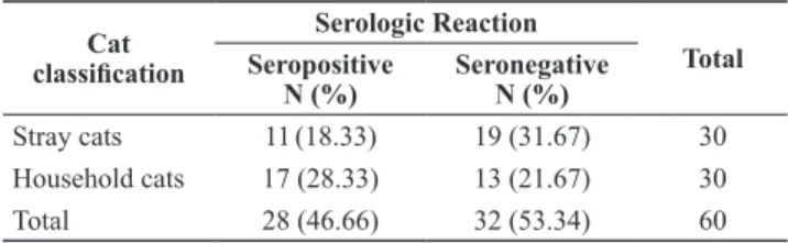
A total of 60 cats were assayed for anti-T.gondii
antibodies. Twenty-eight (46.67%) were seropositive, of these 11 (18.33%) belonged to the stray cat group while 17 (28.33%) were household cats (Table 1). While serologic data suggested greater susceptibility of household cats relative to stray cats, statistical analysis showed no association between the rate of infection by
the parasite and cat classiication.
Table 1. Number of stray and household cats serologically
positive to Toxoplasma gondii infection in the urban communities of Santa Rosa and San Pedro, Laguna, Philippines
Cat
classiication
Serologic Reaction
Total Seropositive
N (%)
Seronegative N (%)
Stray cats 11(18.33) 19 (31.67) 30
Hematologic results
The CBC analysis showed signiicantly higher
number of seropositive cats with abnormalities
(p<0.05) on Hgb level, RBC count, and differential
count on segmenters and monocytes compared to the control (Table 2). The levels of other parameters such as PCV, WBC, eosinophil and lymphocytes showed
insigniicantly different results between seropositive
cats and the control.
Table 3. Number of seropositive cats and control showing
hematologic abnormalities based on blood chemistry analysis
Blood chemistry
Abnormal blood chemistry* seropositive cats
(% based on N=28)
seronegative cats (% based on N=16
Blood uric acid (BUA)
5 a
(17.9)
1 a
(6.3) Fasting blood sugar
(FBS)
8 a
(28.6)
8 a
(50.0) Creatinine 1 a
(3.6)
0 a
(0.0) Blood urea nitrogen
(BUN)
7 a
(25.0)
4 a
(25.0) Potassium 21 a
(89.3)
4 b
(25.0)
Amylase 1 a
(3.6)
1 a
(6.3)
* Values per parameter with different letters were statistically signiicant.
Table 2. Number of seropositive cats and control showing
hematologic abnormalities based on complete blood count analysis
Blood count
Abnormal blood count* seropositive cats
(% based on N=28)
control (% based on N=16)
Hemoglobin 25 a
(89.3)
5 b
(31.3) Red blood cell
(RBC)
28 a
(100)
3 b
(18.8) Packed cell volume
(PCV)
13 a
(46.4)
6 a
(37.5) White blood cell
(WBC)
17 a
(60.7)
8 a
(50.0) Eosinophil 5 a
(17.8)
3 a
(18.7) Lymphocytes 16 a
(57.1)
6 a
(37.5) Monocytes 21 a
(75.0)
2 b
(12.5) Segmenters 18 a
(64.3)
5 b
(31.3)
* Values per parameter with different letters were statistically signiicant.
In blood chemistry analysis, signiicantly higher
number of seropositive cats show irregularities in potassium level as compared to the seronegative cats (Table 3). Other parameters such as amylase, blood sugar,
BUA, creatinine and BUN were statistically insigniicant
in terms of the number of affected seropositive and seronegative cats manifesting abnormalities.
Signiicantly affected hematologic parameters
were noteworthy implication of active toxoplasmosis in seropositive cats (Table 4). This study further determined the type of abnormality as to whether the value range was abnormally high or low. The result of
hematologic analysis showed signiicantly low levels of Hgb with a value ranging from 6.8-9.0 g/dL as against
the normal 9.5 to 15 g/dL, RBC count with a value range of 3.3x10⁶-5.7x10⁶/mm3 compared to the normal
value of 6.0x10⁶-10x106/mm3, and monocytes with no
traces or very few (<1-4%) were seen. Abnormally
Table 4. Number of seropositive stray and household
cats manifesting abnormally high or low values on hematologic analyses
Hematologic parameters
Abnormalities (%)*
Stray cats (N=11) Household cats (N=17)
High range Low range High range Low range
Potassium 8a
(100)
0 b
(0.0)
13a
(100)
0 b
(0.0)
Hemoglobin 1a
(10.0) 9b (90.0) 2a (13.3) 13b (86.7) Red blood cell (RBC)
0 a
(0.0) 11b (100) 0a (0.0) 17b (100) Monocytes 0 a
(0.0) 8b (100.) 0a (0.0) 13b (100) Segmenters 7 a
(100)
0b
(0.0)
11a
(100)
0 b
(0.0)
* Values per parameter with different letters were statistically signiicant
across same range level/cat classiication, and across different ranges within cat classiication
high levels of segmenters (neutrophils) were observed with a value range of 79-96% (normal=35-75%), and potassium with a slightly higher value ranging 5.9-6.1 mg/dL (normal=3.7-5.8 mg/dL). The types of abnormalities manifested by both stray and household seropositive cats were the same. Furthermore, the number of affected household and stray seropositive
cats revealed no signiicant differences as regards abnormalities on segmenters, monocytes, RBC, Hgb
DISCUSSION
The relatively high number of seropositive cats is consistent with earlier documented studies in domestic cats.1, 17, 21 The high infectivity of T. gondii to cats can be
attributed to the relatively wide range of portal of entry
of the parasite to its deinitive host. The ingestion of any
of the three infective stages (oocysts, tachyzoites and bradyzoites) of T. gondii through preying on infected wild animals like rats and mice, or eating contaminated raw meat or dairy products from infected sources, is probably the most common route of T. gondii infection in cats.2, 22
The seropositivity of cats to T.gondii is indicative of an outcome of shedding episode of oocysts in cats.16,23,24 This suggests that seropositive cats already
posed lesser risk of exposure to infection, and thereby shedding oocysts to potentially transmit the infection to humans and other possible intermediate hosts is minimal, unless they are immunocompromised. In this study, however, the oocyst shedding episodes of cats were not recorded and thus, the likely transmission of oocysts from seropositive cats to humans and other animals during the active shedding episodes was not established.
Although lesser infectivity was observed in stray cats, the uninfected stray cats are still considered a potential risk of infection because these cats, being unowned, are more exposed to free environment where they have their uncontrolled activities. This makes them more exposed to T. gondii infection by ingesting infective oocysts or by ingesting tissues cysts from intermediate hosts, such as small rodents which they prey on.7, 25 The rate of T. gondii infection in small
rodents in some parts of the world was estimated up to 73%.26 In the Philippines, high population of small
rodents has been reported.14 These rodents had been
established to be one of the most available intermediate hosts of T.gondii because of their close proximity with
cat habitation, thus, they play a signiicant role in
infecting stray cats.
Although in some studies hematologic values usually remain unaltered during the course of uncomplicated toxoplasmosis,27 this study implicated
otherwise. In fact, signiicantly affected hematologic
parameters were noteworthy implication of active toxoplasmosis in seropositive cats.
Cats with clinical toxoplasmosis show variety of clinocopathologic abnormalities associated with irregularities in blood components either their morphological characteristics or the number of cells present.27 Studies had established the development of
anemia in cats as shown in low percentage of PCV, Hgb
concentration, and RBC as compared to the reference range.27,28 In this study, PCV did not show critical
value, but the RBC count and Hgb concentration were
below the reference range, which may be suggestive of possible development of anemia in seropositive cats.
Low RBC count and Hgb, among other combined
factors, could cause anemia which has been recorded in toxoplasmic cats and even in humans.29,30 These results
were suggestive of T. gondii-infection induced effect on cats.
In totality, the presence of WBC in the blood
relects immunologic condition of seropositive cats.27, 28 Any changes in the WBC may relect serious
abnormalities on the health condition of the cats. The differential counts on neutrophils and monocytes revealed appreciable results in this study as earlier mentioned.
Defects in neutrophil functions can be due to a reduction of neutrophil count at a critical level due to impaired immunity or high levels may indicate an active infection.31,32 Both conditions can be present
in T.gondii infection, depending on the underlying health conditions of the host or aggravating factors
that could inluence the condition of the host.31,33 In
the present study, neutrophil count was signiicantly
higher in seropositive cats. Such result agreed with previous studies on T. gondii infected cats showing diseases associated with increased neutrophil which may be indicative of an active infection. On the other hand, this study showed that seropositive cats lacked or had very few monocytes. Ironically, most toxoplasmic conditions in humans were associated with monocytosis (increased monocyte counts) rather than low monocyte which when progresses, develops into monocytopenia (a form of leukopenia associated with a deiciency of monocytes).33 The absence or low monocytes level in
the blood can occur in response to the release of toxins into the blood by certain microorganisms, specially bacteria. In retrospect, leucopenia is often associated with acute and chronic forms of toxoplasmosis.29
Potassium levels that are too high or too low can increase the risk of an abnormal physiologic condition. Abnormal potassium level posed serious problems together with underlying factors such as increased BUN and creatinine which is a manifestation of renal dysfunction.34 In this study the slightly high level of
potassium may not implicate serious effect as other factors such as creatinine, BUA and BUN did not
pose signiicant indings in this study. Other studies
however reported that creatinine was observed to be elevated during Toxoplasma infection.27 And high level
This study also showed that amylase was not affected by seropositivity of the cats. Such result is in congruence with an earlier report that amylase is unreliable determinant of T. gondii infection.35,36
The fasting blood sugar analysis, which tested the carbohydrate metabolism through measurement of blood glucose levels after the cats fasted, showed
insigniicant association with T. gondii infection. Based on our knowledge, no study has directly established the association between T.gondii infection and abnormal blood sugar level in cats.
The development of toxoplasmosis may be quiescent or produce many signs and symptoms resulting from the parasite’s invasiveness of a wide-range of tissues. The reports documented through this study on the hematologic changes in cats for both stray and household cats supported the fact that toxoplasmosis is a multi-systemic disease which affects almost all major organs of the body, manifesting a wide range of symptoms and abnormalities. Because of this, the importance of cats
as the only deinitive hosts of T.gondii parasite must be underscored. It is therefore recommended that further studies on the chronicity of infection through serologic, hematologic and histopathologic investigations be
done. Hence, these will provide more basic and clinical
research information that would improve cat health management.
ACKNOLWLEDGEMENT
The authors wish to acknowledge Carlos Veterinary clinic (Sucat, Paranaque City, Philippines) and Mother Savior Polyclinic and Laboratory (San Pedro, Laguna, Philippines) for the technical assistance; and Dr. Johnny A. Ching, Faculty, College of Science for his assistance on statistical analysis of this study.
REFERENCES
Dubey JP, Zhu XQ, Sundar N, et al. Genetic and biologic 1.
characterization of Toxoplasma gondii isolates of cats from China. Vet Parasitol. 2007;145:352-6.
Omata Y, Oikawa H, Kanda M, et al. Transfer of antibodies 2.
to kittens from mother cats chronically infected with Toxoplasma gondii. Vet Parasitol. 1994;52:211-8.
Frenkel JK. Pathology and pathogenesis of congenital 3.
toxoplasmosis. Bull NY Acad Med. 1974;50:182-91. Dubey JP, Beattie CP. Toxoplasmosis of animals and man. 4.
Boca Raton: CRC Press; 1988.
Unno A, Suzuki K, Xuan X, et al. Dissemination of 5.
extracellular and intracellular Toxoplasma gondii tachyzoites
in the blood low. Parasitol Int. 2008,57:515-18.
Cabanacan-Salibay C, Claveria FG.
6. Toxoplasma gondii
infection in Philippine Rattus spp. conirmed through
bioassay in Mus musculus. Vet arh. 2006;76:351-61.
Salibay CC, Claveria FG. Serologically conirmed
7.
Toxoplasma gondii infection in Philippine Rattus spp. and its histopathology in Mus musculus. Phil J Sci. 2005;134: 95-104.
Kikuchi T, Furuta T, Kojima S. Kinetics of the nucleotide 8.
Triphosphate hydrolase of Toxoplasma gondii in mice with acute and chronic toxoplasmosis. Ann Trop Med Parasitol. 2002;96:35-41.
Manuel MF. Prevalence of
9. Toxoplasma gondii antibodies in swine in the Philippines. Phil J Vet Med. 1982;20:71-7. Marbella CO.
10. Toxoplasma gondii: antibody titer determination, histopathologic and isolation studies from naturally infected swine [thesis]. Philippines, Los Baños,Laguna: University of the Philippines; 1980. Mendoza CB. A serological survey on the incidence of 11.
toxoplasmosis in swine. [thesis]. Philippines, Los Baños, Laguna: University of the Philippines; 1974.
Kawashima T, Khin-sane-win, Kawabata M, et al. Prevalence 12.
of antibodies to Toxoplasma gondii among urban and rural residents in the Philippines. Southeast Asian J Trop Med
Public Health. 2000;31:742-6.
Salibay CC, Dungca JZ, Claveria FG. Serological survey 13.
of Toxoplasma gondii infection among Urban (Manila) and Suburban (Dasmariñas, Cavite) Residents, Philippines. J Protozool Res. 2008;18:26-33.
Salibay CC, Claveria FG. Serologic detection of
14. Toxoplasma
gondii infection in Rattus spp. collected from three different sites in Dasmariñas, Cavite, Philippines. Southeast Asian J
Trop Med Public Health. 2005;36:46-9.
Dans BK. A study on the presence of
15. Toxoplasma gondii
antibodies in the blood serum of stray cats in the Ninoy Aquino Parks and Wildlife Nature Center using enzyme-linked immunosorbent assay (ELISA) [thesis]. Philippines, Los Baños, Laguna: University of the Philippines; 2002.
Minervini N. A study on the presence of
16. Toxoplasma gondii
antibodies in the Philippines raised cats using the enzyme-linked immunosorbent assay test [thesis]. Philippines, Los Baños, Laguna: University of the Philippines; 1985. Molina EC, Ridley-Dash LS. Toxoplasmosis in cats at 17.
Kabacan, Cotabato, Philippines. USM Res Dev J. 2008; 16:53-5.
Feral Cat Coalition. Humane Trapping Instruction. [homepage
18.
on the Internet] San Diego: Feral Cat Coalition [cited 2009 Nov 30]. Available from: http://www.tomahawklivetrap.au/
Humane Trapping Instructions.htm.
Philippine Association for Laboratory Animal Science 19.
(PALAS). Code of practice for the care and use of laboratory animals in the Philippines. Philippines: Committee on PALAS; 2002.
Tilley LP, Smith FWK Jr. editors. The 5 minute veterinary 20.
consult: canine and feline. Maryland: Lipincott Williams & Wilkins; 2000.
Dubey P, Su C, Cortés JA, et al. Prevalence of
21. Toxoplasma
gondii in cats from Colombia, South America and genetic characterization of T. gondii isolates. Vet Parasitol. 2006;141:42-7.
Frenkel JK. Biology of
22. Toxoplasma gondii. In:
Ambroise-Thomas P, Petersen E. Congenital toxoplasmosis: scientiic
background, clinical management and control. Paris: Springer; 2000. p. 9-25.
Summer B, Ackland ML.
Haddadzadeh H
24. R, Khazraiinia P, Aslani M, Rezaeian
M, Jamshidi S, Taheri M, Bahonar A. Seroprevalence of
Toxoplasma gondii infection in stray and household cats in Tehran. Vet Parasitol. 2006;138:211-6.
Tenter AM, Heckeroth AR, Weiss LM.
25. Toxoplasma
gondii: from animals to humans. Australian Soc Parasitol. 2000;3:34-46.
Jackson MH, Hutchison WM. The prevalence and source
26.
of Toxoplasma infection in the environment. Adv Parasitol. 1989;28:55-105.
Lappin MR. Feline toxoplasmosis: interpretation of 27.
diagnostic results. Semin Vet Med Surg.1996;11:154-60. Okewole EA, Akpan MO. Clinical feline toxoplasmosis: para- 28.
sitological, haematological and serological indings in
retro-viral infected and uninfected cats. Vet Arhiv. 2002; 72:67-79.
Smart ME, Downey RS, Stockdale PHG. Toxoplasmosis in a
29.
cat associated with cholangitis and progressive pancreatitis. Can Vet J. 1973;14:313-6.
Alford CA Jr, Stagno S, Reynolds DW. Congenital toxo- 30.
plasmosis: clinical laboratory, and therapeutic considerations, wih special reference to subclinical disease. Bull N YAcad Med. 1974;50:160-81.
Bliss SK, Gavrilescu LC, Alcaraz A, et al. Neutrophil 31.
depletion during Toxoplasma gondii infection leads to impaired immunity and lethal systemic pathology. Infect Immunol. 2001;69:4898-905.
Stites DP, Stobo JD, Fudenberg HH, editors. Basic and 32.
clinical immunology. 5th ed. California: Lange Medical Publications; 1984.
Rajantie J, Siimes MA, Taskinen W, et al. White blood cells in 33.
infants with congenital toxoplasmosis: transient appearance of cALL antigen on reactive marrow lymphocytes. Scan J Infect Dis. 1992;24:227-32.
Chrisman CL. Acute weakness in the cat [Internet]. Florida: 34.
Waltham. The world’s leading authority on pet care and nutrition [cited 2009 Nov 30]. Available from: http://www.
walthamusa.com/articles/CHRISMAN.pdf.
Barr SC. Toxoplasmosis [Internet]. Westbrook: IDEXX 35.
Laboratories [cited 2009 Nov 30]. Available from: https:// www.vetconnect.com.au/5min/data/11101111.htm.
Dubey JP, Greene CE, Lappin MR. Toxoplasmosis and 36.