Nitrated Fatty Acids Reverse Cigarette
Smoke-Induced Alveolar Macrophage
Activation and Inhibit Protease Activity via
Electrophilic S-Alkylation
Aravind T. Reddy1,2, Sowmya P. Lakshmi1,2, Ramamohan R. Muchumarri1,2, Raju C. Reddy1,2*
1Department of Medicine, Division of Pulmonary, Allergy and Critical Care Medicine, University of Pittsburgh School of Medicine, Pittsburgh, PA, 15213, United States of America,2Veterans Affairs Pittsburgh
Healthcare System, Pittsburgh, PA, 15240, United States of America
*reddyrc@upmc.edu
Abstract
Nitrated fatty acids (NFAs), endogenous products of nonenzymatic reactions of NO-derived reactive nitrogen species with unsaturated fatty acids, exhibit substantial anti-inflammatory activities. They are both reversible electrophiles and peroxisome proliferator-activated recep-torγ(PPARγ) agonists, but the physiological implications of their electrophilic activity are poorly understood. We tested their effects on inflammatory and emphysema-related biomark-ers in alveolar macrophages (AMs) of smoke-exposed mice. NFA (10-nitro-oleic acid or 12-nitrolinoleic acid) treatment downregulated expression and activity of the inflammatory tran-scription factor NF-κB while upregulating those of PPARγ. It also downregulated production of inflammatory cytokines and chemokines and of the protease cathepsin S (Cat S), a key media-tor of emphysematous septal destruction. Cat S downregulation was accompanied by
decreased AM elastolytic activity, a major mechanism of septal destruction. NFAs downregu-lated both Cat S expression and activity in AMs of wild-type mice, but only inhibited its activity in AMs of PPARγknockout mice, pointing to a PPARγ-independent mechanism of enzyme inhibition. We hypothesized that this mechanism was electrophilic S-alkylation of target Cat S cysteines, and found that NFAs bind directly to Cat S following treatment of intact AMs and, as suggested byin silicomodeling and calculation of relevant parameters, elicit S-alkylation of Cys25 when incubated with purified Cat S. These results demonstrate that NFAs’electrophilic activity, in addition to their role as PPARγagonists, underlies their protective effects in chronic obstructive pulmonary disease (COPD) and support their therapeutic potential in this disease.
Introduction
Chronic obstructive pulmonary disease (COPD) is the third leading cause of death in the US [1], characterized by chronic inflammation of small airways and destruction of alveolar septa
a11111
OPEN ACCESS
Citation:Reddy AT, Lakshmi SP, Muchumarri RR, Reddy RC (2016) Nitrated Fatty Acids Reverse Cigarette Smoke-Induced Alveolar Macrophage Activation and Inhibit Protease Activity via Electrophilic S-Alkylation. PLoS ONE 11(4): e0153336. doi:10.1371/journal.pone.0153336
Editor:Yunchao Su, Augusta University, UNITED STATES
Received:December 24, 2015
Accepted:March 28, 2016
Published:April 27, 2016
Copyright:This is an open access article, free of all copyright, and may be freely reproduced, distributed, transmitted, modified, built upon, or otherwise used by anyone for any lawful purpose. The work is made available under theCreative Commons CC0public domain dedication.
Data Availability Statement:All relevant data are within the paper and its Supporting Information files.
Funding:This work was supported by a Merit Review award from the U.S. Department of Veterans Affairs and National Institutes of Health grant HL093196 to RCR. The funder had no role in study design, data collection and analysis, decision to publish, or preparation of the manuscript.
with consequent emphysema [2]. Such septal destruction reduces the surface area available for pulmonary O2and CO2exchange, and combines with airway inflammation and mucus pro-duction to impair respiratory mechanics. Because COPD is progressive and often fatal, and no effective therapies are available to impede its course, identification of new potential therapeutic targets is needed.
Among the many cell types that contribute to COPD pathophysiology, alveolar macro-phages (AMs) are especially critical [3]. They are directly exposed to cigarette smoke, the major risk factor for COPD, and other noxious airborne agents. Such agents activate AMs to produce pro-inflammatory cytokines that activate other cells, and chemokines that attract neu-trophils and T lymphocytes, prominent in COPD-associated inflammation. Such activated and recruited cells likewise secrete mediators that further activate AMs in a vicious positive-feed-back cycle. Yet AMs also exert an inflammation-dampening influence by phagocytosing apo-ptotic neutrophils and epithelial cells that, if allowed to become necrotic, would further feed the inflammatory cycle. Such phagocytotic activity of AMs is reduced in COPD, thereby exac-erbating pathogenesis [4].
AMs are also the primary sources of elastin-degrading proteases that largely drive septal destruction [5]. Most recent attention has focused on the Zn-containing matrix metalloprotei-nases (MMPs), but cathepsins, including cathepsins K, L, and S, are also important mediators of septal degradation [6]. Production of inflammation–associated oxidants by AMs can also contribute to septal destruction.
Inflammation-associated oxidants include NO and derived reactive nitrogen species that react nonenzymatically with unsaturated fatty acids to produce nitrated fatty acids (NFAs), including 10-nitro-oleic acid (OA-NO2) and 12-nitrolinoleic acid (LNO2) [7], the most preva-lent NO reaction products in the human bloodstream [8]. NFAs are potentially important endogenous modulators of inflammatory processes [9–11], acting at least in part as agonists of the nuclear hormone receptor peroxisome proliferator activated receptor-γ(PPARγ) [12]. They are also reversible electrophiles [13]. As such, they can react with thiol groups including those of cysteine residues within proteins to produce an S-alkyl bond that is cleavable (thus reversible) by reaction with free intracellular thiols such as glutathione (GSH). It has long been observed that specific cysteines are preferentially alkylated, but the factors that render such spe-cific cysteines more readily or stably alkylated are unknown. Known cysteine targets of NFA S-alkylation include one within the ligand-binding site of PPARγthat contributes significantly to agonist potency [14].
Reversible Cys S-alkylation provides a unique regulatory mechanism for biologically active proteins. For example, reversible electrophiles regulate oxidative stress by reversibly S-alkylat-ing the inhibitory protein Keap-1, allowS-alkylat-ing activation of the antioxidant transcription factor Nrf2 [15]. NFAs may also alleviate inflammation by S-alkylating the pro-inflammatory tran-scription factor NF-κB [10]. No such influence of reversible electrophiles in general or NFAs in particular has been recognized in the pathophysiology of COPD. We hypothesized that anti-inflammatory and related actions of NFAs may mitigate COPD pathogenesis, including secre-tion of the proteases that mediate septal destrucsecre-tion, and tested this idea and the mechanisms involved in AMs from smoke-exposed mice.
Materials and Methods
Animals
performed according to protocols reviewed and approved by the VA Pittsburgh Healthcare System Institutional Animal Care and Use Committee.
Smoke Exposure
Mice were exposed to cage-air cigarette smoke (CS) or to filtered air for 14 days. CS was gener-ated by burning five 3RF4 research cigarettes (Tobacco Research Institute, University of Ken-tucky, Lexington, KY) according to the Federal Trade Commission (FTC) protocol, each puff being of 2-second duration and 35-ml volume, in an automated TE-10 smoking machine (Tea-gue Enterprises, Davis, CA). The machine was adjusted to produce 89% sidestream and 11% mainstream smoke. The chamber atmosphere was monitored to maintain TPM at 250 mg/m3. Twenty-four hours following the last exposure, mice were euthanized and BAL fluid and lungs were collected for further analysis.
BAL Fluid Collection, Cell Count and AM Isolation from Mice
BAL fluid was collected by flushing 3 × 1 ml of PBS containing 0.1 mM EDTA into the lung via a tracheal cannula. The pooled BAL fluid was centrifuged at 500 × g at 4°C for 5 min. Pelleted cells were then resuspended in 1 ml of PBS. Total cell number was counted by hemocytometer and differential cell count was performed using cytospin preparations stained with Diff-Quik (Siemens, Newark, DE). AMs were isolated as described previously [17].
Measurement of Cytokine, Chemokine and Cathepsin S Levels
Cell culture medium was collected and stored at–80°C. Levels of TNF-αand KC were mea-sured using ELISA kits (R&D Systems, Minneapolis, MN) according to the manufacturer’s instructions. Cat S levels in media samples were measured using an ELISA kit (MyBioSource, Inc, San Diego, CA) according to the manufacturer’s instructions.
Western Blotting
Total protein extracts were prepared and Western blotting was performed as described previ-ously [18]. Primary antibodies against NF-κB p65 (372), PPARγ(7196), lamin B1 (20682), bio-tin (57636), Cat S (6505), andβ-actin (1616) were from Santa Cruz Biotechnology (Santa Cruz, CA). The secondary antibodies donkey anti-mouse IR-680RD (926–68072), donkey anti-rabbit IR-680RD (925–68073), donkey anti-goat IR-680RD (926–68074), goat anti-rabbit IR-800CW (925–32211), and goat anti-mouse IR-800CW (926–32210), were from LI-COR (Lincoln, NE). The infrared signal was detected using an Odyssey Infrared Imager (LI-COR).
Immunoprecipitation
Total protein extracts were immunoprecipitated using the Dynabeads Protein G Immunopre-cipitation kit (Invitrogen, Carlsbad, CA). Antibodies were bound to Dynabeads Protein G, and the Dynabeads-Ab complex was used to precipitate target proteins from the total protein extracts. Precipitates were washed to remove unbound proteins and complexes were eluted. All samples were separated by electrophoresis on SDS-polyacrylamide gels, transferred to mem-branes, and Western blotting was performed.
EMSA
Nuclear proteins (5μg) were incubated with 2.5 nM infrared dye (IRDye) 700 end-labeled
EDTA), 25 mM DTT and 2.5% Tween 20. Samples were then separated on 5% non-denaturing polyacrylamide gels in 1× Tris-Borate EDTA buffer (130 mM Tris, pH 8.3, 45 mM boric acid, 2.5 mM EDTA). The infrared signal was detected using an Odyssey Infrared Imager (LI-COR).
Monocyte Chemotaxis Assay
Mouse blood monocytes were isolated using an EasySep mouse monocyte enrichment kit (Stem-Cell Technologies Inc, Vancouver, BC, Canada). Briefly, leukocyte pools from blood were incu-bated with an antibody mixture against non-monocytes followed by a biotin selection mixture and magnetic particles. Labeled cells were removed using DynaMag-2 magnet (Invitrogen), resulting in highly significant enrichment of monocytes in the remaining cell preparation.
Blind-well chemotaxis chambers separated with 8-mm pore polycarbonate membrane (Neuro Probe Inc., Gaithersburg, MD) were used to assess migration of monocytes toward cell culture media. The upper wells contained monocytes isolated as described above; the lower wells were loaded with culture media supernatant from macrophages isolated from mice exposed to ciga-rette smoke for 14 days and then treated either with vehicle or with OA-NO2for 6 h. The cham-bers were incubated at 37°C for 1 h, after which the membranes were fixed and stained with Diff-Quik stains. The membranes were retrieved and mounted on glass slides, covered with a cover-slip, and examined under a microscope. The number of cells that migrated through the mem-brane into the lower chamber and cells that were attached on the lower side of the memmem-brane were counted and transmigration was expressed as % of monocytes added in upper chamber.
Elastolytic Activity Assay
Intracellular and extracellular elastin degradation studies were performed using soluble and insoluble fluorescein (FITC) labeled elastin respectively (Elastin Products Company, Inc, Owensville, MO). To determine extracellular elastolytic activity insoluble FITC-elastin was added to culture media or BAL fluid and treated as indicated. After incubation, insoluble elas-tin was removed by centrifugation and fluorescence in supernatants was determined using excitation at 490 nm and emission at 520 nm. Intracellular elastolytic activity was determined by incubating AMs treated as indicated above with soluble FITC-elastin. After incubation cells were washed and mounted with Vectashield mounting medium with DAPI (Vector Laborato-ries, Burlingame, CA). The slides were viewed by an Olympus Fluoview FV1000 confocal microscope (Olympus, Center Valley, PA) using a 60× objective lens along with Fluoview con-focal software (FV10-ASW v1.7, Olympus).
ChIP Assay
The ChIP assay was performed using SimpleChIP Enzymatic Chromatin Immunoprecipitation kit with magnetic beads (Cell Signaling Technology, Beverly, MA, USA). Briefly, cellular
Table 1. Consensus oligonucleotides employed.
Binding site Prime Sequence End labelling
κB Site F 50-AGTTGAGGGGACTTTCCCAGGC-30 IRDye 700
R 30-TCAACTCCCCTGAAAGGGTCCG-50 IRDye 700
PPRE Site F 50-AGACAAGTCAGAGGCCACGGT-30 IRDye 700
R 30-TCTGTTCAGTCTCCGGTGCCA-50 IRDye 700
Nonspecific Control F 50-AGACTGGGGCTGGAGTGCGGTT-30 IRDye 700
R 30-TCTGACCCCGACCTCACGCCAA-50 IRDye 700
chromatin was crosslinked with 1% formaldehyde for 10 min at room temperature, the cross-linking was stopped with 0.125 M glycine, and cells were washed twice with ice cold PBS. Nuclei were pelleted and digested by micrococcal nuclease. Following sonication and centrifu-gation, equal amounts of sheared chromatin were incubated overnight at 4°C with antibodies, IgG as negative control, or RNA polymerase II as positive control. Protein G magnetic beads were then added and the chromatin was incubated with rotation for 2 h at 4°C. An aliquot of chromatin that was not incubated with any antibody was used as the input control sample. Antibody-bound protein/DNA complexes were eluted and subjected to real-time PCR as described previously [18] with specific primers for Ctss (Cat S), CD36, andα-satellite (Table 2).
Molecular Modeling and Computer Simulations of Binding of OA-NO
2with Cat S
Molecular modeling and covalent docking analysis was carried out as described previously [19]. The X-ray structure of human Cat S {PDB ID 1NPZ; [20]} and its C25S mutant {PDB ID 1GLO; [21]} were obtained from the RCSB Protein Data Bank. Covalent docking was per-formed with OA-NO2(ligand) using Discovery Studio 2.5 (Accelrys Inc., San Diego, CA).
Cat S Enzyme Inhibition Assay
Cat S enzyme inhibition assay was performed using the Cathepsin S Drug Discovery Kit (BML-AK431-0001; Enzo Life Sciences, Farmingdale, NY) according to the manufacturer’s instructions.
Cat S Enzyme Activity Assay
Cat S enzyme activity in cell culture media samples was measured by incubating these samples with Cat S-specific fluorescent substrate (Ac-KQKLR-AMC; 600742, Cayman Chemical) according to the manufacturer’s instructions.
Cat S Cys Mutants
A plasmid containing the human Cat S gene was obtained from Addgene (#11251) and sub-cloned into a pGEX 6P1 plasmid. Cysteine residues at positions 12, 25 and 110 or all three (12, 25 and 110) were mutated to serine using site directed mutagenesis (custom cloning core facil-ity at Emory Universfacil-ity, Atlanta, GA). Recombinant proteins were expressed in BL21-DE3 cells (Sigma-Aldrich, St. Louis, MO), purified using GST SpinTrap columns (GE Healthcare Life Sciences), and the GST tag was removed with PreScission Protease (GE Healthcare Life Sciences) according to the manufacturer’s instructions.
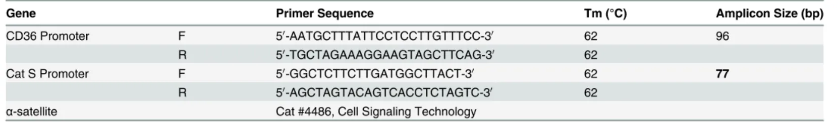
Table 2. Oligonucleotide primers employed.
Gene Primer Sequence Tm (°C) Amplicon Size (bp)
CD36 Promoter F 50-AATGCTTTATTCCTCCTTGTTTCC-30 62 96
R 50-TGCTAGAAAGGAAGTAGCTTCAG-30 62
Cat S Promoter F 50-GGCTCTTCTTGATGGCTTACT-30 62 77
R 50-AGCTAGTACAGTCACCTCTAGTC-30 62
α-satellite Cat #4486, Cell Signaling Technology
Cat S S-alkylation
To demonstrate S-alkylation of Cat S, human recombinant Cat S (EMD Millipore Billerica, MA) and variants−Cat S WT, C12;25;110S mutant, C12S mutant, C25S mutant, and C110S mutant−were incubated with test compounds for the indicated time periods and S-alkylation was analyzed by running these samples in a SDS-PAGE gel under non-reducing conditions and performing Western blots using antibody to biotin (Santa Cruz Biotechnology).
Electrospray Ionization-Liquid Chromatography-Mass Spectrometry/
Mass Spectrometry (ESI-LC-MS/MS)
ESI-LC-MS/MS analyses were performed on a Q Exactive Hybrid Quadrupole-Orbitrap mass spectrometer (Thermo Scientific, Pittsburgh, PA). The electrospray system employed a 3.5 kV spray voltage and a capillary temperature of 260°C.
Statistical Analysis
Data are presented as mean ± SD. Differences between groups were analyzed using an unpaired t-test or analysis of variance, followed by Bonferroni multiple comparison correction. Analyses were performed using GraphPad Prism 5.03 (GraphPad Software, La Jolla, CA). Differences withP<0.05 were considered significant.
Results
OA-NO
2reduces cigarette smoke-induced AM activation in mice
Considering the pivotal role of AMs in the response to smoke inhalation and COPD pathogen-esis, we tested the effects of NFAs on AM activities relevant to inflammation and septal destruction in a mouse model of COPD. We exposed mice to CS delivered via cage air for 14 days. Smoke exposure greatly increased the numbers of macrophages in BAL fluid, with lesser increases in the number of neutrophils (Fig 1A and 1B). Because AMs represented the majority of inflammatory cells found in BAL fluid of smoke-exposed mice, we assessed them further.
In vitroOA-NO2treatment (no toxicity was observed,S1 Fig) of AMs isolated from BAL fluid
of smoke-exposed mice reduced both nuclear expression and DNA-binding activity of the pro-inflammatory transcription factor NF-κB (Fig 1C and 1D), but increased those of the anti-inflammatory nuclear hormone receptor PPARγ(Fig 1C and 1E). Based on these changes in activity of pro- and anti-inflammatory transcriptional regulators, we predicted that expression of pro-inflammatory genes would be reduced, and indeed found that secretion of TNF-α(Fig 1F) and KC (Fig 1G) by AMs was suppressed. Secretion of chemokines was also reduced, as seen using a monocyte transmigration assay (Fig 1H and 1I). Assessing emphysema-causing protease secretion and elastin degradation, we found that OA-NO2treatment markedly reduced secreted Cat S levels and activity in culture medium (Fig 1J and 1K). Degradation of elastin by culture medium of OA-NO2-treated AMs (Fig 1L) and intracellular elastin degradation by whole AMs
(Fig 1M) were similarly reduced. Thus, OA-NO2suppressed both the inflammatory and
proteo-lytic activities induced by smoke exposure in AMs. As these cells play a prominent role in COPD pathophysiology [3], these effects carry significant implications for modulation of this disease.
OA-NO
2Effects on Cathepsin S Expression, but not Activity, are
PPAR
γ
-dependent
and activity using AMs from wild-type (WT) and PPARγknockout (KO) mice following 14-dayin vivosmoke exposure and 6-hin vitroexposure to OA-NO2or vehicle. Absence of PPARγin AMs from KO mice was confirmed by Western blotting, which also revealed modest
Fig 1. NFAs mitigate cigarette smoke-induced alveolar macrophage activation.(A, B) C57BL/6 mice were exposed to cigarette smoke (CS) or to filtered-air for 14 days. Twenty-four h after the final exposure, BAL samples were collected. (A) Photomicrographs (40× objective lens) of Diff-Quik-stained cells and (B) total (Tot), neutrophil (PMN), and macrophage (MAC) counts in BAL fluid from the indicated treatment groups. (C-M) Alveolar macrophages (AMs) were isolated from BAL fluid of mice exposed to CS as described above and were then treated with 1μM OA-NO2or vehicle (Veh) for 6 h. (C) Western blots for PPARγand p65 in nuclear extracts. (D, E) DNA-binding activity of (D) NF-κB, and (E) PPARγ, in OA-NO2or Veh treated AM extracts measured by EMSA using IRDye oligonucleotides. (F, G) Release of (F) TNFα. and (G) KC. (H, I) Mouse monocytes were isolated and transmigration was assayed using a chemotaxis chamber in which they were separated by an 8-mm pore polycarbonate membrane from chemoattractant culture media from the above treatment groups. (H) Percentage transmigration, and (I) photomicrographs (40× objective lens) of the lower side of the polycarbonate membrane. (J) Cat S expression and (K) activity in culture media from above treatment groups. (L, M) AMs were collected and treated as indicated above, then incubated with FITC-elastin for further 1 h to determine (L) extracellular and (M) intracellular elastolytic activity (60× objective lens). Data are representative of two to three independent experiments with cells obtained fromn= 14–18 mice/group (cells from 3 mice pooled together).**P<0.01,***P<0.001.
increases in Cat S (Fig 2A). OA-NO2treatment reduced Cat S expression in AMs from WT but not PPARγKO mice (Fig 2B), indicating that NFA-induced suppression of Cat S expression requires PPARγactivation. In contrast, NFAs reduced Cat S activity in culture media of AMs to approximately the same extent irrespective of the presence or absence of PPARγ(Fig 2C). Therefore, inhibition of Cat S activity does not depend on PPARγ, and we thus hypothesized it reflects S-alkylation by NFAs. Supporting this hypothesis, we found that NFA bound directly to Cat S following incubation of Cat S with biotin-labeled OA-NO2, immunoprecipitation, and Western blotting using anti-biotin antibody (Fig 2E). Conversely, supporting a PPARγ -depen-dent mechanism of NFA effects on Cat S expression, we found by chromatin immunoprecipi-tation (ChIP) that OA-NO2treatment increased PPARγbinding to both theCd36promoter, which is known to be upregulated by PPARγactivation, and also theCtss(Cat S) promoter (Fig
2D). This was consistent with prior findings that under certain circumstances binding of PPARγto DNA inhibits rather than stimulates transcription [22]. Thus, the observed NFA effects on Cat S expression and activity were mediated by distinct, complementary mechanisms in which electrophilic addition plays a prominent role.
OA-NO
2Inhibits Cathepsin S by S-alkylating Cys25
We next sought to specifically identify the site of NFA-Cat S binding and the type of bond involved. Molecular docking studies revealed covalent bond formation between the electro-philic carbon of OA-NO2and sulfur atom Cys25 of Cat S (Fig 3A). No covalent bond Fig 2. NFAs’inhibition of Cat S expression but not activity, is PPARγ-dependent.(A) Western blots for PPARγand Cat S in whole cell extracts from AMs isolated from BAL fluid of C57BL/6 (WT) and Tie2 Cre-PPARγflox/flox(KO) mice. (B, C) Mice were exposed to cigarette smoke (CS) for 14 days, AMs were isolated and then treated with 0.5 and 1μM OA-NO2or Veh for 6 h. (B) Cat S expression and (C) activity in culture media. (D) Following AM isolation and OA-NO2(1μM) treatment as above, chromatin was crosslinked and immunoprecipitated with PPARγantibody; the antibody-bound DNA-protein complexes were then subjected to real-time PCR with primers specific for PPRE sites in Cat S and CD36 promoter regions,α-satellite was used as control. (E) As above, AMs were collected from CS-exposed mice and treated with 5μM biotin-labeled OA-NO2or Veh for 6 h. Total protein extracts were prepared, Cat S was immunoprecipitated and Western blotting was performed with anti-biotin and -Cat S antibodies. Data are representative of two to three
independent experiments with cells obtained fromn= 14–18 mice/group (cells from 3 mice pooled together).*P<0.05,**P<0.01,***P<0.001, n.s. = non-significant.
Fig 3. NFAs inhibit Cat S enzymatic activity by S-alkylation.(A-C) Binding of OA-NO2to Cat S was modeledin silicoand evaluatedin vitro. (A) Schematic representation showing covalent interaction of OA-NO2with Cat S Cys25 residue. (B) Schematic representation showing absence of any covalent interaction of OA-NO2with Cat S Cys25Ser mutation. (Left panels) Cat S is shown as a flat ribbon colored according to secondary structure; OA-NO2(yellow) is shown as a stick model and interacting amino acids are shown in scaled ball-and-stick representation. (Right panels) Cat S is shown with interacting amino acid residues as a scaled ball-and-stick model with labels and OA-NO2(yellow) as a stick model. In both figures the covalent bond is indicated bywhite arrowand hydrogen bonds are indicated bygreen dotted lines. (C) Human recombinant Cat S in active form (200 ng) was incubated for 30 min with the indicated compounds. Following incubation, S-alkylation of Cat S by biotin-labeled OA-NO2in test samples was assessed by Western blotting under non-reducing conditions. (D) Human recombinant Cat S in active form (6μU/μl) was incubated for 1 h with the indicated compounds and enzyme activity determined by cleavage of fluorescent substrate peptide. Data are representative of three independent experiments withn= 4/group.*P<0.5,***P<0.001.
formation was seen when Cat S with Cys25 to Ser mutated was used, as the oxygen in serine is not sufficiently nucleophilic to bond with the NFA’s electrophilic carbon (Fig 3B).
We confirmed that NFA binds to Cat S protein by incubating recombinant human Cat Sin vitrowith biotin-labeled OA-NO2followed by Western blotting (Fig 3C). Incubation with
either OA-NO2or LNO2dose-dependently inhibited human Cat S activity, and at the highest dose tested exhibited inhibition similar to that seen with the classic Cat S inhibitor E-64 (Fig 3D). This inhibition was blocked by the thiol exchange inhibitor bacitracin, thus confirming S-alkylation as the mechanism of inhibition.
We next sought to specifically identify the site of NFA-Cat S binding and the type of bond involved. We modeled Cat Sin silico(Fig 4) to 1) determine the presence or absence of disul-fide bonding for each Cys residue and the S atom’s partial positive charge (Fig 4A); and 2) cal-culate solvent accessibility (Fig 4B), H-bonding potential (Fig 4C), and pKa (Fig 4D) for each of its cysteine residues. The results suggested that Cys25 is the most nucleophilic and thus the most likely site of S-alkylation. This conclusion was supported byin silicomodeling of
OA-NO2bonded to Cys25, indicating appropriate solvent accessibility and the presence of sta-bilizing H-bonds (Fig 4B–4D).
To test whether Cys25 was the specific Cat S residue to which NFAs bond covalently, we incubated biotin-labeled OA-NO2with WT Cat S or Cat S with Cys residues mutated to Ser [either individually at Cys12 (C12S), Cys25 (C25S), or Cys110 (C110S), or at all three positions, (C12;25;110S)]. OA-NO2binding was then determined by Western blotting (Fig 5). OA-NO2 was bound to Cat S by S-alkylation only in Cat S WT, C12S, and C110S mutants; the interac-tion was absent when Cys25 was mutated to Ser (C12;25;110S and C25S). These results indicate that the S-alkylation of Cat S by OA-NO2occurs specifically at Cys25.
To further confirm the covalent interaction we incubated OA-NO2(or vehicle) with a syn-thetic Cat S peptide (Cat S23-29) containing either the native Cys25 (sequence GACWAFS) or a Cys25Ser substitution (GASWAFS), and analyzed the products by mass spectrometry, which indicated that NFA was S-alkylated to the Cys25-containing fragment (Fig 6Band table). As predicted, Cys25Ser mutation abolished NFA S-alkylation (Fig 6Dand table) and no addition was seen in the absence of NFA (Fig 6A and 6Cand table). Our initial prediction, based on the theoretical calculations shown inFig 4, that Cys25 would be the alkylated residue was thus confirmed.
Discussion
We find that NFA treatment of activated AMs from CS-exposed mice downregulates expres-sion and activity of Cat S, a protease heavily involved in the septal destruction that leads to emphysema. Because NFAs are known PPARγagonists [23], we tested whether PPARγ medi-ates their suppressive effects on Cat S, finding that NFAs downregulate both Cat S gene expres-sion and its enzymatic activity. These actions are mechanistically distinct, however, as PPARγ
Bonnaci and colleagues similarly observed PPARγ-dependent decreases in expression of MMPs, another class of COPD-related proteases, in conjunction with PPARγ-independent effects on enzyme activity [24]. Mechanisms of the apparent MMP inhibition were not investi-gated, however. Reversible electrophiles, including NFAs, were found earlier to S-alkylate other regulatory proteins, including the p65 subunit of NF-κB [10], protein kinase CB[25] and Keap-1[15,26], but these actions have not previously been linked to mechanisms of disease
Fig 4.In silicomodeling shows Cat S Cys25 is the preferred target for NFA S-alkylation.In silicomodeling of Cat S showing potential for electrophilic S-alkylation. (A) Cysteine residues are indicated by yellow spheres and disulfide bonds by short yellow lines. Each residue is colored according to its calculated pKa. The protein surface is colored to indicate solvent accessibility and the surface of the potential OA-NO2binding pocket is colored according to its H-bonding potential. The bar scales indicate the values corresponding to each color used. Numerical values of these parameters are shown in the
accompanying table. (B-D)In silicomodels of Cat S with OA-NO2bound to Cys25. Protein surface is color coded to indicate: (B) solvent accessible surface area (SASA); (C) potential for H-bond formation; (D) pKa of OA-NO2–Cat S binding pocket. OA-NO2is rendered as a scaled ball-and-stick model and Cat S as a stick model. The covalent bond between OA-NO2and Cys25 is indicated by a red arrow.
pathogenesis. Indeed, despite NFAs’recognized ability to electrophilically alkylate targeted protein cysteines via Michael addition, the physiological and pathogenetic relevance of these actions has remained largely unknown prior to our current investigations.
The biological roles of endogenous NFAs are likewise uncertain, but available evidence sug-gests that they may be important inflammatory modulators. They are produced by nonenzy-matic reaction of unsaturated fatty acids with NO-derived reactive nitrogen species [7], production of which is upregulated during inflammation. Inflammation also upregulates cho-lesteryl hydrolase activity [27], which drives release of free, active NFAs from their esters. Even in the absence of inflammation, the combined concentrations of free and esterified fractions of NFAs in blood are within the range required to activate PPARγ[8]. Endogenous NFAs may also downregulate inflammation by S-alkylating NF-κB, and may alleviate inflammation-pro-duced oxidative stress by S-alkylating Keap-1 [26]. Definitive proof of NFAs’endogenous role has remained elusive, however, because traditional techniques are not applicable: Endogenous NFA production cannot be effectively blocked and their multiple mechanisms of action, as illustrated by our present results, render it difficult to block their effects. Indeed, it will be chal-lenging to block S-alkylation experimentally in intact cells.
Beyond Cat S regulation, we found that NFAs downregulate activation of AMs obtained from smoke-exposed mice, as assessed by a variety of inflammatory markers including NF-κB activity, cytokine/chemokine production, and transmigration. AMs are pivotal contributors to inflammation, particularly in COPD [3]. Through their production of inflammatory cytokines and chemokines [28], activated AMs serve to activate and recruit other cells, complementing their own direct pro-inflammatory actions. Our finding that treating AMs with OA-NO2 reduces ability of their conditioned medium to recruit additional macrophages reveals a poten-tially powerful pathway via which NFAs can act to alleviate inflammation. As key sources of the principal elastolytic proteases, MMPs and cathepsins [5], AMs also contribute crucially to emphysema-causing septal destruction. Thus, our finding that NFAs downregulate total Cat S activity via both PPARγ- and electrophile-dependent mechanisms, while others found NFAs downregulate MMP expression [24], points to a potentially important protective role of endog-enous or therapeutically-aimed NFAs in COPD (Fig 7).
In summary, our findings demonstrate that NFAs act as both electrophiles and PPARγ ago-nists to suppress the pathophysiology of CS-induced COPD. In particular, NFAs downregulate expression of the septum-destroying protease Cat S while specifically S-alkylating the enzyme’s
Fig 5. NFAs S-alkylate Cat S specifically at Cys 25.Human recombinant Cat S WT and Cys mutants C12;25;110S, C12S, C25S, C110S (200 ng) were incubated for 30 min with biotin-labeled OA-NO2(1μM). Following incubation, S-alkylation of Cat S by biotin-labeled OA-NO2was assessed by Western blotting under non-reducing conditions. Data are representative of two independent experiments.
Fig 6. Mass spectrometric analysis of Cat S Cys25 S-alkylation.Oneμg of synthetic Cat S23-29 containing either native Cys25 (GACWAFS) or Ser25 (GASWAFS) was incubatedin vitrofor 30 min with (A, C) Veh or (B, D) 20μM OA-NO2. Reaction mixtures were resolved by reverse phase chromatography and analyzed by electrospray ionization-tandem mass spectrometry. (A, B) The spectrum for peptide GACWAFS showsB, S-alkylation of OA-NO2on the Cys residue butA, no alkylation by Veh. (C, D) Absence of S-alkylation of OA-NO2with peptide GASWAFS where Cys is replaced by a Ser residue.Tableshowsm/zof fragment ions from peptide GACWAFS, with detected ions highlighted in color. Column AA identifies the corresponding amino acids. Data are representative of three independent experiments.
catalytic Cys25, thus inhibiting its elastolytic activity and ability to promote emphysema. These results provide mechanistic insights into the part NFAs may play in modulating COPD sever-ity, highlighting the novel role of their electrophilic activsever-ity, and support their potential thera-peutic role in this disease.
Supporting Information
S1 Fig. OA-NO2treatment has no toxic effects on mouse AMs.AMs were isolated and
cul-tured as described and were treated with OA-NO2(0.1, 0.5, 1, 5 and 10μM) for 6 h. After
treat-ment AM viability, cytotoxicity and apoptosis were assayed as indicated inMaterials and Methods. Data are representative of two independent experiments withn= 3/group.
(TIFF)
S1 File. Materials and Methods. Viability, Cytotoxicity and Apoptosis.Mouse AMs were isolated and treated as indicated with OA-NO2. Cell viability, cytotoxicity and apoptosis were measured according to manufacturer’s instructions using ApoTox-Glo Triplex Assay (Pro-mega, Madison, WI). Briefly, at the end of the OA-NO2treatment period 20μl of viability/
cytotoxicity reagent containing both GF-AFC substrate and bis-AAF-R110 substrate was
Fig 7. Schematic illustration of effects of S-alkylation by NFAs on COPD.Exposure to cigarette smoke induces COPD with accompanying increases in inflammation (NF-κB), oxidative stress, and release of elastolytic proteases (cathepsins and MMPs). Treatment with NFAs, acting partially through electrophilic S-alkylation of specific cysteines, reverses all these COPD-associated changes.
added to all wells, briefly mixed by shaking, and incubated for 30 min at room temperature. Viability and cytotoxicity were assessed by fluorescence measured using a plate reader (VIC-TOR X, PerkinElmer; Waltham, MA). After measurement, 100μl of Caspase-Glo 3/7 reagent
was added to each well, briefly mixed by shaking, and incubated for 30 min at room tempera-ture. Apoptosis was assessed by luminescence measured using a plate reader.
(DOCX)
Author Contributions
Conceived and designed the experiments: ATR SPL RCR. Performed the experiments: ATR SPL RRM. Analyzed the data: ATR SPL RRM RCR. Wrote the paper: ATR SPL RCR.
References
1. Number of Deaths for Leading Causes of Death. Centers for Disease Control and Prevention. 2011. 2. Chung K, Adcock I. Multifaceted mechanisms in COPD: inflammation, immunity, and tissue repair and
destruction. European Respiratory Journal. 2008; 31(6):1334–56. doi:10.1183/09031936.00018908 PMID:18515558
3. Barnes PJ. Alveolar macrophages as orchestrators of COPD. COPD: Journal of Chronic Obstructive Pulmonary Disease. 2004; 1(1):59–70. PMID:16997739
4. Hodge S, Hodge G, Scicchitano R, Reynolds PN, Holmes M. Alveolar macrophages from subjects with chronic obstructive pulmonary disease are deficient in their ability to phagocytose apoptotic airway epi-thelial cells. Immunology and cell biology. 2003; 81(4):289–96. PMID:12848850
5. Russell RE, Thorley A, Culpitt SV, Dodd S, Donnelly LE, DeMattos C, et al. Alveolar macrophage-medi-ated elastolysis: roles of matrix metalloproteinases, cysteine, and serine proteases. American Journal of Physiology-Lung Cellular and Molecular Physiology. 2002; 283(4):L867–L73. PMID:12225964 6. Barnes P, Shapiro S, Pauwels R. Chronic obstructive pulmonary disease: molecular and
cellularme-chanisms. European Respiratory Journal. 2003; 22(4):672–88. PMID:14582923
7. Schopfer FJ, Baker PR, Giles G, Chumley P, Batthyany C, Crawford J, et al. Fatty acid transduction of nitric oxide signaling Nitrolinoleic acid is a hydrophobically stabilized nitric oxide donor. Journal of Bio-logical Chemistry. 2005; 280(19):19289–97. PMID:15764811
8. Baker PR, Lin Y, Schopfer FJ, Woodcock SR, Groeger AL, Batthyany C, et al. Fatty acid transduction of nitric oxide signaling: multiple nitrated unsaturated fatty acid derivatives exist in human blood and urine and serve as endogenous peroxisome proliferator-activated receptor ligands. Journal of Biologi-cal Chemistry. 2005; 280(51):42464–75. PMID:16227625
9. Rudolph V, Rudolph TK, Schopfer FJ, Bonacci G, Woodcock SR, Cole MP, et al. Endogenous genera-tion and protective effects of nitro-fatty acids in a murine model of focal cardiac ischaemia and reperfu-sion. Cardiovascular Research. 2010; 85(1):155–66. doi:10.1093/cvr/cvp275PMID:PMC2791055. 10. Cui T, Schopfer FJ, Zhang J, Chen K, Ichikawa T, Baker PR, et al. Nitrated fatty acids: endogenous
anti-inflammatory signaling mediators. Journal of Biological Chemistry. 2006; 281(47):35686–98. PMID:16887803
11. Liu H, Jia Z, Soodvilai S, Guan G, Wang M-H, Dong Z, et al. Nitro-oleic acid protects the mouse kidney from ischemia and reperfusion injury. American Journal of Physiology-Renal Physiology. 2008; 295(4): F942–F9. doi:10.1152/ajprenal.90236.2008PMID:18753300
12. Li Y, Zhang J, Schopfer FJ, Martynowski D, Garcia-Barrio MT, Kovach A, et al. Molecular recognition of nitrated fatty acids by PPARγ. Nature structural & molecular biology. 2008; 15(8):865–7.
13. Baker LM, Baker PR, Golin-Bisello F, Schopfer FJ, Fink M, Woodcock SR, et al. Nitro-fatty acid reaction with glutathione and cysteine Kinetic analysis of thiol alkylation by a Michael addition reaction. Journal of Biological Chemistry. 2007; 282(42):31085–93. PMID:17720974
14. Geisler AC, Rudolph TK. Nitroalkylation—a redox sensitive signaling pathway. Biochimica et Biophy-sica Acta (BBA)-General Subjects. 2012; 1820(6):777–84.
15. Kansanen E, Bonacci G, Schopfer FJ, Kuosmanen SM, Tong KI, Leinonen H, et al. Electrophilic nitro-fatty acids activate NRF2 by a KEAP1 cysteine 151-independent mechanism. Journal of Biological Chemistry. 2011; 286(16):14019–27. doi:10.1074/jbc.M110.190710PMID:21357422
17. Reddy AT, Lakshmi SP, Reddy RC. The nitrated fatty acid 10-nitro-oleate diminishes severity of LPS-induced acute lung injury in mice. PPAR research. 2012;2012.
18. Lakshmi SP, Reddy AT, Zhang Y, Sciurba FC, Mallampalli RK, Duncan SR, et al. Down-regulated per-oxisome proliferator-activated receptorγ(PPARγ) in lung epithelial cells promotes a PPARγ agonist-reversible proinflammatory phenotype in chronic obstructive pulmonary disease (COPD). Journal of Biological Chemistry. 2014; 289(10):6383–93. doi:10.1074/jbc.M113.536805PMID:24368768 19. Reddy AT, Lakshmi SP, Zhang Y, Reddy RC. Nitrated fatty acids reverse pulmonary fibrosis by
dedif-ferentiating myofibroblasts and promoting collagen uptake by alveolar macrophages. The FASEB Jour-nal. 2014; 28(12):5299–310. doi:10.1096/fj.14-256263PMID:25252739
20. Pauly TA, Sulea T, Ammirati M, Sivaraman J, Danley DE, Griffor MC, et al. Specificity determinants of human cathepsin S revealed by crystal structures of complexes. Biochemistry. 2003; 42(11):3203–13. PMID:12641451
21. Turkenburg JP, Lamers MB, Brzozowski AM, Wright LM, Hubbard RE, Sturt SL, et al. Structure of a Cys25!Ser mutant of human cathepsin S. Acta Crystallographica Section D: Biological Crystallogra-phy. 2002; 58(3):451–5.
22. François M, Richette P, Tsagris L, Raymondjean M, Fulchignoni-Lataud M-C, Forest C, et al. Peroxi-some proliferator-activated receptor-γdown-regulates chondrocyte matrix metalloproteinase-1 via a novel composite element. Journal of Biological Chemistry. 2004; 279(27):28411–8. PMID:15090544 23. Schopfer FJ, Lin Y, Baker PR, Cui T, Garcia-Barrio M, Zhang J, et al. Nitrolinoleic acid: an endogenous
peroxisome proliferator-activated receptorγligand. Proceedings of the National Academy of Sciences of the United States of America. 2005; 102(7):2340–5. PMID:15701701
24. Bonacci G, Schopfer FJ, Batthyany CI, Rudolph TK, Rudolph V, Khoo NK, et al. Electrophilic fatty acids regulate matrix metalloproteinase activity and expression. Journal of Biological Chemistry. 2011; 286 (18):16074–81. doi:10.1074/jbc.M111.225029PMID:21454668
25. Guo C-J, Schopfer FJ, Gonzales L, Wang P, Freeman BA, Gow AJ. Atypical PKCζtransduces electro-philic fatty acid signaling in pulmonary epithelial cells. Nitric Oxide. 2011; 25(3):366–72. doi:10.1016/j. niox.2011.07.003PMID:21871968
26. Tsujita T, Li L, Nakajima H, Iwamoto N, Nakajima‐Takagi Y, Ohashi K, et al. Nitro‐fatty acids and cyclo-pentenone prostaglandins share strategies to activate the Keap1‐Nrf2 system: a study using green fluo-rescent protein transgenic zebrafish. Genes to cells. 2011; 16(1):46–57. doi:10.1111/j.1365-2443. 2010.01466.xPMID:21143560
27. Lindhorst E, Young D, Bagshaw W, Hyland M, Kisilevsky R. Acute inflammation, acute phase serum amyloid A and cholesterol metabolism in the mouse. Biochimica et Biophysica Acta (BBA)-Protein Structure and Molecular Enzymology. 1997; 1339(1):143–54.