Mestrado Integrado em Medicina Veterinária
Ciências VeterináriasComparison of canine sperm quality under
different temperature storage
Paulo Alexandre Paulos Borges
Orientador:
Professora Doutora Rita Maria Payan Martins Pinto Carreira Co-orientador:
Professor Alain Fontbonne
UNIVERSIDADE DE TRÁS-OS-MONTES E ALTO DOURO VILA REAL, 2011
ii
Not everything that can be counted counts and not everything that counts can be counted.
iii
ABSTRACT
Successful gamete cryopreservation is essential to preserve the genetic pool of several species. The canine semen is known to possess a certain resilience to chilling procedures, which has a limited interest for long term gamete preservation; however, when frozen, canine sperm suffers an important decrease in quality. This feature determined the development of different extenders aiming the protection of the cell and to increase the success of canine sperm freezing/thawed procedures. In parallel, development of new techniques allowing a more objective evaluation of putative sperm fertility were also developed. However, those methods do not explain why some stud males are “good freezers” while other are “bad” freezers, when both were selected at start as adequate for freezing. Further, no clear definition exists on the cut-off values for specific seminal parameters to distinguish between a fertile and infertile stud, although the analysis of semen is regularly performed to assess the male fertility.
In this study we plan to evaluate cold treatment associated changes on sperm cells by using two different approaches. For that, semen was collected from 8 dogs frequently used as semen donors. Freshly ejaculated samples were assess (Control group) before its use for chilling (Treatment A) or freezing (Treatment B), using routine procedures. For treatment A, three different times were considered: at 1.5h, at 4h and at 24h of chilling (respectively times 1, 2 and 3). Sperm characteristics were assessed by the convention methods, CASA and hypoosmotic test (HOST) for motility and movement parameters, velocity fractions, cell morphology and membrane integrity. Additionally, a molecular approach was tried by the study of heat sock protein 70 (HSP70) immunoexpression in the spermatozoa. HSP70 has protective effects over cells under conditions of thermal or proteotoxic stress.
In the study presented here, the more conservative tests confirmed that chilling is not an aggressive procedure for dog semen in comparison to freezing, provided that the samples have a minimum acceptable quality at start. In frozen/thawed samples it was found a decrease in sperm motility and an increase in the percentage of static cells, along with a loss of the sperm membrane integrity. In parallel, a decrease in HSP intensity of immunostaining and a dislocation of the immunoreaction towards the flagellum were observed in the frozen samples (treatment B), while for treatment A no significant changes were found in the pattern of HSP immunoexpression. The use of molecular marker may reprosent an increase value in the study of the mechanisms underlaying the cenine sperm sensitivity to cryopreservation.
iv
RESUMO
A criopreservação de gâmetas é essencial à conservação do pool genético das espécies. O sémen canino é conhecido por ter uma capacidade de resistência à refrigeração aceitável, mas quando congelado, perde potencial fecundante. Esta sensibilidade do sémen canino à congelação incentivou o desenvolvimento de vários diluidores que potencializassem o sucesso da técnica, tendo sido neste processo uma parte integrante o desenvolvimento de métodos relativamente objectivos para avaliação do potencial fecundante do sémen ou dose seminal. No entanto, estes métodos não conseguem explicar os motivos subjacentes à perda de fertilidade observada nem porque alguns machos se apresentam como “bons congeladores” e outros como “maus congeladores” quando todos eles foram seleccionados como adequados para congelar. E apesar de a análise de sémen ser rotineiramente usada para avaliar a fertilidade do macho, não está claramente definido que valores dentro dos parâmetros de sémen distinguem um macho fértil de um infértil.
Neste trabalho propusemo-nos a avaliar as alterações associadas ao tratamento térmico pelo frio por dois tipos de metodologia. Assim foram obtidas amostras de sémen de 8 animais dadores regulares de sémen. As amostras foram analisadas a fresco (grupo controlo) e depois processadas de forma rotineira para refrigeração (Tratamento A) e congelação (tratamento B). No grupo de tratamento A foram ainda considerados 3 tempos (tempos 1, 2 e 3, respectivamente às 1.5h, 4h e 24h de refrigeração). O estudo comparativo do potencial fecundante foi realizado por intermédio de métodos convencionais, por CASA e pelo teste hipoosmótico(HOST), para a motilidade e parâmetros de movimento, características de velocidade e integridade funcional da membrana. Esta avaliação foi complementada pelo estudo de imunoexpressão de HSP70 (proteína de shock térmico 70), uma molécula com efeitos protectores sobre a célula em situações de stress térmico e proteotóxico,
No trabalho agora apresentado, os testes mais conservadores permitiram confirmar que a refrigeração não é uma técnica muito agressiva para o espermatozóide canino, comparativamente à congelação. Associada a esta última foi observado uma perda na motilidade e na velocidade além de um aumento do nº de células com alteração na função de membrana. Em simultâneo, foi observado um decréscimo na intensidade de marcação para a HSP70, associada a uma deslocação da marcação para a cauda do espermatozóide. A utilização de marcadores moleculares pode revelar-se útil na identificação de mecanismos biológicos subjacentes à sobrevivência do espermatozóide à congelação.
v
CONTENTS
ABSTRACT ... iii
RESUMO ... IV PART I. ASSESSMENT OF CANINE SEMEN AFTER CHILLING AND FREEZING: A REVIEW ... 1
1. SEMEN COLLECTION ... 4
2.1 METHODS OF SEMEN COLLECTION ... 7
2.THE SEMEN ... 8
3. THE SEMEN QUALITY ... 9
3.1 SEMEN ASSESSMENT ... 10
3.1.1 CLASSICAL METHODOLOGY ... 10
3.1.2 ADVANCED METHODOLOGY ... 15
3.2 SPERM MOTILITY ASSESSMENT ... 16
3.3 HYPO-OSMOTIC SWELLING TEST ... 17
4. HEAT SHOCK ... 19
4.1 HEAT SHOCK PROTEINS ... 20
PART II. COMPARISON OF CANINE SPERM QUALITY UNDER DIFFERENT TEMPERATURE STORAGE: EXPERIMENTS ... 21
1. INTRODUCTION AND AIMS ... 22
2. COMMON MATERIAL AND METHODS ... 23
2.1 ANIMALS... 23
2.2 SAMPLE PREPARATION ... 23
2.3 EXTENDERS... 24
2.4 CHILLING PROCEDURE ... 25
2.5 FREEZING/THAWING PROCEDURES... 26
3. EXPERIMENTS... 29
3.1 EXPERIMENT 1- EVALUATION OF DOG SEMEN QUALITY AFTER CHILLING AND FREEZING . 29 3.1.1 GOALS ... 29
3.1.2 SPECIFIC METHODS ... 29
3.1.3 STATISTICAL ANALYSIS ... 30
3.1.4 RESULTS ... 30
3.2 EXPERIMENT 2- IDENTIFICATION OF HSP70 CHANGES IN CHILLED AND FROZEN SPERM SAMPLES ... 39 3.2.1 GOALS ... 39 3.2.2 MATERIALS ... 39 3.2.3 METHODS ... 39 3.2.4 STATISTICAL ANALYSIS ... 41 3.2.5 RESULTS ... 41 4.DISCUSSION ... 45 5.FINAL CONSIDERATIONS ... 51 REFERENCES ... 52 ANNEXES ... 59 ANNEX 1 ... 60 ANNEX 2 ... 61
vi
LIST OF FIGURES
Figure 1 – Morphological structure of the canine spermatozoon ... 9
Figure 2 – Sperm cell abnormalities (vetmed.lsu.edu) ... 14
Figure 3 - Semen collection material ... 23
Figure 4 – For preservation, either for chilling or freezing, the collected semen is centrifuged to obtain a pellet with spermatozoa. ... 24
Figure 5 – Sequential procedure of egg yolk addition ... 25
Figure 6 – From left to right, removal of the supernatant, addition of half of the extender; addition of the second half of the extender ... 25
Figure 7 – Process of storage in the refrigerator ... 26
Figure 8 – From left to right, sequential process of supernatant removal, extender addition and storage in the refrigerator ... 26
Figure 9 - Addition of the second extender ... 26
Figure 10 - On the left image, equipment for casing the straws; on the right, procedure of filling up the straws. ... 27
Figure 11 - From left to right, procedure of casing the straws... 27
Figure 12 - From left to right, procedure with the liquid nitrogen ... 28
Figure 13 - Storage of the straws ... 28
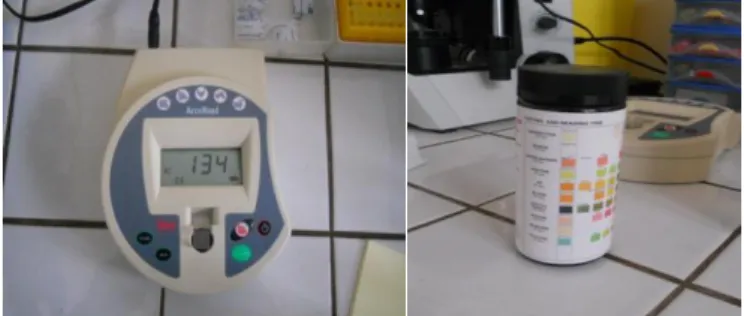
Figure 14 - Concentration determination with a spectrophotometer and pH analysis ... 30
Figure 15 – Immunocytochemical evidence of HSP70 in the spermatozoa head from the control group spermatozoa . ... 42
Figure 16 – Immunocytochemical evidence of HSP70 in chilled spermatozoa ... 43
Figure 17 - Immunocytochemical staining for HSP70 in frozen/thawed samples ... 43
LIST OF GRAPHS Graph 1 – Individual variation of the total motility with treatment ... 32
Graph 2 – Individual variation of the progressive motility with treatment . ... 33
Graph 3 - Representation of the sperm total motility (in the left) and progressive motility (on the right) for the group of samples according to the treatment... 33
Graph 4 – Individual variation of the velocity of the spermatozoa with treatment . ... 36
Graph 5 - Individual variation of the hypo-osmotic swelling test (HOST) of the spermatozoa with treatment. ... 38
Graph 6 – Effect of the cryopreservation process on the coiling of spermatozoa... 38
Graph 7 – Distribution of HSP 70 intensity in the spermatozoa’s head ... 44
vii
LIST OF TABLES
Table 1 - The most common causes leading to semen collection in dogs ... 2
Table 2- CASA parameters... 17
Table 3 – Composition of the extenders used for chilling and freezing/thawing procedures ... 24
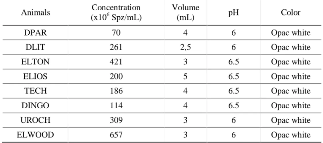
Table 4 – General characterization of the individual semen samples used in this study. ... 31
Table 5 – Spermatozoa morphology for the freshly ejaculated samples ... 31
Table 6 - Total and progressive motility ... 32
Table 7 - Velocity of spermatozoa movement ... 35
Table 8 – Hypo-osmotic swelling test ... 37
Table 9 – Immunocytochemical classification of the HSP70 in the spermatozoa ... 45
Table on Annex 1 – Total and progressive motility and consequent treatment losses ... 60
viii
List of abbreviations, symbols and units
% - percent
® - registered brand AI – artificial insemination
ALH - amplitude of lateral head displacement ARTs - artificial reproductive techniques AV – artificial vagina
BCF - beat cross frequency CA – California
CASA – computerized assisted semen analysis
CERCA - Centre d'Étude en Reproduction des Carnivores C-FDA - 6-carboxyfluorescein diacetate
DAB - 3,3’-diaminobenzidine tetrahidrocloret DHS – dihydrostreptomycin
ENVA - École Nationale Vétérinaire d’Alfort EthD – 1-ethidium homodimer
h - hour
HOST – hypo osmotic swelling test HSP - heat shock proteins
Hz – hertz IL - Illianois
IVF – in vitro fertilization KG – kilogram
L – liter
LIN - linearity of sperm movement ML – mililiter ng – nanogram p – significance level PBS – phosphate-buffered saline PGF2α – prostaglandin F2α PM – progressive motility rpm – rotations per minute s - second
STR – straightness SP – seminal plasma TM – total motility UK – United Kingdom
USA – United States of America
UTAD - University of Trás-os-Montes and Alto Douro VAP – velocity average pathway
VCL – curvilinear velocity VSL – velocity straight line
WHO – World Health Organization WI – Wisconsin
x - average
ZBA – zona pellucida binding assay ZP - zona pellucida
μm – micrometer μl – microliter
ix
ACKNOWLEDGEMENTS
To both, the University of Trás-os-Montes and Alto Douro (UTAD) and the École
Nationale Vétérinaire d’Alfort (ENVA) for all the support given, which allowed the development of this study.
To Professor Rita Payan Carreira, my coordinator, I recognize all the dedication applied,
the long hours of work, the patience, the friendship, all the wise advices given and the
opportunities provided, along with the priceless knowledge she transmitted me throughout my,
still short, career.
To Doctor Alain Fontbonne, who provided me everything required for the elaboration of
my work, along with very important knowledge and opportunities to evolve in the area of small
animal reproduction. Within the ENVA and specially the CERCA (Centre d'Étude en
Reproduction des Carnivores), I thank Fernando Mir, Emeline Leblond, Cindy Maenhoudt,
Natalia Santos and Emmanuel Fontaine for all the attention, knowledge, advices and tender that
they offered me during my internship. Also I would like to thank Karine Reynaud and all the
laboratory staff that received me very well and helped with everything I needed.
To Professor Maria dos Anjos Pires I leave a special acknowledgment, for the special
attention and care that she always showed towards me, being available every instant needed and
offering all the help and means required.
To the Histology and Pathological Anatomy Laboratory of UTAD, Professor Anabela
Alves and Professor Maria dos Anjos Pires as its directors along with its technician and staff,
Mrs. Lígia Lourenço, Ms. Ana and Mrs. Glória, I thank all the help provided.
To my laboratory partners, who were of extreme importance, Inês Santana and Inês
Carvalho, I leave a very special thanks for making me embrace the work every day with a smile,
x
To all my great friends in Vila Real, Badano, Luis Moreira, Rapper, Verguinhas, Diana,
Tatiana, Afilhada, Francisco, Rui, Marta, Ana Margarida, Xami, Zero, João Pires, Magda and
Ana Andrade for sharing and giving me so much throughout this so long, though short years.
To Professor Wojtek Nizanski who induced and enhanced in me this reproduction
thematic and to Natalia Mikolajewska who has been a great support and a very good friend in all
the occasions, I thank all the care and preoccupation shown and most importantly the friendship.
To Nuno Escudeiro, Vitor Lopes and Ana Lúcia a special thanks for telling me what I
need to hear, no matter what, making them true very best friends.
The last, but definitely not the least, I would like to thank my family, specially my
mother, my father and my little sister, with whom I shared all the tears and joys along my path
and who made me the person who I am today most importantly, because without them this would
mean nothing.
1
P
ART
I.
A
SSESSMENT OF CANINE SEMEN AFTER CHILLING AND
2
FUNDAMENTALS
The collection of semen from a male dog may represent an extended procedure during reproductive examination technique. Most frequently, it is used for three major purposes: artificial insemination (AI), semen cryopreservation or diagnostic purposes (Kustritz, 2007). Artificial insemination may be needed in the eventuality of vaginal anomalies of the bitch (such as vaginal septum, vaginal hyperplasia or ptosis, narrowed vagina or vaginal-vestibular stricture), whenever a stud dog is required to produce a seminal dose for breeding at distance or due to other sort of problems, such as behavioral issues between the male and the female (Kutzler, 2005). Canine semen may also be collected in a regular basis for cryopreservation, allowing dog owners to preserve the genetics of their male specimens, thus enabling breeding, even, when the male is no longer able to copulate, if it becomes infertile or if it is physically absent due to the distance or to its death (Johnston et al., 2001; Kutzler, 2005). The most common factors that usually lead to semen collection are described in Table 1.
Table 1 - The most common causes leading to semen collection in dogs
MOST COMMON CAUSES FOR SEMEN COLLECTION
FOR MEDICAL OR OTHER REASONS ASSOCIATED WITH THE REPRODUCTIVE ACTIVITY
- Evaluation of abnormal clinical signs (i.e. prepuce discharge)
- Following the treatment of different reproductive tract diseases
- For research
- Breeding soundness examination
- Males kept from copulation for several years - History of infertile breedings
- AI procedures with fresh or preserved semen - Behavioral issues
- Female opposition to the mounting
Nowadays, semen cryopreservation and artificial insemination are two of the major biotechnological techniques currently applied in animal breeding (Foote, 2002). Its large-scale introduction in cattle breeding around the 1940’s had the purpose of preventing genital infections transmissible through natural mating (Wilmot, 2007). Semen cryopreservation has also been seen for a long time as a way to improve the breeding of animals of greater farm importance, as a contributor to the conservation of endangered species and also as a way to overcome some aspects of the male infertility (Watson, 2000).
Successful preservation of semen is also important to improve the results of the main artificial reproductive techniques (ARTs), such as in vitro fertilization and, most commonly, AI (Luvoni, 2006). Semen cryopreservation eases the transport and exchangeability of genetic
3
material across short or long distances, contributing effectively to widen the restricted gene pool of some species (Johnston et al., 2001; Kutzler, 2005; Kustritz, 2007). Chilling and freezing can also turn into reality the creation of gene banks for particular wild species, which are for the moment maintained under controlled hunting, but may eventually become endangered in the future, as in the case of the Eurasian lynx (Lynx lynx) in Northeastern Europe, or the wild felids in Africa or South America (Swanson, 2006). Cryopreservation of spermatozoa has been well studied in different species, including the dog, having the first pregnancies been successfully achieved with frozen/thawed spermatozoa, using lactose and Tris-(hydroxymethyl)-aminomethane (Tris)-based extenders (Seager, 1969; Andersen, 1972).
Canine AI with cryopreserved semen has received increased interest from dog breeders in the past years due to its versatility and to the possibilities that it brings to genetic exchange between distant geographical areas (Thomassen et al., 2009). Furthermore, interest for in vitro technologies for canine species is increasing worldwide and to accomplish the aimed outcomes it is demanded good quality standards for preserved semen (Larsson et al., 2000; Johnston et al., 2001; Kutzler, 2005).
The chilling process of dog spermatozoa has been reported to induce less damage in the spermatozoa than freezing (Oettlé, 1986; England et al., 1996). When compared to the observed for the frozen spermatozoa, chilled sperm quality measured by motility, sperm morphology, acrosome status, hypo-osmotic swelling test and longevity, at a temperature of 39ºC, has been reported to be superior, for up to 4.9 days of chilling, despite some deterioration may occur during cold storage (England et al., 1996). Along with motility, the acrosome reaction in dogs seems to be more affected by freezing and thawing than by chilling (Oettle, 1986; Burgess et al., 2001). Recently, few studies have been developed in dogs to determine if the spermatozoa can be chilled and then successfully frozen, having in the end similar post-thawing viability to the semen that follows the normal freezing/thawing procedure (Hermansson, 2006).
Cryopreservation of semen, taking into account the process from cooling to thawing, has been proposed to present several negative effects on sperm viability, which could be related to injury of the plasma membrane, associated to changes in lipid phase transition, mechanical stress, efflux of water and high salt solutions and, possibly, by interfering with intracellular ice crystals formation (Woelders, 1997).
The type of semen extender and the freezing rates are determining factors on sperm survival during cryopreservation procedures and might explain the variability in the response of canine semen to freezing and thawing, between studies (Peña et al., 2000; Yildiz et al., 2000;
4
Nöthling et al., 2005). The major side effect of the freezing/thawing is a loss of sperm fertilizing ability, mainly associated with reduced motility, promotion of the capacitation and loss of viability (Yildiz et al., 2000). Some of these parameters are associated with sperm membrane damages, which are considered to be the primary cause of freezing induced injuries to canine sperm (Ström Holst, 1998). Furthermore, the same authors also report the existence of significant changes in the elemental composition of post-acrosomal region of the canine spermatozoa after freezing/thawing. In addition, important individual variations were found between stud dogs on what concerns sperm quality after freezing procedures, leading to the common designation of “good” and “bad” freezers (Silva et al., 2003).
1. Semen collection
Prior to semen collection a thorough and complete historical review of the dog’s previous health and breeding experiences should be obtained; in addition, information regarding the medication or supplements administered over the previous 6 months (at the minimum) and on the genetic or familiar background could be important (Johnson, 2006). There should also be collection of information about the status of vaccinations, dewormings and heartworm protection history, as well as on the duration of the ownership and accuracy of the history of the animal even before the establishment of the ownership (Freshman, 2002; Ettinger et al., 2010).
The anamnesis should be completed with a physical examination, which must consist of a visual, an auscultatory and a palpable examination of the entire animal, with special focus on the parameters or characteristics known to be heritable (CBRA, 1998; Johnson, 2006). This is the moment to talk with the owners and to advise them whether the animal should or should not be bred (Johnson, 2006). This evaluation should be followed by the examination of the reproductive system and it should include the palpation of all the reproductive structures, such as, the scrotum and its contents, the penis and the prepuce and finally the prostate (CBRA, 1998; Simpson et al., 1998). The order of the examination, like the palpation, can and should be postponed till after the collection of the semen, depending on the male’s behavior (Ettinger et al., 2010). The examination of the scrotum and its contents should start with the location of both testes within the scrotum and if that is not the case, the history should confirm a reliable information about the missing testis in order to consider the animal proper for breeding or not (Ettinger et al., 2010). The size and consistency of the testis should be evaluated and registered, along with the position of the tail of the epididymis (to check for example, for a possible torsion of the testicle), as well as the evaluation of the vas deferens, the spermatic cord, the thickness of the scrotal skin and the
5
state of the skin tissues, correlating them with a possible cause of temperature imbalance (CBRA, 1998). The palpation of the prostate should also be included in the physical examination of every male dog, considering its size and shape (Ettinger et al., 2010). Brucella screening tests should also be done in all breeding males (Wanke, 2004).
Following the physical examination, it is important to assess the libido, using or not a bitch in heat to facilitate the procedure, but always taking into account that the libido is influenced by several factors, such as the physical conditions of the collection site, the existence of olfactory cues or other specific and unknown reasons related to the bitch used for the effect (Simpson et al., 1998; Johnson, 2006).
Many factors influence semen quality, including the animal’s age, the size of the testicles, the degree of sexual arousal, the frequency of ejaculation, the collection procedure and the amount of seminal fluid collected (Johnson, 2006). It is expectable that males may react differently to the surroundings during semen collection and the procedures that precede it (Kutzler, 2005). The collection area should be calm, isolated and interruptions during the procedures should be prevented; the soil should be of secure footing for the animal, using for example a rubber-backed mat that besides providing good footing can also provide olfactory cues to the stud dog (Simpson et al., 1998; Freshman, 2002). Furthermore, in case of a toy dog it could be of benefit to position him on a grooming table (Freshman, 2001; Nelson et al., 2009). The presence of the owner is contradictory, as some dogs can be more comfortable with the owner standing next to them, whilst others can show reluctance to their master’s presence in the room (Purswell et al., 1992; Freshman, 2001). If there are any, the owner should bring the toys or other accessories that the dog associates with the breeding. The collector should avoid any “doctor” paraphernalia, like the white coat or the stethoscope (Freshman, 2002). It is important to remember that the collection of semen should be performed prior to the physical examination, injections, venipunctures or other stressful procedures. As last resort the physical or complementary exams can be even postponed to a different session (Feldman et al., 1996; Johnston et al., 2001). The equipment needed for the procedure should be promptly available and warmed approximately to body temperature (Seager, 1986). Some concerns have been raised on the effect of the usage of latex materials, on the canine spermatozoa motility, for example in the use of artificial vaginas (AV) (Althouse et al., 1991). There are some studies that correlate a decrease in the motility of spermatozoa and the prolonged time of contact with latex, not only with AV but also with gloves; however, this is not an issue when the different techniques are properly chosen and performed (England et al., 1992; Johnston et al., 2001). Another
6
inconvenience of the use of latex collection cones, is the need for a careful cleaning between collections, including the removal of all disinfectant residues, soap and water prior to the next use, taking into account that these are spermicidal (Kutzler, 2005). A sterile non-spermicidal lubricant can be used in minimal quantities in the AV, as so it decreases the friction with the penile mucosa, reducing the risk of injury of the superficial vessels and the penile mucosa (Froman et al., 1983).
Previously to the collection procedure, the stud dog should be walked, allowing him to urinate; afterwards, ideally, he should be trotted, to help the cleaning of the urethra from residual urine (Freshman, 2002). The presence of a teaser bitch is usually of benefit, especially if she is in proestrus or estrus and has the same proportions as the stud dog, although a calm bitch can also be used (Freshman, 2002). The typical scent from estrus can either be obtained from frozen swabs or sponges in plastic bags, taken from vaginal secretions (Purswell et al., 1992; Kutzler, 2005) or through the use of a chemical pheromone, methyl p-hydroxybenzoate (Aldrich Chemical, Milwaukee, WI), having them used in the vulvar area and in the tail head of a teaser bitch (Johnston et al., 2001; Kutzler, 2005). It has also been described that administration of prostaglandin F2α (PGF2α; Lutalyse®, Pfizer) in a dosage of 0.1mg/kg, given subcutaneously 15 minutes before the collection, enhances the concentration of the ejaculate, having also a positive effect on the libido. However secondary effects such as salivation, defecation and vomiting can be present due to the use of this drug (Nelson et al., 2009), and the sensitivity of individual males should be tested prior to collection. Allying the additive effects of both a teaser bitch and PGF2α the number of spermatozoa may increase in 300% (Kustritz, 2007). The bitch should be restrained and a muzzle may be used if she seems to be aggressive. The stud dog should be walked behind her, allowing him to sniff and make himself at ease before the collection procedure begins (Schubert et al., 1991).
Regarding the collection itself, dog’s prepuce should be vigorously massaged at the region of the bulbus glandis until a partial erection develops, time at which the bulb should be extruded from the prepuce to avoid possible pain for the animal (Freshman, 2001; Kutzler, 2005; Ettinger et al., 2010). If the bulbus glandis becomes too enlarged to be extruded from the preputial opening, removal of the bitch is indicated so that the erection can subside. Once the erect penis is completely extruded, a gentle, firm, circular pressure should be maintained, so that the male dog starts to ejaculate (Ettinger et al., 2010). After the collection, the bitch should be withdrawn from the room and the dog should be monitored till the penis returns to its normal position, inside the prepuce (Freshman, 2002).
7 2.1 Methods of semen collection
Semen collection in animals became easier due not only to the development of the AV in 1914 (Gordon, 2004), but also due to the use of the electroejaculator in 1936 (Gunn, 1936). However, nowadays, those methods have been discarded in dogs, in favor of the digital manipulation technique. Besides the digital manipulation of the penis, there are other methods, such as pharmacological ones, that can be used. Nevertheless, the studies that have been done are not very complete, regarding the parameters needed to privilege one method to the others; as an example, an essay has been done consisting on collecting male dogs using pilocarpine hydrochloride dissolved in saline solution, that showed better results than digital manipulation, according to the concentration of spermatozoa, however, no data has been registered relatively to the quality of the collected semen (Juniewicz et al., 1989).
Depending on the intended use for the semen, there are some particularities on the way the semen sample should be collected from a dog, taking into account the different fractions that compose the semen. Nevertheless, when there is the need to perform a breeding soundness examination on a dog or if there is suspicion of a reproductive disease, all the three fractions should be collected and evaluated (Kutzler, 2005; Lagishetty et al., 2011). Briefly, if there is the likelihood of risk regarding the fertility, it is important that a complete ejaculation occurs, which can be verified by the analysis of the seminal plasma alkaline phosphatase that should be superior to 10.000 U/L in the combined fractions (Kutzler et al., 2003).
For preservation reasons, either semen freezing or chilling, or for fresh semen AI, it may be of benefit to perform two collections, with an interval of 45 to 75 minutes among them. Although the number of spermatozoa is rather low in the second collection comparing with the first, the amount of both collections is in average, 70% more than if the collection is performed only once (England, 1999).
The total volume of a dog’s ejaculate may vary from 1.0 mL up to 30.0 mL (England et al., 2010; Ettinger et al., 2010). The canine ejaculate is composed of 3 distinct fractions (Johnston et al., 2001; Kustritz, 2007). The first or pre-spermatic fraction is composed of clear seminal plasma, devoided of sperm cells, originates from the prostatic gland and its main function is to flush the urethra (England et al., 2006; Nelson et al., 2009; Ettinger et al., 2010). The volume of the pre-spermatic fraction usually varies between 0.5 and 2.0 mL (Feldman et al., 1996; Freshman, 2001). The second fraction, also denominated sperm-rich fraction, has a cloudy
8
and opalescent appearance with an opaque consistency, varying in volume between 0.5 and 5.0 mL, depending on the testicular size and on the individual variation; moreover, in its composition there should be no cellular components besides sperm cells (Boucher et al., 1958; England, 1999; Nelson et al., 2009). It originates from the storage in the tail of the epididymis as from the daily sperm output (Johnston, 1989). The dog can take up to 2 minutes to achieve the emission of the sperm-rich fraction (Kutzler, 2005). Ideally there should be a separation between the first and the second fractions, especially for cryopreservation, as it is described that the sperm’s contact with both the first and third fractions may diminish the motility after 2 hours (England et al., 1992). The last portion, the third or prostatic fraction, is usually collected in order to increase the volume of the ejaculate for fresh semen AI and to perform a cytology or a culture (Ettinger et al., 2010). This last fraction can reach up to a total volume of 30 mL, depending on the duration of the pressure maintained around the bulbus glandis (Johnston, 1989).
The penile erection can remain for up to 10 minutes and it should be supervised till it wears off, in order to avoid complications (Freshman, 2002; Ettinger et al., 2010).
2. The semen
Whenever a semen sample is collected from a dog it should be assessed in order to determine its quality (Kutzler, 2005).
At the time of semen collection, information regarding the sexual abstinence should be checked and ideally the stud dog should have 4 to 5 days of sexual rest prior to collection (Freshman, 2002). Nevertheless, it has been reported that more than 10 days of sexual rest can result in an increase of morphologic abnormalities and a decrease in the motility related not only with the ageing of the spermatozoa but also with the increase of debris (Purswell et al., 1992; Johnston et al., 2001).
The dog’s ejaculate is composed of cells - spermatozoa (Spz)-, suspended on a fluid - the seminal plasma (SP). Among its constituents, diverse glandular products, proteins and electrolyte were identified (Kustritz, 2007). Several analyses have been performed in order to create a pattern within the normal values of SP mineral contents, specifically zinc, iron, calcium, magnesium and copper (Johnston et al., 2001). Their amounts usually drop till minimum values after the semen collection, due to the metabolism of the spermatozoa and its enzymatic
9
degradation. These parameters can also be influenced by the time interval of the last ejaculation, the sexual arousal of the male and the physiological status of the sexual accessory glands (Kustritz, 2007).
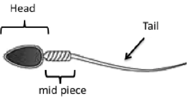
Structurally, a spermatozoon can be divided in 3 parts (Figure 1): the head, the midpiece and the tail (England et al., 2010). The sperm head contains a nucleus, which is covered proximally by the acrosome (Cunningham et al., 2007). The midpiece has approximately 1.5 times the length of the spermatozoon’s head (Feldman et al., 2004). A normal dog spermatozoon has approximately 7.0 µm, a midpiece with 1.1 µm and a tail with the length of 5.0 µm divided in its main piece and an end piece (Johnston et al., 2001).
Figure 1 – Morphological structure of the canine spermatozoon
3. The semen quality
Considering that semen contains live cells, the collector should handle it with special care while executing the procedures, since the interval from the collection till the evaluation should be as brief as possible, in order to prevent cellular modifications or even death of the cells (Feldman et al., 2004). The semen should be maintained between 20ºC to 30ºC (room temperature or water bath) and kept away from direct sunlight (England et al., 2010). Some authors defend that for the analysis of chilled semen, the tubes containing the samples should be kept in a glassed warm water recipient in order to prevent thermal shock during the chilling process, along with temperature variations during the period when the samples stand outside the fridge, for analysis (Iguer-Ouada et al., 2001). Immediately after the end of the collection procedure, the ejaculate should be assessed, since some parameters start to change soon after the collection (Kutzler, 2005).
10 3.1 Semen assessment
Each time that a dog is collected with the purpose of insemination, the sample should be assessed in order to determine its quality and to evaluate the reproductive and fertility potential for both the male and the seminal dose (Kutzler, 2005). Usually, available assessment methods vary mainly according to the use of an in-house or a research laboratory on what concerns its profundity or subjectivity (Kutzler, 2005).
Although several methods have been proposed for semen evaluation in dogs, the analysis of conception rates should remain as the ultimate test to assess male fertility (Oettle, 1993).
The assessment techniques used to determine semen quality may be divided into:
- a classical methodology, which include the evaluation of the physical parameters of the collected sample and the assessment of motility, concentration and morphology (Simpson et al., 1998);
- a variety of sophisticated assessment techniques, less subjective than the classical, that can be used to assess the motility (like the CASA system) and quality of sperm cells, including the evaluation of the viability, the integrity of the plasma membrane, the capacitating status and the acrosome reaction, among others (Nelson et al., 2009).
3.1.1 Classical methodology
The macroscopic parameters in the evaluation of semen are an important component of the classical method and include the volume, the color and the pH (Johnston et al., 2001).
The volume, by itself, is not a parameter that defines the quality of the semen, once it depends on the amount of prostatic secretion collected, the age and size of the animal and also the frequency of ejaculations of the dog (Feldman et al., 2004). Nevertheless, it is an essential parameter in order to calculate the total number of spermatozoa in the ejaculate, being of outmost relevance for the semen quality (Simpson et al., 1998). From a laboratorial point of view, the small volume obtained in each ejaculation consists of a great challenge in the semen processing, limiting the number of experiments possible to be done per dose (Farstad, 2009).
Usually the appearance of dog’s semen is white, cloudy or opaque and the intensity of its opacity depends on the concentration of spermatozoa (Feldman et al., 2004). The samples that possess a slightly cloudy coloration should be examined under the microscope, to check for the presence of spermatozoa, once that the cloudy color can characterize a sample with no spermatozoa, but can also be associated with a great amount of adipose tissue, bacteria or
11
inflammatory cells (Johnston et al., 2001). The yellow color can indicate the presence of urine, pus or some inflammatory exudates, while the green color normally indicates the presence of pus. The red or brown color can traduce the presence of fresh or haemolyzed blood, respectively (Johnston et al., 2001).
The pH should be expected to be within the range of 5.5 to 8.0. Specifically, the normal pH values of the third fraction should vary between 6.0 and 7.4 (Freshman, 2002). As an example, pH measurement may be important regarding the choice of an antimicrobial drug in the eventuality of a prostatitis (Johnston et al., 2001; Freshman, 2002).
The microscopic characteristics that should be taken into consideration during the assessment of dog’s semen are included in the protocol used by many andrologic centers and laboratories where light microscopic techniques are routinely used to evaluate the three conventional sperm parameters: concentration, motility and morphology (Johnston, 1991).
Semen concentration, along with the volume, is not per se, an indicator of the quality of the semen, as it also depends on the amount of the collected seminal secretion (Johnston et al., 2001; Freshman, 2002). Despite all the technological investment, the method still considered to be the gold standard is also the most traditional one, which uses a counting chamber, such as the Neubauer or the Bürker chambers (Kustritz, 2007). The total number of spermatozoa is achieved multiplying the concentration (millions per mL) by the ejaculate’s volume (mL); the value can vary between 300 million and 2 billion spermatozoa (Ettinger et al., 2010). This wide range of normal values enhances the theory that semen production is directly influenced by the quantity of testicular mass (Johnston et al., 2001; Freshman, 2002; Feldman et al., 2004; Kustritz, 2007).
The total and progressive motility can be assessed subjectively on a pre-warmed glass slide and analyzed under a light microscope (Johnston, 1991; Strom et al., 1997). The method should include five different fields of the microscope, in a total of 200 spermatozoa (Kustritz, 2007). The evaluation of the motility can become extremely difficult because of the high concentration of the sample, making it necessary to dilute the semen with a solution that can be the prostatic secretion (natural extender) or a synthetic extender (Feldman et al., 2004; Martinez, 2004). Some saline solutions used as extenders can lead to a decrease on the progressive motility of spermatozoa, due to its pH (Johnston et al., 2001). Other extenders, either due to the presence of viscous substances in their composition (such as egg yolk), the dilution factor or even the temperature at which the analysis is made, may induce a change in the velocity of the spermatozoa, along with the linearity of its movements (Schafer-Somi et al., 2007). The reference value for the progressive motility of dog semen is ≥ 70% (Johnston et al., 2001). The
12
progressive motility of a dog’s semen sample is not affected by the frequency of collections performed (Kustritz, 2007). Nonetheless, the first ejaculate after a long period of abstinence may present a great number of older spermatozoa that have been stored in the epididymis, resulting in a significant decrease in the total spermatozoa with progressive motility in the sample (Feldman et al., 2004). The percentage of morphologically normal spermatozoa is posit ively correlated with the percentage of spermatozoa with progressive motility (Johnston et al., 2001; Kustritz, 2007), once the motility is considered to be a manifestation of the structural and functional competences of the spermatozoa (Martinez, 2004; Volpe et al., 2009). Although it might not be the best parameter to define the capacity of fertilization of the spermatozoa, motility is an evaluation parameter fairly easy to assess and it can be used as a guideline for the evaluation of fresh and cryopreserved semen (LeFrapper, 2010). Even with all the development around this area and the improvement of more objective techniques for semen evaluation, the progressive motility is still the most used indicator for the spermatic function, once that it is unquestionable that the motility is essential for the progression of the spermatozoa throughout the oviducts (Martinez, 2004).
The abnormal motility can be associated with morphologically abnormal spermatozoa and with decreased fertility; however, there can be morphologically abnormal spermatozoa with a normal motility, making it important to evaluate the morphology (Ettinger et al., 2010).
The percentage of morphologically normal spermatozoa in a semen sample is considered acceptable over a value of 70% (Feldman et al., 2004). The sperm morphology can be evaluated through the use of different staining techniques, such as eosin/negrosin, Diff Quick or trypan blue (Bangham et al., 1955; Dott et al., 1972; Johnston et al., 2001; Freshman, 2002; Risopatron et al., 2002; WHO, 2010) and it is regarded as an important parameter in the conventional semen analysis (Johnston, 1991; Oettle, 1993). The two most used staining techniques are eosin-negrosin and modified Giemsa (Johnston et al., 2001; Freshman, 2002). The eosin-eosin-negrosin as a vital staining has the purpose of emphasizing the borders of the spermatozoa than the cell itself. It also allows, through the use of a contrast phase microscope, the evaluation of the acrosome of live and dead spermatozoa, having the live and acrosome intact spermatozoa a white color with round, regular and defined borders, whilst the live Spz with a reacted acrosome show the acrosomic region in a dark coloration with an unidentifiable apical extremity (Martinez, 2004). This method of coloration allows the differentiation between live and dead spermatozoa, considered as dead ones, the spz that become stained due to the absorption of eosin (staining),
13
once that they are considered to have lesions at the level of the cellular membrane (Kustritz, 2007).
It has been described the use of Spermac®. This staining allows a quick staining with unique characteristics, marking the spermatozoon’s nucleus in red, the acrosome, midpiece and tail in green and the equatorial region of the acrosome in pale green (Feldman et al., 2004); this makes this technique a very interesting staining to evaluate the morphology of the acrosome (Martinez, 2004; Monteiro et al., 2009).
Apart from the staining technique used, there are always some changes in spermatozoa, resulting from the cytology execution, called artifacts, such as detached heads, coiled or folded tails and folded intermediate pieces, so it can be concluded that the consistency used in the technique is essential for the accuracy and precision of the semen evaluation (Johnston et al., 2001). The use of stainings that are osmotically not similar to the spermatic samples, result in the detachment of the acrosome of some spermatozoa. This osmotic effect is more pronounced in the frozen/thawed than in the fresh semen (Martinez, 2004). Besides the Spz alterations caused by the processing of the semen samples, there are other factors such as testicular traumas, fever or infection of the reproductive tract, that can origin seminal changes, due to the increase of temperature in the testes (Martinez, 2004). However, these changes should only be noted some time after the initial problem, as the spermatogenesis on the dog has a length of 62 days to which the time for epididymal transit should be added (Johnston et al., 2001; Freshman, 2002). Other possible causes for the morphological changes of spermatozoa can be associated with the decrease of LH, a disturbed testosterone secretion or even iatrogenic causes (Martinez, 2004). Healthy dogs that change either of owner or environment can show an increase in the number of abnormal spermatozoa, possibly due to the increase of endogenous corticosteroids (Martinez, 2004).
The morphological changes of spermatozoa can be classified according to the cellular location where they occur: head, acrosome, intermediate piece and tail (Figure 2). Furthermore, they can be divided in primary, occurring during spermatogenesis, and secondary if occurring during maturation and for some authors also as tertiary, if during the preparation of the sample (Johnston et al., 2001). Another classification that tries to associate the sperm morphology to the sperm capacity for fertilization, divides the abnormalities of the spermatozoa in minor defects, as those which do not influence the fertility, and major defects, as those which are negatively correlated with the ability to fecundate the oocytes (Johnston et al., 2001).
14
Figure 2– Sperm cell abnormalities (vetmed.lsu.edu)
The morphological abnormalities that have been linked to infertility include defects in the connection with the intermediate piece, microcephalic spermatozoa and spermatozoa that possess proximal cytoplasmatic droplets (Feldman et al., 2004).
Two main problems that subsist with the light microscopic methods are the subjectivity and the variability (Oettle, 1993; Hewitt et al., 1998). Visual sperm motility assessment is difficult and may be influenced by both the temperature and the evaluator’s skills, leading to high variability among laboratories and observers (Verstegen et al., 2002). On the other hand, the evaluation of the morphology is also problematic due to the fact that it not only depends on the fixation, on the staining technique and on the quality of the microscope, but most importantly it further depends on the observer’s experience and skills (Pena et al., 1999; Pena et al., 1999).
The low number of spermatozoa assessed with the conventional techniques is also a factor for inter-laboratory variation (Pena et al., 1998).
15 3.1.2 Advanced methodology
Aiming to overcome the principal disadvantages of the current methods of canine semen assessment, other techniques have been proposed, such as fluorescence microscopy, flow cytometry, computerized sperm analysis systems and zona pellucida binding and penetration assays (Gunzel-Apel et al., 1993; Hewitt et al., 1998; Pena et al., 1999; Strom Holst et al., 2000b). One advantage of some of these techniques, such as the use of fluorophore combinations is to evaluate live and dead cells at the same time, either by fluorescent microscopy or by flow cytometry, the last one allowing the evaluation of large numbers of spermatozoa in a short period of time (Rijsselaere et al., 2005). The use of these methods allows also the simultaneous evaluation of several sperm characteristics, relatively to different organelles or sperm domains (Rijsselaere et al., 2005).
Wide variations have been described on what concerns the analysis of sperm motility (Chong et al., 1983; Jequier et al., 1983; Mortimer et al., 1986) along with the morphological parameters (Baker et al., 1987; Kruger et al., 1995) of the same ejaculate. The need for higher objectiveness in the methods used, recurring to its standardization for both practical and research purposes, was one of the major concerns in the past two decades (Smith et al., 2001; Iguer-Ouada et al., 2001a). The gathering of these factors opened a window for the development of several semi-computerized (England et al., 1990; Iguer-Ouada et al., 2001b) and computerized measuring devices (Gunzel-Apel et al., 1993; Smith et al., 2001; Iguer-Ouada et al., 2001a; Iguer-Ouada et al., 2001b) for the evaluation of canine sperm motility, morphology and concentration.
In order to fertilize an oocyte, a sperm cell should possess the ability to play several functions, including the cascade of reactions that allow the sperm/oocyte interaction at fertilization (Rijsselaere et al., 2005). A great deal of evolution has been gathered around the domains of the assessment of several sperm functions, allowing a more detailed evaluation of dog semen quality (Hewitt et al., 2001). There are some functional assays that are able to examine the interaction between the spermatozoa and the oocyte, thus giving a good estimation of sperm fertilizing capacity, by testing for example the ability of the spermatozoon to bind and penetrate the ZP and fertilize in vitro (i.e. achieved in vitro fertilization, IVF). The ZP binding assay (ZBA) has been used in several species, including the dog (Hay et al., 1997; Strom Holst et al., 2000a; Strom Holst et al., 2000b). In general, the techniques available to assess the semen can be resumed in fluorescent staining, computer-assisted sperm analysis, zona pellucida-binding assay, oocyte penetration assay/in vitro fertilization, and sperm-oviduct interaction (Rijsselaere et al., 2005).
16 3.2 Sperm motility assessment
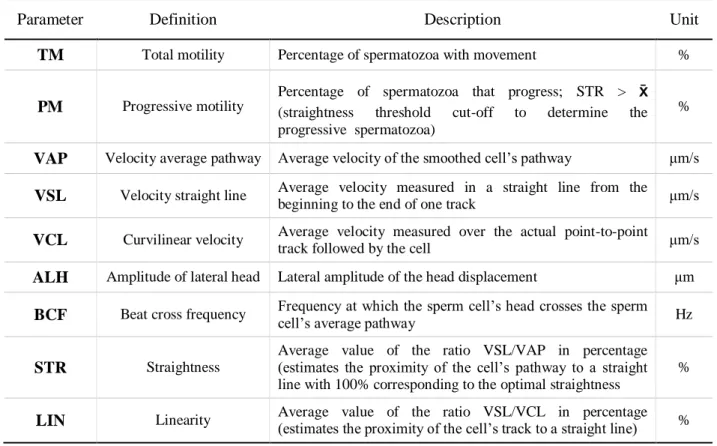
Several commercial computer assisted sperm analysis systems, generally named CASA, such as Strömberg-Mika Cell motion analyzer, CellSoft computer videomicrography; Hobson Sperm Tracker and Hamilton-Thorne, base predominantly on individual spermatozoon assessment and have been validated for use in dogs (Gunzel-Apel et al., 1993; Smith et al., 2001; Iguer-Ouada et al., 2001a; Iguer-Ouada et al., 2001b). These kind of devices allow an accurate and quick calculation of various semen parameters such as the total and progressive motility, the static, slow, medium and rapid moving spermatozoa, the linearity of sperm movement (LIN), the beat cross frequency (BCF), the amplitude of the lateral head displacement (ALH) and quite a few other velocity parameters (Table 2) (Gunzel-Apel et al., 1993; Smith et al., 2001; Iguer-Ouada et al., 2001a; Iguer-Ouada et al., 2001b). There are several studies reporting the existence of high correlations among the computer-calculated motility, progressive motility and concentration, and the conventional light microscopic evaluation (Gunzel-Apel et al., 1993; Iguer-Ouada et al., 2001a). In addition, these systems have proved to be useful when assessing various semen characteristics simultaneously and objectively and are endowed to detect subtle changes in the sperm movement, unnoted with the conventional semen analysis (Gunzel-Apel et al., 1993; Rota et al., 1999; Verstegen et al., 2002). Also, a very important characteristic is that a high number of spermatozoa can be analyzed in a short period of time (Iguer-Ouada et al., 2001a).
The main disadvantages pointed out for those devices are the need for high investments and the notorious need for standardization and validation of the software and the system previously to its practical use (Smith et al., 2001; Iguer-Ouada et al., 2001b; Verstegen et al., 2002). Moreover, to avoid a new source of subjectivity among laboratories, associated to the definition of the computer parameters to be applied, the selection of the software and the microscope conditions, the standardization of the technical settings became in focus (Smith et al., 2001; Iguer-Ouada et al., 2001b). In addition, it still needs to be determined which sperm movement characteristics are of best clinical value for the prediction of in vivo fertility in dogs, along with the standardization of the parameters, which differ between users or laboratories and the different commercial systems (Verstegen et al., 2002).
17
Table 2 - CASA parameters (modified from Iguer-ouada et al., 2001a; Verstegen et al., 2002; Rijsselaere et al., 2005)
Parameter Definition Description Unit
TM Total motility Percentage of spermatozoa with movement %
PM Progressive motility
Percentage of spermatozoa that progress; STR > x (straightness threshold cut-off to determine the progressive spermatozoa)
%
VAP Velocity average pathway Average velocity of the smoothed cell’s pathway μm/s VSL Velocity straight line Average velocity measured in a straight line from the
beginning to the end of one track μm/s VCL Curvilinear velocity Average velocity measured over the actual point-to-point
track followed by the cell μm/s
ALH Amplitude of lateral head Lateral amplitude of the head displacement μm BCF Beat cross frequency Frequency at which the sperm cell’s head crosses the sperm cell’s average pathway Hz
STR Straightness
Average value of the ratio VSL/VAP in percentage (estimates the proximity of the cell’s pathway to a straight line with 100% corresponding to the optimal straightness
%
LIN Linearity Average value of the ratio VSL/VCL in percentage (estimates the proximity of the cell’s track to a straight line) %
The CASA is basically a computer analyzer for sperm motility and morphometry. Its use has become more popular, but due to its high cost it is still not available to the majority of the veterinarians (Verstegen et al., 2002; Kustritz, 2007). With a contrast microscope integrated, this equipment allows a quick and objective evaluation of many parameters, some of them described in Table 2.
As this equipment allows to identify even very small variations in the spermatozoa motility, that would, otherwise be impossible to detect (Rijsselaere et al., 2005), it allows the classification of small populations of spermatozoa in a sample that may react differently when submitted to processes, such as cryopreservation (Kustritz, 2007).
3.3 Hypo-osmotic swelling test
The morphological and functional integrity of the sperm membrane has been intensely studied, given its importance as a cellular barrier and in the cell to cell interaction (Rodríguez-Martínez, 2000). The integrity of the plasma membrane is crucial for the fertilizing capacity of spermatozoa. As described to the morphology, in the classical methods, the membrane of dog’s
18
spermatozoa was routinely assessed by means of light microscopic stainings, such as eosin/negrosin (Bangham et al., 1955; Dott et al., 1972) or trypan blue (Risopatron et al., 2002). An indirect method to evaluate the membrane integrity is by exposing spermatozoa to hypo-osmotic conditions, for example through the hypo-hypo-osmotic swelling test (Kumi-Diaka, 1993), once that the number of swollen spermatozoa was shown to be inversely proportional to the number of membrane damaged spermatozoa (England et al., 1993).
This test is considered, by many laboratories, as an appropriate method to evaluate the integrity of the membrane and due to its simplicity, it can be introduced into the routine semen analysis (Pinto et al., 2008). This technique consists of mixing the semen sample in a hypo-osmotic solution and incubating it at 37ºC, for 45 to 60 minutes (Rodríguez-Gil et al., 1994). When submitted to a hypo-osmotic solution, a viable spermatozoa possessing an intact, active and functional plasmatic membrane, will react, allowing the fluid to enter the cell, causing the swelling and coiling of the tales of spermatozoa (Kustritz, 2007). It has been proved that no significant differences exist between incubation times of 1 or 60 minutes, and also that this test is easily performed by any veterinarian (Pinto et al., 2008). The solution used in this test should be enough hypo-osmotic to cause the swelling, without causing lysis to the cell (Bencharif et al., 2008). The hypo-osmotic solutions described for this test include sodium citrate, fructose in distilled water and a solution of sacarose (Kustritz, 2007).
The swelling effect is more easily identifiable on the tails of the spermatozoa for two main reasons: the membrane of the tail is more flexible and is less adherent to the internal structures comparing to the membrane of the head, and at the spermatozoon’s head the intracellular fluid compartment is rather small making it difficult to observe the variation in the volume when the water enters the cell (Jeyendran et al., 1984). Previously to the realization of the hypo-osmotic swelling test, it is of high relevance that the percentage of spermatozoa with tail abnormalities is measured, in order to subtract this value to the counting made after incubation, obtaining the true value of spermatozoa with an intact plasmatic membrane (Kustritz, 2007).
This test shows a good correlation among the spermatozoa that suffer swelling and the percentage that were able to fertilize the oocytes in vitro. In the same study, the correlations obtained between this parameter, the percentage of progressive motility, the percentage of morphologically normal spermatozoa and the percentage of live spermatozoa (negative with vital staining) was not so relevant (Jeyendran et al., 1984). There is no relation described between the
19
hypo-osmotic swelling test results and the fertilization capacity of dog spermatozoa (Martinez, 2004), however there is a correlation between these results and the motility and viability for both fresh and frozen dog semen (Pinto et al., 2008).
4. Heat shock
A reaction to an induced heat shock is inherent to the procedure of semen cryopreservation, which is accompanied by the irreversible loss of viability when the spermatozoa rapidly reach neutral temperatures. This co-exists with a decrease in sperm motility (Quinn et al., 1966), a reduction in the energetic ability and an imbalance of the membrane’s permeability (Watson, 1981). Thus, the refrigeration of the semen during the freezing process should be done with extreme precaution, in a slow procedure, even if it does not prevent all the damage to the membrane (Watson, 2000).
It has been reported that the spermatozoa become susceptible to the heat shock in the proximal region of the body of the epididymis, during the epididymal maturation process; more precisely it occurs when the cytoplasmatic droplet moves towards the distal portion of the midpiece. It has been concluded that acquisition of motility and heat shock susceptibility may occur at the same time (White, 1993). The freezing-associated heat shock and the irreversible lesions that it causes, seem to result from the changes in the lipid organization in the sperm cell membrane (Drobnis et al., 1993).
The heat shock has different impact according to the species, being the sperm cells of both swine and bull the most susceptible. There are relevant changes in the ion exchanges, with Ca2+ increase and loss of K+ and Mg2+. On the other hand, the sperm of humans, dog and rabbit seems not to show so intense changes (Quinn et al., 1966). Nevertheless, freezing/thawing processes in canine semen are usually referred as being detrimental to the sperm fertility, mainly due to reduced longevity (England et al., 1993; Bessa, 2005) and to an increase in the proportion of capacitated cells after thawing (Rota et al., 1999; Bessa, 2005). These issues were at the origin of intense scientific work aiming to identify the most adequate extender for dog semen.
20 4.1 Heat Shock Proteins
Heat shock as other stressing factors stimulate every cell to synthesize a small group of protective proteins, which are named heat shock proteins (HSPs) (Schlesinger et al., 1982; Craig, 1985; Schlesinger, 1986). HSPs are classified into several families, named according to their approximate molecular weight (Sonna et al., 2002).
Among several stress stimuli, elevated temperature or heat stress have been described as inducers of the HSPs production in certain tissues (Nollen et al., 1999). The concentration of stress proteins increase, responding to stress factors, therefore acting as cell protectors and facilitating cell survival, proportioning renaturation of partially denaturated proteins (Parcellier et al., 2003).
There have been important and relevant studies about the heat shock proteins throughout the last years. These studies have been more incisive in a special set of evolutionary conserved heat inducible and related proteins of molecular weight of around 70 kDa (Lawson et al., 1984; Craig, 1985; Schlesinger, 1986).
HSPs, and specially HSP70, under physiological conditions act as molecular chaperones as well as assisting protein folding, transport and degradation; on the other hand, subjected to stress conditions, they prevent aggregation and promote refolding after denaturation, protecting cells of stress or other hazardous conditions (Georgopoulos et al., 1993). Induction of heat shock response is related to the physiological temperature range (Liu et al., 1994) and can occur either when the temperature increases upwards a threshold or when it decreases below a specific value. Responding to heat stress, heat shock genes are stimulated and new molecules of HSP are produced within the cells, in an attempt to prevent or to correct denaturing of cell proteins. Thus, induction of the heat shock response should be read as a manifestation of temperature-induced cell damage (Liu et al., 1994). One important implication of this finding in sperm post-thawing fertility is that events involved in the expression of the heat shock response require active cell metabolism, such as ATP production or macromolecule synthesis, which are of limited capacity in sperm cells due to their highly specialization.
21
P
ART
II.
C
OMPARISON OF CANINE SPERM QUALITY UNDER
22
1. Introduction and aims
The specific thematic for this work was chosen accordingly to the work developed throughout the 3.5 months of practical training in the École Nationale Vétérinaire d’Alfort, in the area of small animal reproduction, CERCA (Centre d'Études en Reproduction des Carnivores), which is a highly regarded centre for reproduction and specifically cryopreservation.
In the process of cryopreservation, the sperm cells, in particular dog sperm cells are highly susceptible and suffer substantial alterations, especially on what concerns sperm motility. As proposed aims for this study, we ought to:
1 – Evaluate the sperm quality upon chilling and freezing/thawing procedures and to compare it to the freshly ejaculated sperm.
2 – Characterize the pattern of immunolocalization of HSP70 in the freshly ejaculated canine spermatozoa and to study putative changes after being chilled and frozen/thawed.
We further will attempt to study eventual association between data gathered in those two studies, and to ascertain possible influences of the temperature stress induced by the two cold treatments on sperm quality.
To achieve the proposed objectives we performed two different studies where the same samples were used. For the first experiment, freshly ejaculate samples were obtained from 8 dogs. Those samples were processed in straws as for routine seminal doses and submitted to chilling for 24h and to freezing in liquid nitrogen with posterior thawing. The quality of each sample was assessed by the use of both classical and advanced methods, for each temperature-challenge tests, compared to controls (fresh ejaculates) and differences among treatments and individual dogs were analyzed. In this study, the following parameters were evaluated: concentration, pH, volume, color, morphology and HOST, as well as CASA characteristics (total and progressive motility, VAP, VSL, VCL, ALH, BCF, STR, LIN).
For the second experiment, cytological specimens were prepared from each sample, fixed at 95% ethanol and processed for immunocytochemistry. The characterization of the normal expression pattern of HSP70 was studied in the control samples (fresh ejaculates). The intensity of immunostaining as well as the sub-cellular location of the protein was recorded. Comparison between controls and the cold-treated groups (chilling and freezing/thawing) was evaluated on what concerns the intensity of immunoreactions and any possible changes in the sub-cellular location of HSP70. With this approach we ought to test the possible involvement of HSP70 in some treatment-associated changes found in more conventional sperm assessment.
23
2. Common material and methods
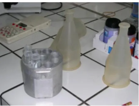
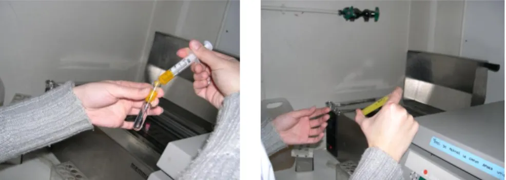
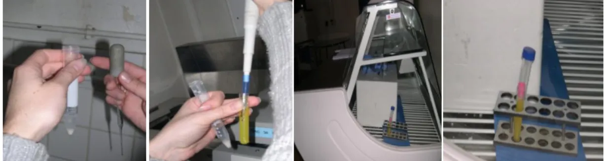
2.1 AnimalsIn this work, fresh ejaculates were obtained by digital manipulation, according to the method described by Linde-Forsberg (Linde-Forsberg, 1991), from 8 dogs, with ages ranging between 3 to 4 years, that either belonged to the École Nationale Vétérinaire d’Alfort, Paris or were received in CERCA (Centre d'Études en Reproduction des Carnivores) for consultation. There was no specificity neither according to the breed chosen, nor the age of the dogs, besides that all of them were considered to be healthy and reproductively within the accepted fertility parameters, since a reproductive exam and a prostatic ultrasound were previously performed. The collected sample included the sperm-rich (second) fraction and a small amount of the third fraction Figure 3.
Figure 3 - Semen collection material
2.2 Sample preparation
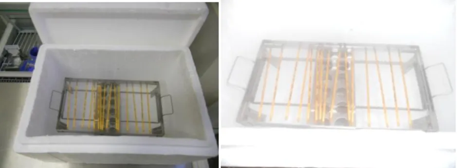
For chilling procedures, after dilution the samples were stored in test tubes in the refrigerator for up to 24h. For frozen samples, 0.50 mL straws were used and stored in liquid nitrogen for a period of 2 days.
From fresh ejaculates, and after semen evaluation, the straws were prepared in order to obtain a concentration of 150 x 106 spermatozoa in each straw. The following formula was applied:
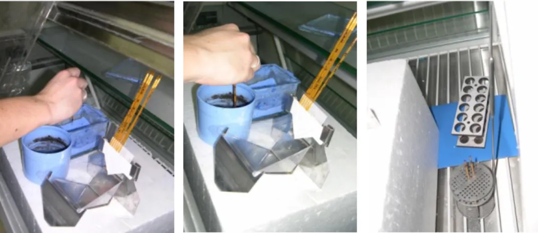
24
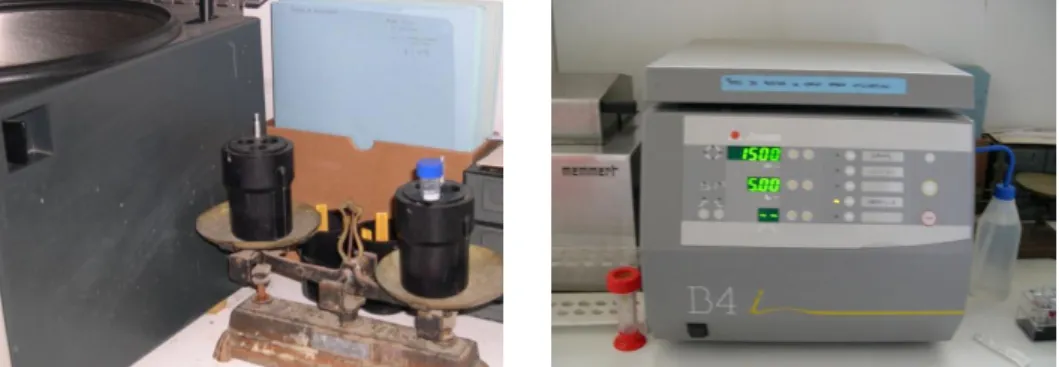
Fresh samples were centrifuged for 10 minutes at 1.500 rpm, as displayed in figure 4. The exceeding fluid was withdrawn and only the sperm pellet was kept. The pellet was then re-suspended in the adequate extender, according to the temperature treatment to be followed: chilling or freezing.
Figure 4 – For preservation, either for chilling or freezing, the collected semen is centrifuged to obtain a pellet with spermatozoa.
2.3 Extenders
The extenders used in the study, for both chilling and freezing/thawing of the semen, correspond to the method of Uppsala, (Linde-Forsberg et al., 1999). The extenders were prepared with Tris, citrate, glucose, antibiotics, glycerol, Equex and distilled water (Table 3) and were frozen.
Table 3 – Composition of the extenders used for chilling and freezing/thawing procedures
Components Chilling extender Freezing extender 1 Freezing extender 2 Thawing
extender TRIS 6.025 g 3.025 g 3.025 g 3.025 g Citrate 3.4 g 1.7 g 1.7 g 1.7 g Glucose - 1.25 g 1.25 g 1.25 g Fructose 2.5 g - - -Peniciline 200 000 UI 0.06 g 0.06 g 0.06 g DHS 0.2 g 0.1 g 0.1 g 0.1 g
Distilled water Till 200 mL 77 mL 72 mL Till 100 mL
Glicerol 12 mL 3 mL 7 mL
-Equex - - 1 mL