2
.º CICLO FCUP2019
Dissecting formin´s molecular
mechanisms of action during
embryonic cell division
Jaime Daniel Leite Pereira
Dissertação de Mestrado apresentada à
Faculdade de Ciências da Universidade do Porto em
Bioquímica
2019
D iss ec ting f o rm in´s mo lec ular mech an is ms o f ac tion du ri ng e mbryon ic ce ll divi s ion Ja ime Danie l Leit e P e rei raDissecting formin´s
molecular
mechanisms of action
during embryonic
cell division
Jaime Daniel Leite Pereira
Mestrado em Bioquímica
Departamento de Química e Bioquímica 2019
Orientadora
Ana Xavier de Carvalho, Group Leader
Instituto de Investigação e Inovação em Saúde (i3S)
Coorientador
Pedro Nuno Simões Rodrigues, Professor Associado Instituto de Ciências Biomédicas Abel Salazar (ICBAS)
Todas as correções determinadas pelo júri, e só essas, foram efetuadas.
O Presidente do Júri,
Autor
Jaime Daniel Leite Pereira
Rua do Campo Alegre, 4169-007, Porto, Portugal, [email protected]
Orientadora
Ana Costa Xavier de Carvalho, Group Leader, i3S
R. Alfredo Allen, 4200-135 Porto, Portugal, [email protected]
Coorientador
Pedro Nuno Simões Rodrigues, Professor Associado, ICBAS
Acknowledgments
Após a finalização das atividades laboratoriais no âmbito do projeto de dissertação de mestrado em Bioquímica, gostaria de agradecer e deixar uma referência a todas as pessoas envolvidas no meu percurso académico e na realização deste meu objetivo pessoal.
Começo por prestar um agradecimento ao diretor do mestrado em bioquímica, Professor Doutor Paulo Correia de Sá pelo acolhimento, integração e ingresso no mestrado em bioquímica e às instituições: Faculdade de Ciências da Universidade do Porto (FCUP); ao Instituto de Ciências Biomédicas Abel Salazar (ICBAS) e ao Instituto de Investigação e Inovação em Saúde (i3S), onde decorreram as minhas atividades durante o mestrado.
Posteriormente, gostaria de agradecer à minha orientadora Ana Xavier de Carvalho, por me conceder a oportunidade de incorporação no seu grupo de investigação, que me permitiu poder evoluir tecnicamente como cientista, bem como por todos os seus conselhos, apoio, disponibilidade e ensinamentos transmitidos ao longo deste projeto de dissertação. Do mesmo modo, transmito também o meu agradecimento à minha supervisora de laboratório Elaine Chan, pela sua orientação e ensinamento de técnicas laboratoriais, pela sua paciência, amizade, companheirismo, por me oferecer o seu apoio durante os momentos difíceis, por todo o seu empenho e dedicação para me transmitir os seus conhecimentos e exigir de mim sempre a minha melhor versão.
Adicionalmente, agradeço às restantes pessoas que constituem o grupo Cytoskeletal Dynamics e aos membros do grupo Cell Division Mechanisms e Tiago Dantas Lab, por me receberem de braços abertos, estarem sempre disponíveis para me ajudar a ultrapassar as minhas dificuldades, por terem sempre uma palavra amiga nos momentos difíceis e usarem toda a sua experiência para me indicar sempre as melhores decisões a tomar. Foi um prazer compartilhar todos os momentos convosco e poder estar rodeado de um ambiente cheio de um positivismo contagiante.
Gostaria ainda de agradecer aos meus amigos que conheci na Residência Universitária do Campo Alegre: Cláudia Antunes, Diogo Rodrigues, Rodrigo Silva, Marcelo Loureiro, Sara Mendes, Cláudio Graça e Diogo Ferraz, por todas as experiências e histórias que compartilhamos e pela amizade que desenvolvemos e a todos os meus colegas de curso, com uma palavra de apreço para os meus amigos Flávio Costa, Rita Fonseca, Alexandre Pinto e Karenina Santos.
fazem diariamente para me proporcionarem um futuro melhor, pelo carinho e amor que sempre me demonstraram, pela importância que tiveram na minha educação e formação como pessoa e à minha irmã Sónia Pereira por todo o seu apoio e por ser um modelo para mim pela sua capacidade de trabalho e dedicação e por todos os seus ensinamentos e que aprendi com ela.
Muito obrigado a todos os que contribuíram de alguma forma no meu percurso académico e para o meu bem-estar e satisfação pessoal. Um bem hajam!!!
Resumo
Nas células eucarióticas, a actina desempenha um papel essencial devido à sua participação em diversos processos celulares, entre os quais, a citocinese. A citocinese envolve a formação e posterior constrição de um anel de actomiosina que divide a célula mãe em duas células-filhas. A ação de forminas Diaphanous é essencial para a elongação de filamentos de actina não ramificados que compõem o anel contrátil. Os mecanismos subjacentes a esta regulação espaço-temporal não são totalmente conhecidos. Apesar de alguns mecanismos terem sido propostos, o único mecanismo bem caracterizado é a libertação da autoinibição das forminas por RhoA ligado a GTP. Sabe-se que a citocinese nas células animais é completamente dependente da ativação da RhoA mas em leveduras, a ativação de forminas pela ligação a Rho não é biologicamente relevante. O estudo da regulação de forminas é essencial para entender a citocinese, especialmente porque a sua desregulação tem sido associada a várias doenças incluindo cancro. O principal objetivo do meu projeto, é investigar se a ativação por RhoA (RHO-1 em C. elegans) é o principal mecanismo para a regulação de forminas durante a citocinese embrionária em C. elegans, in vivo.
Existem 6 genes de forminas em C. elegans, mas apenas CYK-1 foi demonstrado ser necessário para a integridade da estrutura do anel contrátil. Assim, nós propusemo-nos a realizar um estudo da estrutura-função de CYK-1 em embriões de C. elegans. Para estudar a regulação de CYK-1 in vivo, nós geramos com sucesso estirpes com a mutação CYK-1(V279D) e uma versão truncada de CYK-1, CYK- 1(∆1250-1437), sem alguns elementos regulatórios do C-terminal, usando a técnica CRISPR/Cas9.
A mutação V279D é suposto impedir a ligação a RhoA. Este resíduo é altamente conservado entre espécies e foi demonstrado que a sua mutação causa uma forte deficiência na ligação de RhoA a forminas em células de mamíferos. Os animais homozigóticos de C. elegans cyk-1(V279D) são estéreis. Para examinar o impacto desta mutação na embriogénese, introduzimos um transgene recodificado de CYK-1::GFPre em animais cyk-1(V279D) que pode ser especificamente deletado por
RNAi. Quando CYK-1::GFPre foi completamente deletado, embriões expressando
apenas o mutante de CYK-1 deficiente para a ligação a Rho falharam a citocinese e não eram viáveis. A geração de animais expressando CYK-1(∆1250-1437), é suposto impedir a formação da interação autoinibitória que mantem a formina numa configuração inativa, incapaz de elongar filamentos de actina. CYK-1(∆1250-1437) é esperado constituir uma versão de CYK-1 constitutivamente ativa. Animais
homozigóticos cyk-1(∆1250-1437) geraram embriões inviáveis. Para examinar o impacto desta mutação na embriogénese, uma vez mais, introduzimos um transgene recodificado de CYK-1::GFPre em animais cyk-1(∆1250-1437) que pode ser
especificamente deletado por RNAi. Após completa deleção de CYK-1::GFPre,
embriões expressando apenas a versão truncada de CYK-1 falharam o completar da citocinese, durante a sua última etapa, a abscisão.
Todos estes resultados, sugerem fortemente que Rho-1 ativa se liga diretamente a CYK-1, regulando a sua atividade durante a citocinese e o desenvolvimento embrionário em C. elegans. Este estudo indica ainda que CYK-1 necessita de ser inativada durante as últimas etapas da citocinese, presumivelmente durante a abscisão, de modo a completar com sucesso a citocinese.
Abstract
In eukaryotic cells, the actin cytoskeleton plays an essential role in driving diverse cellular processes, including cytokinesis. Cytokinesis involves the assembly and subsequent constriction of a contractile actomyosin ring that pinches the mother cell into two daughter cells at the end of mitosis. The action of Diaphanous formins is essential for the elongation of non-branched actin filaments that compose the contractile ring and this formin activity has to be highly regulated in space and time. The mechanisms underlying this spatiotemporal regulation of formin activity are only poorly understood. Although several mechanisms have been proposed to regulate formin activity, the only well-characterized mechanism is RhoA-dependent release of formin autoinhibition. Cytokinesis in mammalian tissue culture cells is known to depend on RhoA activation but limited in vivo studies exist. In fission yeast, for instance, formin activation by Rho binding is not biologically relevant. As alternative mechanisms for formin activation have been proposed, the study of formin´s regulation in vivo is crucial to understand cytokinesis, especially because its deregulation has been linked to various diseases including cancer.
The main goal of my project is to investigate whether activation by RhoA (RHO-1 in C. elegans) is the main mechanism for formin regulation during C. elegans embryonic cytokinesis.
There are six formin genes in C. elegans, but only CYK-1 has been shown to be required for furrow initiation and contractile ring structure integrity. Thus, we proposed to perform a structure-function analysis of CYK-1 in C. elegans early embryos. To study the regulation of CYK-1 in vivo, we successfully generated strains expressing a RhoA-binding deficient mutant, CYK-1(V279D), and a truncated version of CYK-1 missing C-terminal regulatory elements of the protein, CYK-1(∆1250-1437), by directly editing the C. elegans genome using the CRISPR/Cas9 technique. Homozygous cyk-
1(V279D) animals were sterile. To examine the impact of this mutation on
embryogenesis, we introduced a transgene-reencoded wild-type CYK-1::GFPre into
cyk-1(V279D) animals that could be specifically depleted by RNAi. When CYK-1::GFPre
was fully depleted, embryos only expressing RhoA-binding deficient CYK-1 mutant failed cytokinesis and were not viable. Animals expressing CYK-1(∆1250-1437), a version of CYK-1 that is expected to be constitutively active, layed unviable embryos. To examine the impact of this mutation on embryogenesis, we also introduced in these animals a transgene-reencoded wild-type CYK-1::GFPre that could be specifically
depleted by RNAi. After full depletion of CYK-1::GFPre, embryos only expressing CYK-
1 (∆1250-1437) failed to complete cytokinesis during its last step, abscission.
Taken together, our results strongly suggest that active RHO-1 directly binds to CYK-1 regulating its activity during cytokinesis and embryonic development in C.
elegans. Also this study indicates that CYK-1 needs to be inactivated during the last
steps of cytokinesis, presumably during abscission, in order to successfully complete cytokinesis.
KeyWords
Cytokinesis, formins, actomyosin contractile ring, C. elegans, CYK-1; CRISPR/Cas9, Rho-dependent formin activation
Index
Acknowledgments ... v Resumo ... vii Abstract ... ix KeyWords ... xi Index ... xiiList of Tables and Figures ... xiii
Abbreviation list ... xiv
Introduction ... 1
1- Importance of cell cycle to homeostasis in multicellular organisms ... 1
2 – Cytokinesis ... 1
3 - Formins ... 5
3.1 Domain organization of formins ... 5
3.2 Diaphanous Formins and their regulation by Rho-GTP ... 7
3.3 Other regulatory mechanisms of diaphanous formins ... 10
3.4 Diaphanous formins and cytokinesis ... 11
4 - Caenorhabditis elegans: A model organism to study developmental biology... 13
5 - Generation of C. elegans mutants by CRISPR/Cas9 technique ... 16
Aims of this study ... 18
Material and Methods ... 19
C. elegans maintenance ... 19
Freezing C. elegans Stocks ... 21
Generation of new C. elegans strains expressing CYK-1(V279D) and CYK- 1(∆1250-1437) by CRISPR-CAS9-mediated direct editing of the C. elegans genome ... 22
Crossing C. elegans strains ... 26
Embryonic Viability test ... 28
RNA interference ... 28
Results ... 30
1 - Animals expressing CYK-1(V279D) animals are sterile... 30
2 - Embryos expressing CYK-1(V279D) are not viable ... 31
3 - CYK-1(V279D) does not support cytokinesis in 1-cell embryos ... 33
4- CYK-1(V279D) does not elongate actin filaments bundles at the cell equator .. 35
5 - Animals expressing CYK-1(∆1250-1437) generate unviable progeny ... 36
Discussion and Future perspectives ... 40
Conclusion ... 45
List of Tables and Figures
Table 1 - List of C. elegans strains used in this study ... 20
Table 2 - List of CRISPR/Cas9 single guide RNAs (sgRNAs) and repair templates used in this study ... 23
Table 3 - List of CRISPR/Cas9 single guide RNAs (sgRNAs) and repair templates used in this study ... 24
Table 4 - Components and conditions used in the PCR reaction to identify CYK- 1(∆1250-1437) mutation. ... 25
Table 5 - List of dsRNAs used in this study. ... 29
Figure 1 - Overview of animal Cytokinesis ... 3
Figure 2 - Schematic representation of the recruitment and organization of cytokinetic machinery to the formation of the contractile ring during cytokinesis ... 4
Figure 3 - Mechanism of nucleation and elongation of linear actin filaments through the conserved FH1 and FH2 domain of formins ... 7
Figure 4 - Schematic representation of the architecture of families of mammalian formins ... 9
Figure 5 - Schematic representation of the regulatory mechanism of DRF mDia by active Rho GTPase ... 9
Figure 6 - C. elegans life cycle ... 15
Figure 7 - Adult C. elegans hermaphrodite seen with DIC ... 15
Figure 8 – Expression of CYK-1(V279D) leads to animal sterility ... 31
Figure 9 – CYK-1::GFPre probe localizes in the contractile ring and rescues embryonic viability upon depletion of endogenous CYK-1 by RNAi. ... 32
Figure 10 - CYK-1 RHO-binding deficient mutant causes embryo lethality. ... 33
Figure 11 - CYK-1(V279D) leads to failure of cytokinesis in 1-cell embryos. ... 34
Figure 12 - CYK-1(V279D) causes cytokinetic failure due to incapacity to recruit and nucleate F-actin linear filaments at the cell equator... 35
Figura 13 – Cyk-1(∆1250-1437) causes embryo lethality due to inability to proceed cytokinesis after first division ... 37
Figure 14 - Expression of CYK-1(∆1250-1437) causes furrow regression after complete furrow ingression. ... 39
Abbreviation list
BWM - Body Wall Muscle
C. elegans - Caenorhabditis Elegans
CRISPR - Clustered Regularly Interspaced Short Palindromic Repeats DAD - Diaphanous Autoregulatory Domain
DD - Dimerization Domain
DIC - Differential interference contrast DID - Diaphanous Inhibitory Domain DNA - Deoxyribonucleic Acid
DRFs - Diaphanous Related Formins FH1 - Formin Homology domain 1 FH2 - Formin homology domain 2 FH3 - Formin homology domain 3 Fli-I - Flightless-I
GAP - GTPase Activating Proteins GBD - GTPase Binding Domain GDP - Guanosine Diphosphate
GEF - Guanine nucleotide Exchange Factor GFP - Green Fluorescent Protein
GTP - Guanosine triphosphate LB - Luria Broth medium
MLCK - Myosin light chain kinase MosSCI - Mos Single Copy Insertion NGM - Nematode Growth Medium NHEJ - Non-homologous End Joining NMY-2 - Non-muscle myosin 2
PAM - Protospacer Adjacent Motif PCR - Polymerase Chain Reaction PH - Pleckstrin homology domain
PTEN - Phosphatase and tensin homolog RNA - Ribonucleic acid
RNAi - RNA interference
ROCK - Rho-associated Kinase sgRNA - single-guide RNA SH3 - SRC Homology domain 3 SIN - Septation initiation network
Introduction
1- Importance of cell cycle to homeostasis in multicellular
organisms
Since the establishment of the cell as a concept by the mid-nineteenth century, the study of the cell division has become crucial to understand the basis of growth and development of eukaryotic organisms that ensure the continuity of life across generations (Nurse 2000). Cell division is the last step of the cell cycle. Indeed, the goal of the cell cycle is to provide proper duplication of the genome and complete cell division by separation of duplicated DNA, cytoplasm and organelles into two individual daughter cells. Cell division is of major importance during embryonic development to allow the formation of different tissues and organs. In adults, most somatic cells are maintained in a nonproliferative, quiescent G0 phase. Cells in G0 are metabolically active and may be stimulated to initiate cell cycle in situations of injury or disease (Figel and Fenstermaker 2018; Salomoni and Calegari 2010).
In eukaryotes, the cell cycle is composed of two major and distinct phases: interphase and mitosis (or M phase). During interphase, the cell is under a state of development that promotes their growth, the replication of DNA and repair of DNA. This phase is the longest phase of the cell cycle and it is divided into G1, S, and G2 stages. The G1 and G2 stages constitute gap stages that occur before and after DNA replication, respectively. During G1, the cell synthesizes mRNA, enzymes, and nutrients that are essential for their development and preparation for DNA, which occurs in phase S. G2 is characterized by rapid cell growth and protein synthesis that garnishes the cell with resources for mitosis (M phase) (Vermeulen, Van Bockstaele, and Berneman 2003). During mitosis, the separation of the duplicated genome occurs in five stages: prophase, prometaphase, metaphase, anaphase, and telophase. Cell division only completes with the physical separation of the cytoplasm and organelles, in a process designated cytokinesis (Figel and Fenstermaker 2018).
2 – Cytokinesis
The last step of cell division, designated cytokinesis, is responsible for the physical separation of the mother cell into two daughter cells. Failure of cytokinesis suppressed the success of previous mitotic events and leads the cell to progress into
defective mitosis and chromosomal instability. This can lead to the development of several diseases. Thus, the dysregulation of cytokinesis has been related to the appearance of many diseases, including cancer, blood disorders, female infertility, lowe syndrome, and age-related macular degeneration (Lacroix and Maddox 2012).
In animal cells, the process of cytokinesis is initiated during anaphase with the reorganization of the mitotic spindle or de novo polymerization of microtubules to form a structure at the central region of the cell constituted by two parallel arrays of interpolar microtubules known as the central spindle. The central spindle along with the mitotic spindle are essential components for the establishment of the position of the division plane between the two masses of segregated chromosomes, which is crucial to prevent chromosome loss. During the occurrence of this event, the central spindle induces the localization and activation of the small GTPase RhoA in the equatorial region of the cell that will trigger the assembly of an actomyosin contractile ring. This ring is essentially composed of filamentous actin (F-actin), the non-muscle motor protein myosin II, along with other structural and regulatory proteins. Ring contraction causes the ingression of the plasma membrane leading to the formation of a cytokinetic furrow (Miller 2011), until the cytokinetic furrow overlaps with microtubules from the central spindle originating a structure designated intracellular bridge (Douglas and Mishima 2010). This structure connects the two daughter cells at the end of cytokinesis and contains a specific region in its center formed by a dense organization of antiparallel microtubules derived from the central spindle, the mid-body. After this region is formed, occurs the assembly of the machinery of abscission responsible for the physical separation of one mother cell into two daughter cells through procedures that include the removal of cytoskeletal structures from the intercellular bridge, constriction of the cell cortex and plasma membrane fission (Figure 1) (Mierzwa and Gerlich 2014).
During cytokinesis, the event that constitutes the major driving force to allow the ingression of the cleavage furrow is the assembly and contraction of the actomyosin contractile ring, so it is important to understand the mechanisms that are behind its assembly to ensure that cytokinesis occurs. Besides F-actin and non-muscle myosin II, other proteins are required to play functional roles in the contractile ring. Within these proteins, stands out the presence of ADF/cofilin, septins and anillin (Miller 2011). Cofilin is important to regulate the actomyosin dynamics in the ring, due to its ability to sever F-actin and thereby contributing to F-actin dynamics, and for controlling the access of myosin II to F-actin (Wiggan et al. 2012). Septins are GTPases that cooperate with anillin to organize the assembly of the contractile ring (Bridges and Gladfelter 2015). Anillin acts as a scaffold protein that binds to F-actin, non-muscle
myosin II and septins, contributing to the organization and recruitment of these components to the ring, and to the anchorage of the ring to the plasma membrane (Bridges and Gladfelter 2015; Kechad et al. 2012).
Two pathways that involve RhoA are essential to promote actin and myosin II assembly during the formation of the contractile ring. RhoA is a small GTPase that can be maintained into two different configurations: an active (GTP–bound state) and an inactive (GDP-bound conformation). The transition between these two different conformations is regulated by activators and inhibitors. Activators known as guanine nucleotide exchange factors (GEFs) promote the exchange of GDP to GTP and trigger the activation and targeting of RhoA to the plasma membrane while the inhibitors designated GTPase activating proteins (GAPs) stimulate their GTPase activity (Piekny, Werner, and Glotzer 2005).
Ect2, a RhoA GEF, localizes in the equatorial region and promotes RhoA activation. When RhoA is activated it has the ability to interact with Rho-associated Kinase (ROCK) and with members of the formin family proteins in two distinct and parallel pathways. In response to RhoA-GTP binding, formins become active and functional having the ability to nucleate the formation of unbranched actin filaments (Chircop 2014).
On the other hand, the interaction of RhoA with ROCK stimulates this kinase, which along with other kinases, the citron kinase and the myosin light chain kinase (MLCK), phosphorylate several residues on the myosin regulatory light chain. This phosphorylation regulates the activity of myosin II inducing the interaction with actin and the activation of the myosin ATPase to promote contraction (Figure 2) (Amano et al. 1996).
Figure 1 - Overview of animal Cytokinesis. Animal cytokinesis begins after anaphase onset after separation of sister
spindle signals to the cell cortex to localize and activate Rho GTPase at the cell equator that promotes the assembly of a contractile ring at the equator position composed by linear actin filaments (red) that is a component of the cytoskeleton and other proteins like non-muscle myosin II, a motor protein (green) that interacts with actin and it's able to hydrolyze ATP that allows myosin protein slides actin filaments during contraction. Anillin (yellow) and septin are other contractile ring components that bind F-actin and myosin II to the membrane. The contraction of the contractile ring will lead to the formation of an intracellular bridge that contains bundles of antiparallel microtubules derived from the central spindle, which at its center forms a dense structure termed midbody that directs abscission of a single cell into two cells (Adapted from Mierzwa et al, 2014).
Figure 2 - Schematic representation of the recruitment and organization of cytokinetic machinery to the formation of the contractile ring during cytokinesis. To assemble the contractile ring, Rho is activated at the
equatorial cortex by ECT2-GEF. Active Rho activates the downstream protein, formin, to nucleate linear actin filaments at the equator. In a parallel pathway, active Rho also has the ability to promote myosin II assembly by activating ROCK and citron kinases that directly phosphorylate the myosin light chain leading to the activation of its motor activity. Finally, actin filaments and myosin II along with additional components form a contractile ring that is the major driving force of cytokinesis (Adapted from Miller,2011).
3 - Formins
3.1 Domain organization of formins
Formins are a family of large multidomain proteins very well characterized with their size varying between 120 and 220 kDa, that were initially identified in flies, mice, and yeast. The term “formin” was applied for the first time in 1990 to designate protein products of the limb deformity gene that were suggested to participate in the formation of organ systems (Chesarone, DuPage, and Goode 2009; Schönichen and Geyer 2010). It was observed that mutations in those genes cause defects in cytokinesis, polarity, and morphogenesis of tissues and cells (Breitsprecher and Goode 2013). Formins are conserved among eukaryotes and participate in diverse cellular processes. Besides the capacity of reorganizing the actin and microtubule cytoskeletons, formins also play crucial roles in cell polarity, cytokinesis, vesicular trafficking, the formation of adherens junctions, embryonic development and signaling to the nucleus (R. Liu et al. 2010).
Bioinformatic studies show that different eukaryotic species have multiple genes that encode for formins. The yeast S. cerevisiae and S. pombe have 2 and 3 formins respectively; the fruit fly Drosophila melanogaster and the nematode Caenorhabditis
elegans contain 6 formins each, while mammals have 15 formins (Goode and Eck
2007). Most of the formins are characterized by two regions of sequence homology termed formin homology domain 1 (FH1) and formin homology domain 2 (FH2) (Figure 3). Both are essential for the nucleation and elongation of linear F-actin (Breitsprecher and Goode 2013; Frazier and Field 1997). Another formin homology domain (FH3) was identified in the N-terminus of a formin isoform Fus 1, present in fission yeast S.
pombe. However, there is no evidence of the presence of this domain in other formins,
which leads this to be considered the less conserved domain of the formin homology domains (Petersen et al. 1998; Wallar and Alberts 2003).
The FH2 domain is the best conserved domain of formins among species. The flanking regions of this domain vary between isoforms, which leads to different isoforms of formins to have different roles in cellular processes and different types of regulatory mechanisms (Schönichen and Geyer 2010).
The FH1 domain is also very conserved among formins of different organisms and the only formin that is known to lack this domain is the isoform ForC of
Dictyostelium discoideum (Breitsprecher and Goode 2013; Kitayama and Uyeda 2003).
FH1 domain is predicted to be rope-like and its major structural characteristic is the presence of multiple polyproline stretches that form rigid type II polyproline helices that can bind to profilin. Profilin is usually associated with actin monomers and is
responsible to suppress the inefficient process of spontaneous nucleation of actin (Courtemanche 2018; Chesarone and Goode 2009). FH1 is able to recruit profilin associated with actin. Besides the interaction of the proline-rich regions of FH1 with profilin-actin complexes, these regions also have the capacity to interact with diverse SH3 and WW2 domains of proteins that participate in signal transduction. In the structure of the FH1 domain, between polyproline stretches, there are regions that are not conserved and are predicted to be disordered, conferring flexibility to FH1 domains. This flexibility is essential for the function of this domain which is based on the delivery of profilin-actin complexes to the FH2 domain, to increase the rates of elongation and actin nucleation. The FH1 domains of most formins have similarities in structure and organization, which means that the delivery of profilin-actin complexes to the FH2 domain occurs through a general mechanism (Chesarone, DuPage, and Goode 2009; Higgs 2005; Courtemanche and Pollard 2012; Courtemanche 2018). Studies in vitro with the two formin isoforms present in budding yeast, Bni1p and Bnr1p, first demonstrated the role of the FH2 domain noting that constructs with only FH1 and FH2 domains were able to nucleate actin filaments through the association with growing barbed ends of actin filaments. This association also functions as processive capping in which the FH2 domain remains associated with the filament barbed end, for rapid addition of actin monomers. The deletion of the FH2 domain abolished nucleating activity, while the deletion of the FH1 domain diminished the nucleation activity demonstrating that the FH2 domain constitutes the minimal and sufficient domain that is necessary for the nucleation of actin filaments (Pruyne et al. 2002; Sagot, Klee, and Pellman 2002). A later study with similar constructs of the formin cdc12p in fission yeast demonstrated similar results that led to the conclusion that this formin had similar mechanisms of action (Kovar et al. 2003). After these studies in yeast, FH2 and FH1- FH2 fragments from a variety of formins of different organisms were characterized and it was confirmed that in general, the FH2 domain is the domain responsible for actin polymerization (Goode and Eck 2007). The crystallographic structure of the FH2 domain of yeast Bni1p and mammalian mDia1, DAAM and FMNL3 formins were solved and revealed that this domain dimerizes forming a tethered ring-shaped dimer composed of two rod-shaped subunits that are connected by a flexible linker (Xu et al. 2004; Lu et al. 2007; Thompson et al. 2013; Shimada et al. 2004).
A crystallographic structure of the yeast Bni1p FH2 domain in complex with actin shows that each subunit of the FH2 domain has two different binding sites to actin monomers. A model proposes that the nucleation of actin occurs through the FH2 domain either by stabilization of an actin dimer formed by spontaneous nucleation or by the sequential binding of two actin monomers (Otomo, Tomchick, et al. 2005). The
generated model was consistent with in vitro studies of fragments of FH1-FH2 domains of the Bnr1p formin that additionally indicate that filament nucleation by the FH2 domain is inefficient and the nucleation rate increases with the presence of profilin- actin complexes (Pring et al. 2003).
This suggests that it is the FH1 domain, which has the ability to bind profilin- actin, that donates these complexes to FH2, which in turn promotes the nucleation and elongation of non-branched actin filament.
Figure 3 - Mechanism of nucleation and elongation of linear actin filaments through the conserved FH1 and FH2 domain of formins. Two formin molecules dimerize and associate to the barbed end of actin filaments to
promote their rapid elongation (left). The FH1 domain of formin binds profilin-actin complexes and deliver actin subunits to the dimeric head-to-tail doughnut-shaped FH2 domain to nucleate and elongate unbranched linear actin filaments (right) (Adapted from Chesarone et al, 2009 and Campellone et al, 2010).
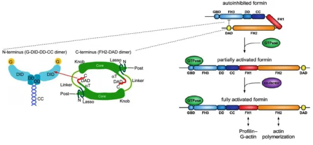
3.2 Diaphanous Formins and their regulation by Rho-GTP
Formins can be classified into two different subgroups: formin homology proteins, also designated as non diaphanous related formins and Diaphanous related formins. The diaphanous related formins (DRFs) are the best-characterized formins and besides the presence of FH1 and FH2 domains in the C-terminus, they also contain domains that are characteristic of this subgroup. In the N-terminus, this type of formins contains a GTPase-binding domain (GBD), a Dia inhibitory domain (DID) and a dimerization domain (DD) followed by a coiled-coil domain (CC). In the C-terminus, they have the FH1 and FH2 domains and a Diaphanous autoregulatory domain (DAD) (Bogdan, Schultz, and Grosshans 2013). Non diaphanous related formins are diverse and composed of several alternative domains. In the N-terminus, this group of formins can contain GTPase binding domains, PDZ domains, Pleckstrin domains (PH) or PTEN domains that regulate the activity of these formins (Pruyne 2016).
subfamilies based on the protein domains they have. These subfamilies of formins are designated as: Dia (Diaphanous), FRL (formin-related proteins in leukocytes), DAAM (Dishevelled-associated activators of morphogenesis), FHOD (formin-homology domain proteins), FMN (Formin), Delphilin, and INF (inverted-formin). Four of the seven mammalians subfamilies, mDia, Daam, FMNL and FHOD are DRFs with similar domain organization (Figure 4) (Kühn and Geyer 2014). The mammalian DRFS are mainly found in a resting state maintained by autoinhibition that is dependent on the intramolecular binding between the N-terminal DID domain and the C-terminal DAD domain. This intramolecular binding keeps the formin in a closed conformation inhibiting the polymerization of linear actin filaments. The intramolecular binding is disrupted after the binding of GTP-Rho to the N-terminal GBD, promoting the change to an open conformation that allows the polymerization of linear actin filaments (Alberts 2001; Schönichen et al. 2006; Vaillant et al. 2008; Li and Higgs 2005; W. Liu et al. 2008).
The formin mDia1, one isoform of the mDia family, constitutes the formin where this process is best characterized (Lammers et al. 2005; Otomo, Otomo, et al. 2005; Rose et al. 2005). The formin mDia1 was shown to interact with Rho A, B and C but only Rho A is specific to bind to the GBD. The interaction between DID-DAD occurs through the central portion (GVMDxLLEALQS) of DAD, which forms an amphipathic helix that binds to the concave surface of DID through several hydrophobic and hydrogen bonds (Nezami, Poy, and Eck 2006). To disrupt this interaction, it is necessary that RhoA induces the displacement of DAD. In mDia1, RhoA interacts with GBD and DID through numerous electrostatic interactions. The switch region I is able to interact with GBD, while the switch II region can interact with both GBD and DID. It was demonstrated that DAD binding sites in DID partially overlap with Rho interaction sites, which indicates that RhoA competes with DAD for the binding to DID. In vitro assay, RhoA-GTP seems to only partially dissociate the binding between DID and DAD, which suggests that other mechanisms or proteins can be involved in the activation of Diaphanous related formins (Figure 5) (Rose et al. 2005; Otomo, Otomo, et al. 2005).
Figure 4 - Schematic representation of the architecture of families of mammalian formins. Mammalians
DRFs: mDia, Daam, FMNL and FHOD besides the FH1 and FH2 characteristics of all types of formins contain additional domains. In N-terminus, the regulatory region of mammalian DRFs consists of GTPase-binding domain (GBD) that is important to bind Rho family GTPases; a Diaphanous Inhibitory Domain (DID) that contains tandem armadillo repeats; a Dimerization Domain followed by a coiled-coil domain that are responsible for the dimerization of the protein. In C- terminus, mammalian DRFs contained the FH1 and FH2 domains responsible for nucleation and elongation of linear actin filaments, and the DAD domain for regulation (left). Non DRFs: INF, Delphillin and FMN, besides FH1 and FH2, are composed of alternative domains. In the N-terminus, this group of formins can contain GTPase binding domains, PDZ domains, Pleckstrin domains (PH) or PTEN domains that regulate the activity of these formins (right) (Adapted from Schönichen et al, 2010).
Figure 5 - Schematic representation of the regulatory mechanism of DRF mDia by active Rho GTPase.
mDia1 is usually in a close conformation in a process of autoinhibition which is necessary for the formin not to be continuously nucleating and elongating actin filaments. The autoinhibition is assured by the intramolecular binding between N-terminal DID and C-terminal DAD that occurs due to a central portion GVMDxLLEALQSf of DAD which forms an amphipathic helix that binds to the concave surface of the DID domain through several hydrophobic contacts (left). To activate formin, active RhoA GTPase binds to the GTPase binding domain of formin releasing the interaction between DID and DAD domain. Other factors are reported to fully activate mDia1 that is placed in an open conformation
that allows the FH1 and FH2 domains to be available to nucleate actin linear filaments (right) (Adapted from Otomo et al,2010 and Kühn et al, 2014).
3.3 Other regulatory mechanisms of diaphanous formins
Although multiple evidences indicate that the binding of active Rho-GTPases is necessary to activate mDia formins, the mechanism of activation is not fully understood. It was demonstrated in vitro that nanomolar concentrations of active Rho are sufficient to disrupt the DID-DAD interaction, however increasing the concentration of active Rho only activated mDia1 partially, which suggests that other factors should cooperate with Rho GTPases to fully activate mDia formins (Rose et al. 2005; Li and Higgs 2003).
Other factors may include the Rho-associated protein kinase (ROCK), the protein flightless-1 (Fli-I) and anillin, which have all been demonstrated to help RhoA in activating DRFs in mammalian cells (S. Watanabe et al. 2010; Higashi et al. 2010; Staus, Taylor, and Mack 2011). ROCK and anillin have been suggested to interfere with the activation of mDia2. In vitro and in vivo kinase assays revealed that mDia2 is phosphorylated by ROCK in two conserved residues of threonine and serine localized near the DAD basic region. The phosphorylation of these residues prevents the basic region of DAD from interacting with DID, abolishing the autoinhibitory interaction within mDia2. Phosphorylation of these residues led to an increase of F-actin levels, which indicates that ROCK-dependent phosphorylation enhances RhoA during mDia2 activation (Staus, Taylor, and Mack 2011). Additionally, another study indicates that anillin also can assist mDia2 activation. An N-terminal region of anillin binds to the DID of mDia2. This binding is competitive with DAD and is required for the localization of mDia2 in the cleavage furrow during cytokinesis. As the depletion of anillin by RNAi caused cytokinesis failure and this process can be rescued when anillin full length is expressed is suggested that its interaction with DID domain can be supplementary to the role played by RhoA in mDia2 activation (S. Watanabe et al. 2010). Also, flightless-I (Fli-I), a gelsolin family protein, plays a role in fully activating mDia1 (Higashi et al. 2010). This protein competes with DID for binding to DAD of mDia1. In vitro assays evidenced that Fli-I binds to a conserved residue of leucine present in the DAD of mDia1, which prevents the DID-DAD interaction leading to an increase of F-actin levels in the presence of RhoA. This suggests that Fli-I contributes to RhoA-mediated activation of mDia1 (Higashi et al. 2010).
3.4 Diaphanous formins and cytokinesis
In mammalian cells, it is known that from all the mDia formins, the only isoform required for cytokinesis is mDia2. This formin localizes in the cleavage furrow during cytokinesis. Its depletion by RNAi causes an increase in binucleated cells that failed to assemble the contractile ring due to decreased levels of F-actin. Depletion of mDia1 or mDia3 did not interfere with cytokinesis (S. Watanabe et al. 2008). To check whether RhoA activation of mDia2 is important for cytokinesis, two strategies were used. First, the Ect2 GEF, which activates RhoA, was depleted by RNAi, which caused RhoA to be always inactive. In this situation, the localization of mDia2 was affected during cytokinesis. In control cells, RhoA and mDia2 localized at the cleavage furrow whereas in Ect2-depleted cells RhoA and mDia 2 localized at the interdigitating microtubules of the central spindle. This revealed that the localization of mDia2 at the cleavage furrow is dependent on active RhoA. In a second strategy, a mDia2 Rho-binding deficient mutant was generated (S. Watanabe et al. 2010). The construction of this mutant was based on previous in vitro studies that showed that the V161D mutation in mDia1’s GBD decreases mDia1 affinity to RhoA without affecting the DID-DAD interaction, and abolishes the ability of FH2 domain to elongate actin filaments. This demonstrated that the interaction of RhoA with the GBD domain is necessary to disrupt the DID-DAD interaction and promote activation of mDia1 (Otomo, Otomo, et al. 2005; Seth, Otomo, and Rosen 2006). An analogous Rho-binding deficient mutant of mDia2 was generated: mDia2 (V180D). This mutant was confirmed in vitro to be deficient for Rho- binding, and as expected caused a similar effect to that of inactive RhoA (Ect2 RNAi), leading to mDia2 localization on the central spindle instead of the cleavage furrow and was unable to rescue cytokinesis failure caused by the depletion of mDia2 by RNAi. These experiments revealed that the binding of active RhoA to mDia2 is fundamental and indispensable for the localization and function of mDia2 in mammalian cells to promote proper cytokinesis in mammalian cells (S. Watanabe et al. 2010).
Although the importance of active Rho GTPases has been notorious to activate mammalian formins and hence to promote the assembly of the contractile ring during cytokinesis there is no evidence that a similar mechanism occurs in the fission yeast S.
pombe, which is a popular model organism for cytokinesis studies. S. pombe cells are
rod-shaped and, similarly to mammalian cells, utilize a medially-placed actin-and myosin-based contractile ring. A cell wall division septum is deposited behind the constricting ring, forming the new ends of each daughter cell. The S. pombe contractile ring forms from cortical precursor nodes that form at the cell equator. The anillin like protein Mid1 is the major upstream protein in the node-assembly pathway and is
responsible for the recruitment of other contractile ring proteins and to determine the position of the division plane. The main proteins present in the cytokinesis nodes are the anillin like protein Mid1; the IQGAP protein Rng2, the myosin-II motor (heavy chain Myo2, essential light chain Cdc4, and regulatory light chain Rlc1), the F-BAR protein Cdc15 and the formin Cdc12 (Lee, Coffman, and Wu 2012). During ring assembly, Cdc12 has been shown to play an essential role in nucleation and elongation of actin filaments for contractile ring formation (Pelham and Chang 2002; Chang, Drubin, and Nurse 1997). One way to tightly regulate the formin Cdc12 activity is by multimerization. A domain in Cdc12 C-terminus mediates oligomerization to form puncta of different sizes on the cortex at interphase. At anaphase onset, the septation initiation network (SIN) becomes active to phosphorylate the four residues at the C- terminus of Cdc12 by the SIN kinase Sid2 that inhibits oligomerization. When this phosphorylation does not occur, Cdc12 and other contractile ring proteins cluster abnormally causing contractile ring disintegration and failure of cytokinesis. Thus, the phosphorylation by Sid2 constitutes a mechanism of regulation of the formin Cdc12 (Bohnert et al. 2013; Willet, McDonald, and Gould 2015). Interestingly, there is no evidence that Cdc12 is regulated by an autoinhibition mechanism, as mutations in most conserved residues of DID and DAD domains still allow for cytokinesis to complete with only subtle problems (Yonetani et al. 2008). Rho GTPases, the main regulators of mammalian DRFs, have also been suggested not to impact Cdc12 regulation (Martin et al. 2007). In fact, it was demonstrated that in S. pombe the main conserved Rho GTPases: Cdc42, Rho1, Rho3 and Rho4, are implicated in different processes that include septum formation, cell polarity and cell morphology. Cdc42 is responsible for the activation of formin For3 in a mechanism similar to that of mammalian formins, leading to the nucleation of actin cables that are important for polarized cell growth (Martin et al. 2007; Wei et al. 2016). Rho1 is essential for primary septum formation and must be inactive to allow for cell separation and along with Cdc42 and Rho4 regulates septum morphology (N. Wang et al. 2015). Rho3 and Rho4 participate in the delivery and secretion of specific cell wall glucanases required for septation and Rho4 regulates secondary septum formation (Nakano et al. 2003; H. Wang, Tang, and Balasubramanian 2003; Santos et al. 2005).
4 - Caenorhabditis elegans: A model organism to study
developmental biology
Model organisms could be defined as non-human species that are studied to understand biological processes with the purpose of converting the data and theories generated into knowledge about other organisms that can be much more complex than the original models (Ankeny and Leonelli 2011).
C. elegans is a model organism proposed in 1963 by Sydney Brenner that is
actively studied in laboratories worldwide for understanding questions of developmental biology, neurobiology and studying processes that go awry in human diseases, being an ideal system to tackle these problems. C. elegans is a small soil nematode with adults reaching 1 mm in length and exists primarily as a hermaphrodite (XX), although males (XO) can be generated in progeny with a low percentage of 0.1-0.2 % due to nondisjunction of the X chromosome during meiosis. Generation of males can be achieved through exposure of hermaphrodites to high temperatures for a short time. Males are important because through crossing with hermaphrodites, they allow the generation of progeny with different genetic composition. Through mating, due to competition between male sperm and hermaphrodite sperm a higher frequency of males (up to 50%) can be easily achieved.
C. elegans is a model organism with many advantages and benefits for
eukaryotic genetic studies. First, it is easy to maintain C. elegans in a laboratory. Several features that contribute to this characteristic are their short life cycle and the small size. In the laboratory, worms usually grow on agar plates with a lawn of E. coli bacteria as a food source and usually maintained at 20 ºC. In food, they develop through 4 stages (L1-L4) until reaching adulthood (Figure 6). At 20 ºC, worms need approximately 3,5 days to develop from eggs to adults. When food is depleted and animals are maintained in a starved stage, worms in L2 stage progress into an alternative state of development designated dauer. Worms are able to survive for some months in this stage and can continue their normal development if they are transferred to food. The small size (0,25 mm young worms and 1 mm adults) ensures that worms can grow in a small space. Additional advantages for this to be a convenient model organism to keep in the lab are the predisposition of C. elegans strains to be frozen and revived when needed, the possibility of decontaminating adults and isolating eggs that are resistant to bleach solutions, and lastly a single self-fertilizing hermaphrodite can originate a large number of progeny (Corsi, Wightman, and Chalfie 2015; Tucker and Han 2010).
In addition to its easy maintenance, C. elegans exhibits other advantages that allow it to be a relevant model for eukaryotic biology studies. One fundamental inherent feature that contributes to its importance is transparency. C. elegans animals are transparent, thus enabling individual cells and subcellular details to be visualized using
differential interference contrast (DIC) (Figure 7) (Porta-de-la-Riva et al. 2012). Also, it is possible to use fluorescent protein reporters to label cells or cellular structures and follow its behavior in living animals (Chalfie et al. 1994). Thus, development processes can be followed to monitor the impact of mutations that affect cell development in vivo. Lastly, the conservation of key genes, pathways and similarities with cellular and molecular processes between C. elegans and other organisms makes findings in C.
elegans broadly relevant (C. elegans Sequencing Consortium 1998).
In the specific case of studying cell division processes, the one-cell C. elegans embryo constitutes a powerful system being relatively accessible to introduce precise genetic modifications and quantitative live-cell imaging (Hattersley et al. 2018). To perform these modifications, two processes are essential to promote mechanistic cell division studies. The first process is the RNAi mediated protein depletion (Min and Lee 2007). With this procedure, more than 95% of a specific protein can be deleted promoting a gradual depletion of mRNA that codifies for that specific protein and removal of preexisting protein by continued embryo production (Oegema and Hyman 2006). In addition, it is also possible to insert single copy transgenes, which enable expression of RNAi-resistant versions of essential genes at endogenous levels (Frøkjær-Jensen et al. 2008).
The second process is the CRISPR/Cas9 technique, a novel genome editing tool that has also been successfully applied to C. elegans enabling edition and manipulation of DNA in a rapid, accurate and cost-effective way (Dickinson and Goldstein 2016).
if provided an environment with proper growing conditions to this worm, the larvae can development and
Figure 6 - C. elegans life cycle. After fertilization of a single oocyte, embryogenesis occurs over the next 13
h. Larval development consists of four stages (L1–L4) accompanied by a dramatic increase in size, followed by adulthood. During conditions of stress, including starvation and/or overcrowding, an alternative larval stage called the dauer state can be achieved. These larvae are capable of living for 3–6 months in a dormant state. During or after this period,
continuing the normal life cycle to become fertile adults.
Figure 7 - Adult C. elegans hermaphrodite seen with DIC Due to being a transparent organism its easy to identify the major anatomic regions of the worms and follow individual cells and specific components during the occurrence of biological events in vivo.
5 - Generation of C. elegans mutants by CRISPR/Cas9
technique
The CRISPR (clustered regularly interspaced short palindromic repeats) and Cas (CRISPR-associated proteins) are mechanisms present in the adaptive immune system of bacteria to resist virus invasions. In this system, a specific region denominated CRISPR locus has a crucial role in this process. The CRISPR locus comprises a series of conserved short repeats and adjacent to this region a sequence leader A/T rich exists, which functions as a promoter element and several cas genes. After a virus introduces its genetic material into a bacterial cell, the CRISPR mechanism has the ability to integrate short fragments of viral DNA, known as protospacers, into the CRISPR locus between two adjacent repeat units. The short fragments of viral DNA are recognized due to a short sequence of conserved nucleotides that are part of its constitution named PAM (Protospacer Adjacent Motif) required for its acquisition in the CRISPR locus. Then, the CRISPR locus is transcribed, originating a pre CRISPR RNA (pre-crRNA) that is processed by different types of CRISPR/CAS which differ from each other by the presence of specific cas genes (Wiedenheft, Sternberg, and Doudna 2012; Bhaya, Davison, and Barrangou 2011). The CRISPR/CAS system is currently divided in two classes: class 1 that include types I, III and IV and class II that comprises types II, V and VI (Makarova, Wolf, and Koonin 2018; Makarova et al. 2015). The type II CRISPR/CAS9 is the system most well characterized and is recognized as a powerful system for genome editing in a variety of organisms, including C. elegans (Dickinson et al. 2013; Arribere et al. 2014; Ran et al. 2013).
In this type, is contained a trans-activating RNA (tracrRNA) that is complementary to the short repeat sequences of the pre-crRNA forming an RNA duplex. Posteriorly, the RNA duplex is cleaved by RNase III originating fragments of mature crRNA/tracrRNA hybrids. In case of a new invasion caused by the same virus, the hybrid crRNA/tracrRNA formed a complex with cas9, an endonuclease that constitutes the signature of the type II CRISPR/CAS system. The hybrid crRNA/tracrRNA guides the cas9 to recognize the PAM sequence in viral DNA. Next to the PAM, cas9 causes a double strand break in viral DNA through specific domains (Hidalgo-Cantabrana, Goh, and Barrangou 2019). The CRISPR/cas9 bacterial system can be adapted to C. elegans through the design of a single guide RNAs (sgRNA), that is complementary to the specific site desired to be cleaved and consisting a 20 nucleotides sequence identical to the desirable genome fused at its 3’ end to a PAM
motif specific for Cas9 – 5’ NGG 3’. Once cleaved, the double-strand break generated by Cas9 activity can be repaired by Nonhomologous end joining (NHEJ) if the goal is to generate deletions or small insertions that disrupt the gene’s function, or by Homology- directed recombinational repair that will incorporate a repair template harboring the designed changes (Chiu et al. 2013; Lemmens and Tijsterman 2011; Lo et al. 2013).
Aims of this study
Cell division is a fundamental process that constitutes the basis of growth and development of eukaryotic organisms and allow the continuity of life across generations. Cytokinesis is the last step of cell division and is critical for its success, as it causes changes in cell shape that are dependent on reorganization of cytoskeleton. Defects and dysregulation of this process can lead to several diseases. Successful cytokinesis relies on the assembly and activation of an actomyosin contractile ring in a spatially and temporally precise manner.
During the assembly of the contractile ring, formin proteins take on a crucial role being responsible for nucleating and elongating unbranched actin filaments and its dysregulation leads to cytokinesis failure. Although mechanisms of regulation of formins are well characterized in mammalians, this process is not totally known.
In fission yeast S. pombe, a relevant model system that is used to study basic principles of the cell and to understand biological pathways in more complex organisms like mammals, and in particular humans, a novel mechanism of regulation of formins that differ from the mechanism that occurs in mammalians was identified.
In C. elegans, other important model to study cell development, in which, several discoveries were achieved due to conservation of key genes, there is no knowledge about the regulatory mechanisms of formins during cytokinesis. Thus, this question needs to be studied and clarified. In this project, we aimed to study the regulatory mechanism of formin´s activity during embryonic cytokinesis in vivo using C.
elegans as a model, through the following tasks:
1. Generation a version of formin that does not bind RhoA, the major activator of Diaphanous formins in other systems and characterize cytokinesis phenotype on the 1-cell embryo.
2. If regulation of CYK-1 is mediated by RhoA binding, generate a version of formin that is always active and characterize cytokinesis phenotype on the 1-cell embryo.
Material and Methods
The experimental work described in this thesis was conducted by myself and Fung Yi Chan. The immunoblots, live imaging, characterization of the CYK-1::GFPre
transgenic version and protein alignments that will be included in the Results section were conducted by Fung Yi Chan alone and therefore I am not including the methodology for those in this section.
C. elegans maintenance
C. elegans strains were maintained on Nematode growth medium (NGM) agar
plates, in which E. coli OP50 strain is grown to be used as food source (Brenner 1974). OP50 is a uracil auxotroph that is not resistant to antibiotics and its growth requires uracil in the medium that limits their growth. This strain was initially obtained by streaking out some bacteria from a glycerol stock onto a Luria Broth (LB) agar plate [10 g/L Bacto-tryptone, 5 g/L Bacto-yeast, 5 g/L NaCl and 15 g/L agar, pH 7.5] that was left to grow overnight at 37ºC (Byerly, Cassada, and Russell 1976). A single colony was picked to be inoculated in LB liquid medium that grow overnight at 37ºC. The bacterial suspension was used to seed NGM plates.
The preparation of the NGM plates involved the sterilization of an NGM solution (3 g/L NaCl, 17 g/L agar and 2.5 g/L peptone) at 110 ºC for 30 minutes. This solution was cooled in a 55 ºC water bath for 15 minutes. After this step, 1 M CaCl2, 5 mg/ml
cholesterol in ethanol, 1M MgSO4 and 1M potassium phosphate (pH 6) were added.
Two distinct types of NGM seeded plates were used. Medium sized plates (60 mm diameter) were used for general worm maintenance while large plates (100 mm diameter) were used to maintain a large amount of worms that were posteriorly frozen at - 80ºC. The NGM solution was distributed into plates using sterile procedures through a peristaltic pump that was adjusted to place a constant amount of NGM in each plate. The plates were left at room temperature for 2-3 days to dry and to detect possible contaminants. To complete the preparation of the plates, the plates were seeded with E.coli OP50 strain using sterile procedures. For the preparation of medium plates, we used 0,05 ml of E. coli OP50 liquid culture and for the preparation of large plates we used 0,1 ml of E.coli OP50 liquid culture. The plates were left at room temperature to dry during one day before storage.
C. elegans general stocks were maintained at a temperature range between 16
ºC and 25 ºC, more frequently at 20 ºC. The temperature was adjusted according to the planned experiments to control the speed of growth of animals, as it is known that C.
elegans grow 2.1 times faster at 25 ºC than at 16 ºC and 1.3 times faster at 20 ºC than
16 ºC (Wood 1983). All plates were identified with the strain and date. Eventually, C.
elegans plates can become contaminated with other bacteria, yeast or in more hostile
cases, mites, that make it difficult to visualize phenotypes and transfer worms to other plates. In this case, we used an alkaline bleach protocol to remove the contamination. This protocol consisted in putting several adult hermaphrodites from the contaminated strain into a drop of a hypochlorite solution on a new plate, for 24 hours to clean the worms. During this procedure, the hypochlorite solution kills the contaminants and the hermaphrodites, however the embryos remain intact because they are protected by the eggshell. In the next day, the eggs that hatched have moved onto the OP50 lawn and are transferred to a new clean NGM plate seeded with OP50.
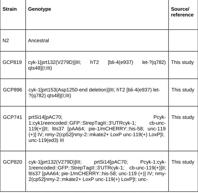
In table 1 are listed all strains used in this study and their respective genotype.
Table 1 - List of C. elegans strains used in this study.
Strain Genotype Source/
reference
N2 Ancestral
GCP819 cyk-1[prt132(V279D)]III; hT2 [bli-4(e937) let-?(q782) qIs48](I;III)
This study
GCP896 cyk-1[prt153(Asp1250-end deletion)]III; hT2 [bli-4(e937) let- ?(q782) qIs48](I;III)
This study
GCP741 prtSi14[pAC70; Pcyk-
1:cyk1reencoded::GFP::StrepTagII::3'UTRcyk-1; cb-unc- 119(+)]II; ltIs37 [pAA64; pie-1/mCHERRY::his-58; unc-119 (+)] IV; nmy-2(cp52[nmy-2::mkate2+ LoxP unc-119(+) LoxP]I; unc-119(ed3) III
This study
GCP820 cyk-1[prt132(V279D)]III; prtSi14[pAC70; Pcyk-1:cyk-
1reencoded::GFP::StrepTagII::3'UTRcyk-1; cb-unc-119(+)]II; ltIs37 [pAA64; pie-1/mCHERRY::his-58; unc-119 (+)] IV; nmy- 2(cp52[nmy-2::mkate2+ LoxP unc-119(+) LoxP]I; unc-
119(ed3) III
GCP880 unc-119(ed3)III; prtSi14[pAC70; Pcyk-1:cyk-
1reencoded::GFP::StrepTagII::3'UTRcyk-1; cb-unc-119(+)]II; zbIs2(pie-1::lifeACT::RFP)
This study
GCP883 cyk-1[prt132(V279D)]III; prtSi14[pAC70; Pcyk-1:cyk-
1reencoded::GFP::StrepTagII::3'UTRcyk-1; cb-unc-119(+)]II; ltIs37 [pAA64; pie-1/mCHERRY::his-58; unc-119 (+)] IV; zbIs2(pie-1::lifeACT::RFP)
This study
GCP928 cyk-1[prt153(Asp1250-end deletion)]III; prtSi14[pAC70; Pcyk- 1:cyk-1reencoded::GFP::StrepTagII::3'UTRcyk-1; cb-unc- 119(+)]II; ltIs37 [pAA64; pie-1/mCHERRY::his-58; unc-119 (+)] IV; nmy-2(cp52[nmy-2::mkate2+ LoxP unc-119(+) LoxP]I; unc-119(ed3) III
This study
GCP929 cyk-1[prt153(Asp1250-end deletion)]III; unc-119(ed3)III;
prtSi14[pAC70; Pcyk-1:cyk-
1reencoded::GFP::StrepTagII::3'UTRcyk-1; cb-unc-119(+)]II; zbIs2(pie-1::lifeACT::RFP)
This study
JK2739 mcm-4(e1466) dpy-5(e61) I/hT2 [bli-4(e937) let-?(q782) qIs48] (I;III)
CGC
Freezing C. elegans Stocks
To perform the freezing of C. elegans strains we picked 15 young adults and placed them into two large plates seeded with OP50. The plates were kept at 20 ºC to allow the development of the worms until the food is completely depleted and the progeny in a starved stage. At this point, the plates had a lot of L1s and L2s that are the worms that are suitable to be frozen. Besides L1 and L2, the plates should also have some unhatched eggs, which ensures that the plates were not without food for a long time. Using sterile procedures, the plates were rinsed twice with 10 mL S-Basal
[100 mM NaCl, 50 mM potassium phosphate (pH 6.0), 5 mg/L cholesterol] and transferred to 50 mL conical tubes. Animals were left until they migrated to the bottom of the tubes. After that, the supernatant was removed until the mark of 2.5 mL and mixed with an equal amount of freezing medium [100 mM NaCl, 50 mM potassium phosphate (pH 6.0), 30 % (v/v) glycerol]. Worms were aliquoted in cryovials (1 mL of worm suspension per cryovial) and then stored at - 80 ºC in a Coolcell container to allow a gradual decrease of temperature required for survival.
Generation of new C. elegans strains expressing CYK-
1(V279D) and CYK-1(∆1250-1437) by CRISPR-CAS9-mediated
direct editing of the C. elegans genome
Regarding this method, the candidate only contributed to the screening of the strains and therefore that part is described in more detail.
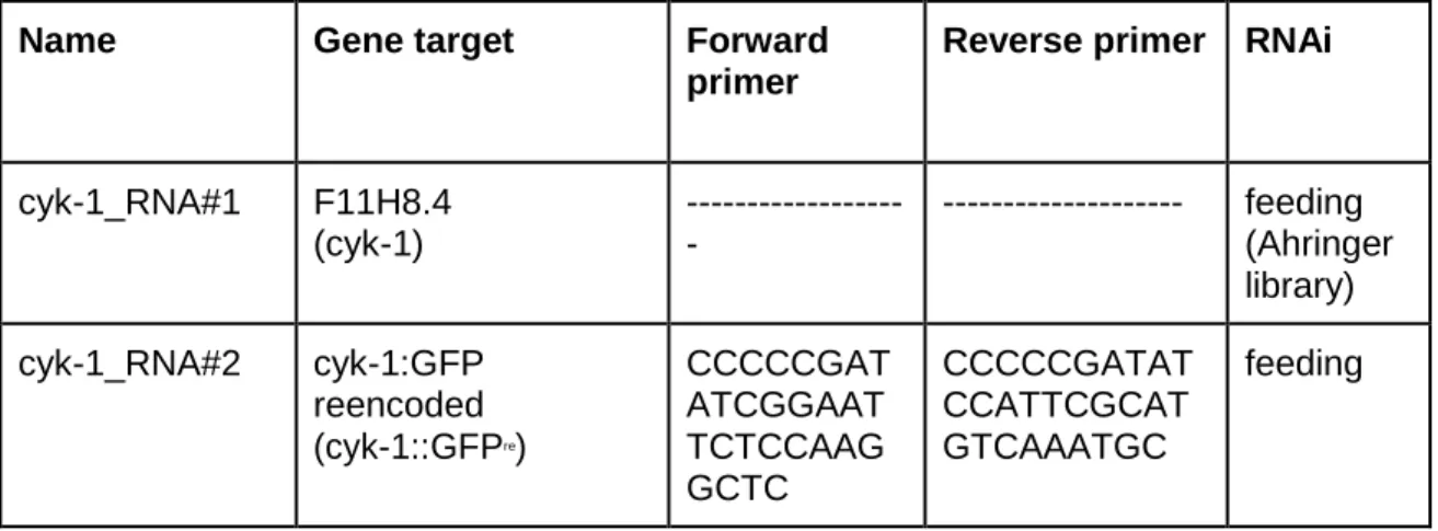
To study the mechanism of regulation of CYK-1 in C. elegans, two different strains were constructed: a strain carrying a point mutation in a conserved residue of valine 279 of the N-terminal GBD of CYK-1(V279D) and a strain expressing a truncated CYK-1 mutant with a deletion of the C-terminal region (∆1250-1437). To generate these strains, we used CRISPR-CAS9 endogenous genome editing co-conversion strategy (Dickinson et al. 2013; Arribere et al. 2014; Ran et al. 2013). Single guide RNAs (sgRNA), complementary to the site desired to be cleaved and repair templates consisting of mutation to be inserted, restriction enzyme site for screening and flanking regions were designed and were already available in the lab (Table 2). A mix of Cas9 and sgRNAs and repair template for the desired mutations was injected in N2 animals. The mix also contained sgRNAs and repair template for a mutation that confers a phenotype of “rolling” to the worms if integration is successful.
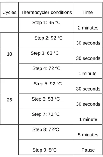
After the injection of the mix, three animals (F0) per plate were left to grow for 3-4 days at 25 ºC. The progeny (F1) from injected mothers that reached the adult stage were observed and the plates that contained worms with the roller phenotype were selected and kept overnight at 20 ºC to allow worms to lay embryos. In the following day, we screened the F1 adult rollers to assess their mutation status. The screening was done using Polymerase Chain Reaction (PCR). First, each worm was lysed in a PCR tube with a 10 µL reaction that contained 0.5 µL of 20 mg/ml Proteinase K and 9.5 µL of a lysis buffer (10 mM Tris pH 8.5 mM KCl, 1.5 mM MgCl2). The reactions were
spin down in a minifuge to assure that the worms settled at the bottom of the tube. The tubes were then placed in a thermocycler with the following conditions: 65 ºC for 90
minutes and 95 ºC for 15 minutes (to inactivate Proteinase K). After the worms were lysed, they were subject to PCR using the oligonucleotides and conditions listed on tables 3 and 4. The PCR product from cyk-1(V279D) mutant screen was digested with SpeI restriction enzyme (digestion reaction 20 µL, 37 ºC for 2 hours: 16.9 µL of distilled H2O, 2 µL 10x Fast Digest Green Buffer (Thermo Scientific), 1 µL of PCR product and
0.1 µL of restriction enzyme SpeI (Thermo Scientific).
If the repair template carrying the mutation was rightly integrated in the genome, a SpeI site must have been integrated also and digestion of the PCR product should detect the generation of fragments of 619 and 340 bp when ran on a 1% agarose gel. In cases where the repair template was not successfully integrated, the band should be of 959 bp when ran on a 1% agarose gel. In the case of proper integration of the repair template for the 1250-1437 truncation the PCR product was just ran on a 1% agarose gel. The mutated version should be 627 bp and the wild-type version should be of 911 bps.
After screening of the F1 progeny, we obtained six positive samples for CYK- 1 (V279D) and one positive sample for CYK-1(∆1250-1437).
Table 2 - List of CRISPR/Cas9 single guide RNAs (sgRNAs) and repair templates used in this study.
Gene/Mutation Repair template* Diagnosis
PCR and restriction enzyme sgRNA sequence cyk-1(V279D) GGGAGAAATATTGAAAACGAAAA ACATTCCGGAATGCAAGCAGGAT ATTGTTACTGTtCGaGaTCAaCTAG TaGGTCAgGGTGTTTCATTTCTTA ATAAGGTTCGTTTTTTAAGATAGA TATTGTTACTATTAAAATAC Forward primer: CCGTTCCT ATGGTGC TAAGATG Reverse primer: GAGAGTT CCGTCGC AATACAA Restriction enzyme: SpeI sgRNA#1 CTTGGGGT TCAGCTAG TTGGTCAA AACTGACC AACTAGCT GAACCC sgRNA#2 CTTGCTGT ACGGGTTC AGCTAGTA AACACTAG CTGAACCC GTACAG sgRNA#3 CTTGCAGG ATATTGTTA CTGTAC