Daniela Helena da Ascenção Loureiro Geraldo
Vaqueirinho
Licenciada em Biologia
The role of Arl13b and the non-muscle
myosin IIA in cancer cell migration
Dissertação para obtenção do Grau de Mestre em
Genética Molecular e Biomedicina
Orientador: Prof. Dr. Duarte Barral, Investigador Principal no
Centro de Estudos de Doenças Crónicas (CEDOC)
Setembro 2017
i
Acknowledgements
Antes de mais, gostaria de agradecer ao Dr. Duarte Barral por me ter aceite como aluna de mestrado no laboratório e, assim, ter tornado possíveis toda a aprendizagem e experiência que adquiri durante este ano. Estou muito grata por poder dizer que tive um excelente orientador, por todo o conhecimento que me transmitiu, todos os conselhos úteis não só para a realização deste trabalho mas também para o meu futuro, toda a disponibilidade e prontidão que sempre demonstrou face a qualquer problema ou questão, todo o acompanhamento que teve o cuidado de realizar continuamente ao longo do ano letivo e ainda pelo esforço que demonstra fazer para manter o grupo unido, dentro e fora do laboratório.
Gostaria de dedicar um agradecimento muito especial à Cristina Casalou, que tive a sorte que me orientasse durante o meu projeto de mestrado. Um enorme obrigada pela excelente orientação que recebi, por todo o conhecimento que me transmitiu desde as bases iniciais aos pequenos detalhes, por todos os conselhos que me ajudaram não só a saber fazer ciência mas também a saber pensar em ciência, por toda a disponibilidade e prontidão que demonstrou para resolver as minhas dúvidas, por toda a paciência com que me ensinou e com que acompanhou também as minhas falhas e ainda por todo o tempo dispensado para esta importante fase da minha formação.
Quero também deixar uma palavra de agradecimento a cada um dos meus colegas de laboratório, que me acompanharam durante este ano. Xana, um grande obrigada por tudo o que me ensinaste, por me ouvires e estares disponível para ajudar e dar sugestões que continuarão a ser úteis daqui para a frente e ainda pela animação e bom humor que trazes ao laboratório. Liliana, um grande obrigada por desde o início me teres feito sentir integrada, pela tua constante prontidão para ajudar, por me ouvires e me aconselhares e pela tua boa disposição. Hugo, muito obrigada por todos os conselhos que sempre estiveste disposto a dar, por teres desde logo contribuído para que sentisse à vontade neste grupo e por tudo aquilo que me ensinaste. Matilde, muito obrigada por toda a prontidão com que te ofereces para colaborar, pelas tuas sugestões sinceras e pelo teu companheirismo. Cristina Escrevente e Cecília, quero deixar-vos também um grande obrigada por estarem sempre disponíveis para me ensinar e ajudar, por todas as sugestões que foram dando ao longo deste ano e que contribuíram para o avanço do meu trabalho e ainda por toda simpatia com que me receberam. Renata e Francisco, apesar de não ter partilhado tanto tempo com vocês, foi também uma honra conhecer-vos e trabalhar com vocês! Finalmente, ao Paul Greiner, com quem foi também uma honra trabalhar e cuja ajuda também quero agradecer.
A todos os membros do laboratório, sem exceção, quero deixar um grande grande obrigada por desde o primeiro dia me terem recebido tão bem e se terem disponibilizado a ensinar-me tudo o que fosse preciso para que me sentisse integrada no laboratório e no CEDOC. Obrigada também por todos os conselhos e por toda a incrível disponibilidade que têm para ajudar. É sem dúvida o vosso
ii exemplo que quero continuar a seguir em qualquer local onde venha a fazer investigação. Foi uma enorme honra trabalhar em equipa e aprender com todos vocês!
A todos os membros que fizeram parte do laboratório 2.7 durante este ano, obrigada pelo fantástico ambiente de trabalho que se sente e por toda a entreajuda que se verifica. Um obrigada especial às vizinhas do lado, Tatiana, Farzaneh e Catarina por serem tão amorosas e por todos os vossos conselhos e ajuda preciosos!
Obrigada ainda a todas as pessoas do CEDOC por me acolherem e por toda a colaboração que torna o trabalho de todos mais fácil.
Gostaria também de agradecer à coordenadora do Mestrado em Genética Molecular e Biomedicina, a professora Paula Gonçalves, por toda a ajuda, disponibilidade e conselhos.
Finalmente, quero deixar um agradecimento muito muito especial à minha família, amigos e namorado por todo o apoio incondicional e toda a força e alegria que me transmitem, direta ou indiretamente, tornando as minhas aspirações possíveis.
Por todo o conhecimento e experiência com que terminarei o meu Mestrado, por todas as bases que adquiri e com as quais irei continuar a minha carreira científica e pela oportunidade que tive de aprender como fazer um bom trabalho em equipa, não tenho dúvidas de que fiz uma excelente escolha de laboratório e de projeto de investigação para a minha tese de Mestrado. A todos, um grande obrigada por me trazerem até aqui!
i
Index of contents
Acknowledgements ... i
Index of figures ... iii
Index of tables ... v Abstract ... vii Resumo ... ix Abbreviations... xi 1. Introduction ... 1 1.1. Cell migration ... 1
1.1.1. Actin-based structures involved in cell migration... 2
1.1.2. Retrograde flow and the “molecular clutch” hypothesis ... 4
1.2. Small GTP-binding proteins and their role in cancer ... 6
1.2.1. The Arf family ... 7
1.2.2. The Arl protein Arl13b ... 8
1.3. NMIIA and cancer cell migration ... 9
2. Objectives... 13
3. Materials and Methods ... 15
3.1. General ... 15
3.2. Cell culture ... 15
3.3. Cell migration ... 15
3.3.1. Stimulation of cell migration stimulation for protein extraction ... 15
3.3.2. Wound healing assay ... 15
3.4. Wound healing assay ... 16
3.5. Cell transfection ... 16
3.5.1. Overexpression ... 16
ii
3.6. RNA extraction, cDNA production and real-time quantitative PCR ... 17
3.7. Immunofluorescence microscopy ... 18
3.8. Protein extraction ... 19
3.9. Immunoprecipitation ... 19
3.10. SDS-Polyacrylamide Gel Electrophoresis ... 20
3.11. Immunoblotting ... 20
3.12. Gel staining ... 21
3.12.1. Coomassie gel staining... 21
3.12.2. Silver nitrate gel staining... 21
3.13. Mass spectrometry ... 22
3.14. Statistical analysis ... 22
4. Results ... 23
4.1. Sub-cellular localization of Arl13b and cytoskeleton proteins ... 23
4.2. Interaction of Arl13b and NMIIA in non-tumorigenic and cancer breast cell lines ... 26
4.3. Influence of NMIIA silencing on breast cancer cells migratory capacity ... 31
4.4. Identification of candidate Arl13b interacting partners that can mediate Arl13b function on cell migration ... 33
5. Discussion... 37
References ... 41
iii
Index of figures
Introduction ... 1
Figure 1.1 – Actin-based structures involved in cell migration. ... 2
Figure 1.2 – Cellular organization of actin in a migrating cell. ... 3
Figure 1.3 – The “molecular clutch” hypothesis. ... 5
Figure 1.4 – The GTP/GDP cycle of small GTP-binding (G) proteins. ... 7
Figure 1.5 – Structure of the non-muscle myosin II (NMII). ... 10
Results ... 23
Figure 4.1 – Arl13b co-localizes with actin in MDA-MB-231 cells. ... 24
Figure 4.2 - Arl13b co-localizes with NMIIA in MDA-MB-231 cells. ... 25
Figure 4.3 – Arl13b does not co-localize with NMIIB in MDA-MB-231 cells. ... 26
Figure 4.4 – Co-immunoprecipitation of non-muscular myosin IIA with Arl13b in MDA-MB-231 cells. ... 26
Figure 4.5 – Co-immunoprecipitation of non-muscular myosin IIA with Arl13b in MDA-MB-231 cells overexpressing Arl13b-GFP. ... 27
Figure 4.6 – Co-immunoprecipitation of non-muscular myosin IIA with Arl13b in distinct breast cell lines. ... 28
Figure 4.7 – Co-immunoprecipitation of non-muscular myosin IIA with Arl13b in MCF7 and MCF10A cells during cell migration. ... 29
Figure 4.8 – Co-immunoprecipitation of non-muscular myosin IIA with Arl13b in GTP bound and unbound conditions. ... 30
Figure 4.9 – Co-immunoprecipitation of non-muscular myosin IIA with Arl13b in MCF7 cells overexpressing Arl13b-wildtype-GFP or Arl13b-R79Q-GFP. ... 31
Figure 4.10 – NMIIA silencing leads to increased migration in MDA-MB-231 cells. ... 32
v
Index of tables
Materials and Methods ... 15
Table 3.1 – DNA overexpression plasmids used for MDA-MB-231 transfection ... 16
Table 3.2 – Sequences of siRNAs used for gene silencing ... 17
Table 3.3 – Primers used in RT-qPCR ... 18
Table 3.4 – Primary antibodies used for immunofluorescence ... 19
Table 3.5 – Secondary antibodies used for immunofluorescence ... 19
Table 3.6 – Primary antibodies used for immunoblotting ... 21
Table 3.7 – Secondary antibodies used for immunoblotting ... 21
Results ... 23
Table 4.1 – Mass spectrometry identification of Arl13b-interacting partners that can be involved in Arl13b function in cell migration. ... 33
vii
Abstract
Cancer metastasis relies on cell migration and invasion of surrounding tissues and is responsible for most cancer-related deaths. Therefore, the study of the molecular mechanisms that govern cell migration and invasion is essential for the development of effective anti-cancer therapies. We previously showed that the small GTP-binding protein Arl13b interacts with the actin cytoskeleton and regulates fibroblast cell migration through the interaction with its effector, the non-muscle myosin heavy chain IIA (NMIIA). Furthermore, we found that Arl13b is required for in vitro and in vivo migration and invasion of breast cancer cells. The main goal of this work was to assess the NMIIA requirement for the function of Arl13b in cancer cell migration. We found that Arl13b-NMIIA interaction is stronger in migrating cells, and more prominently in the non-tumorigenic breast cell line MCF10A than in the breast cancer cell lines MCF7 and MDA-MB-231. We also found that NMIIA silencing in breast cancer cells does not phenocopy the decrease in cell migration that occurs upon Arl13b silencing. Therefore, we set out to identify new Arl13b-interacting partners that may have a direct association with its role in breast cancer cell migration. Thus, our results provide new insights into the molecular mechanisms of Arl13b in cancer cell migration.
ix
Resumo
O processo de metastização depende da migração e invasão de células cancerígenas para os tecidos circundantes e é responsável pela maioria das mortes relacionadas com cancro. Por essa razão, o estudo dos mecanismos moleculares de migração e invasão celulares é essencial para o desenvolvimento de terapias mais eficazes para o tratamento de cancro. O nosso grupo descobriu que a proteína G Arl13b interage com o citoesqueleto de actina e regula a migração de fibroblastos através da interação com a proteína efetora miosina não-muscular IIA. Para além disso, descobrimos que a Arl13b é necessária para a migração e invasão de células de cancro de mama in vitro e in vivo. O principal objetivo deste trabalho foi avaliar a necessidade da NMIIA para a função de Arl13b na migração de células cancerígenas. Descobrimos que a interação entre Arl13b e NMIIA é mais forte em células que estão a migrar, e que isso é mais preponderante na linha celular não-tumorigénica MCF10A do que nas linhas celulares de cancro da mama MCF7 e MDA-MB-231. Descobrímos ainda que o silenciamento da NMIIA nas células de cancro da mama não conduz ao mesmo fenótipo de diminuição da migração celular que se verifica após o silenciamento de Arl13b. Assim, propusemos novas proteínas que interagem com a Arl13b e que possam ter uma relação direta com a sua função na migração de células cancerígenas. Em conclusão, os nossos resultados contribuem para o aprofundamento do conhecimento dos mecanismos moleculares da Arl13b na migração de células cancerígenas.
Palavras-chave: Arl13b, miosina não-muscular IIA, migração celular, cancro, citoesqueleto de
xi
Abbreviations
ANOVA Analysis of variance
Arf ADP ribosylation factor
Arl Arf-like
Arl13bhnn Arl13b hennin
Arp 2/3 Actin-related protein 2/3
ATP Adenosine triphosphate
BSA Bovine serum albumin
cDNA Complementary deoxyribonucleic acid
CDR Circular Dorsal Ruffle
DAPI 4',6-Diamidino-2-phenylindole
DMEM Dulbecco's Modified Eagle Medium
dNTP Deoxyribonucleotide triphosphate
DTT 1,4-Dithiothreitol
E-cadherin Epithelial cadherin
ECL Enhanced chemiluminescence
ECM Extracellular matrix
EDTA Ethylenediaminetetraacetic acid
EGF Epidermal growth factor
EGTA Ethylene glycol-bis(β-aminoethyl ether)-N,N,N',N'-tetraacetic acid
ELC Essential light chain
Erlin Endoplasmic reticulum lipid raft-associated protein
FA Focal adhesion
FBS Fetal bovine serum
xii
GAP GTPase-activating protein
GAPDH Glyceraldehyde-3-phosphate dehydrogenase
GDI Guanine nucleotide dissociation inhibitor
GDP Guanosine diphosphate
GEF Guanine nucleotide exchange factor
GFP Green fluorescent protein
GTP Guanosine triphosphate
HEPES 4-(2-Hydroxyethyl)-1-piperazineethanesulfonic acid
HRP Horseradish peroxidase
IP Immunoprecipitation
MLCK Myosin light chain kinase
mRNA Messenger ribonucleic acid
MYH Myosin heavy chain
Nano LC-MS/MS Nanoscale liquid chromatography coupled to tandem mass spectrometry
NMII Non-muscle myosin II
N-WASP Neural-Wiskott-Aldrich syndrome protein
p(dN)6 Deoxyribonucleotide hexamer random primer sequence
PBS Phosphate buffered saline
PR Peripheral ruffle
Rab Ras-like protein in brain
Ran Ras-like nuclear
Ras Rat sarcoma
Rho Ras homologous
RLC Regulatory light chain
ROCK RhoA-associated kinase
xiii
Sar Secretion associated and Ras-related
SDS Sodium dodecyl sulfate
SDS-PAGE SDS-Polyacrylamide Gel Electrophoresis
Ser19 Serine 19
Shh Sonic hedgehog
siCtrl siControl
siRNA Small interfering RNA
Thr18 Threonine 18
Tris Tris(hydroxymethyl)aminomethane
1
1. Introduction
1.1. Cell migration
Cell migration is a biological process essential in both physiological and pathological conditions. In early embryonic development, cell migration is required for morphogenetic processes, such as gastrulation (Keller, 2005), and colonization of embryonic tissues by neural crest cells (Locascio and Nieto, 2001). In adulthood, cell migration plays a key role in inflammation (Luster et al, 2005) and wound healing (Li, 2013). In fact, the majority of an organism’s cell types is able to and undergoes migration at a certain point in space and time (Friedl, 2004).
When cell migration becomes dysregulated, it can lead to pathological conditions, such as cancer metastasis (Wang et al, 2005). Metastasis is the result of cancer cell spreading from the tumor’s primary site to a distant site in the organism and accounts for the majority of cancer-related cell deaths (Schroeder et al, 2011; Fife et al, 2014). Therefore, understanding the fundamental mechanisms of cell migration and how tumors subvert this process in order to migrate and invade through tissues is key for understanding cancer progression and developing new therapeutic strategies.
Depending on the cell type and the environmental conditions, there are different mechanisms of cell migration, such as single versus collective cell migration or adhesion-dependent versus adhesion-independent migration (Friedl, 2004; Friedl et al, 2012). In every case, cell migration requires the generation of traction forces by the actin cytoskeleton (Pollard and Cooper, 2009; Case and Waterman, 2015). Thus, actin cytoskeleton remodeling, which comprises the assembly, stabilization and organization of actin filaments, is key for cell migration (Le Clainche and Carlier, 2008).
Cell migration involves a series of coordinated steps that allow cells to adapt and move relatively to their surroundings. Initially, a migrating cell has to undergo polarization (Petrie et al, 2009), particularly of the cell cytoskeleton and its associated proteins. This polarization allows dynamic actin structures with protrusive and adhesive functions to form at the cell’s leading edge. Hence, migrating cells create extensions, such as lamellipodia and filopodia, which allow protrusion in the direction of migration. Additionally, migrating cells indirectly anchor the actin cytoskeleton to the extracellular matrix (ECM) through the formation of adhesion structures such as focal adhesions (FAs) at the leading edge of the cell, which ultimately allow the traction of the cell body (Gardel et al, 2010). Therefore, lamellipodial protrusion and adhesion to the ECM act in an orchestrated manner, usually in response to chemical and physical stimuli (Petrie et al, 2009). Lastly, in order to successfully move, the cell has to retract its trailing edge. This is achieved by the simultaneous contractility of the actomyosin network and disassembly of adhesive structures (Le Clainche and Carlier, 2008).
2
1.1.1. Actin-based structures involved in cell migration
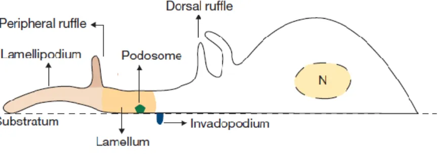
There are several actin-based structures with protrusive and adhesive functions which are considered key players on cell migration (Figure 1.2).
Figure 1.1 – Actin-based structures involved in cell migration. Remodeling of the actin cytoskeleton allows
cells to form several structures with different localizations and functions that result in increased migratory capacities. At the leading edge of the migrating cell, protrusive structures such as lamella and lamellipodia are formed by constant actin polymerization. Dorsal ruffles and peripheral ruffles, collectively called membrane ruffles, are non-adhesive structures that can be found at the cell’s dorsal surface. Podosomes form at the cell’s ventral surface and are characterized by their adhesive and degradative functions. Invadopodia are similar to podosomes, but are characteristic of cancer cells. N, nucleus. Adapted from (Chabra and Higgs, 2007).
The lamellipodium consists of a flat membrane protrusion localized at the migrating cell’s leading edge and it is composed of a dense array of branched actin filaments with barbed ends facing the leading edge (Small et al, 2002; Case and Waterman, 2015) (Figure 1.2). New actin monomers are constantly incorporated on the barbed end of the actin filament, towards the leading edge. At the same time, there is a constant pointed end depolymerization, which contributes with actin monomers for the elongation. This phenomenon, often referred to as “actin treadmilling”, is responsible for the maintenance of the actin filaments of the lamellipodia (Wang et al, 1985). The lamella, which is a loose array of unbranched actin filaments located immediately upstream of the lamellipodia (Figure 1.2), has a slower actin turnover (Le Clainche and Carlier, 2008) and is enriched in myosin II and tropomyosin (Gardel et al, 2010).
Migrating cells also form filopodia, which are finger-like protrusions that overlap but extend beyond lamellipodia (Le Clainche and Carlier, 2008). These structures are composed of aligned actin filaments and their main function is to sense the extracellular environment and direct the cellular movements (Letort et al, 2015) (Figure 1.2). The parallel actin filaments in filopodia also function as tracks for protein intracellular transport (Letort et al, 2015). Despite sharing the same cellular environment, filopodia and lamellipodia have different organization and dynamic properties. This can be attributed to the action of different nucleation mechanisms, involving the Actin-related protein (Arp) 2/3 complex or formins, which control the coordinated formation of the two structures (Firat-karalar and Welch, 2012).
3
Figure 1.2 – Cellular organization of actin in a migrating cell. The organization and dynamics of the actin
cytoskeleton contribute to the formation of protrusive and adhesive structures, which are essential for cell migration. In lamella and lamellipodia, actin is organized in unbranched and branched arrays, respectively. On the other hand, actin filaments in filopodia and stress fibers can be found in bundles of polarized or antiparallel filaments, respectively. Additionally, the filaments in stress fibers are associated with myosin, which gives them contractile properties. Taken from (Letort et al, 2015).
The formation of adhesion structures by the migrating cell is also critical for the process of cell migration. Adhesion structures include nascent adhesions, focal complexes, FAs and fibrillar adhesions. Nascent adhesions and focal complexes are the most dynamic, since they have a high assembly/disassembly rate at the edge of the lamellipodium (Wolfenson et al, 2013). These are similar structures, essentially distinguished by their myosin dependence and size (Choi et al, 2008). Nascent adhesions and focal complexes that do not disassemble undergo maturation, which requires stress fiber assembly and myosin II contractility, and are converted into FAs (Kuo, 2013). These have higher stability and lower adhesion turnover than focal complexes (Le Clainche and Carlier, 2008). Like focal complexes, FAs also localize to the cell periphery. However, FAs have a more central localization and associate with the end of stress fibers, which are contractile bundles of actin, myosin II and bundling proteins such as α-actinin (Le Clainche and Carlier, 2008; Tojkander et al, 2012) (Figure 1.2). Finally, fibrillar adhesions are elongated structures associated with fibronectin fibrils that can originate from FAs (Wolfenson et al, 2013).
To allow rapid migration, adhesion structures have to be formed at the cell’s leading edge and disassembled at the cell’s trailing edge (Le Clainche and Carlier, 2008). Integrins, a family of
4 heterodimeric transmembrane proteins, are an important component of adhesion structures, enabling the connection between the actin cytoskeleton and the ECM (Mayor and Etienne-Manneville, 2016). However, integrins and actin do not bind directly and their association is mediated by the FA, which includes cytoskeletal adaptor proteins such as talin, vinculin and paxillin (Geiger et al, 2001; Case and Waterman, 2015).
Besides these specialized structures, many cell types also form other actin-based structures, which are essential for cell migration and invasion, such as podosomes, invadopodia and membrane ruffles (Figure 1.1). Podosomes and invadopodia are structures formed on the cell’s ventral surface that are mainly involved in the processes of adhesion and matrix degradation for cell spreading and invasion (Linder, 2007). While podosomes are mostly found in invasive monocytic cells, such as macrophages or dendritic cells, invadopodia are typical of cancer cells (Linder, 2007). Membrane ruffles are non-adhesive structures that extend from the cell’s dorsal surface and are subdivided into peripheral ruffles (PRs) and circular dorsal ruffles (CDRs) (Chhabra and Higgs, 2007). PRs extend at the cell’s periphery, usually at the leading edge, and move rearward (Abercrombie et al, 1970). After assembling at the dorsal surface, CDRs typically expand and then constrict towards their center, maintaining a circular appearance, until they disappear (Krueger et al, 2003; Itoh and Hasegawa, 2013). Both types of membrane ruffles have been associated to the process of cell migration (Buccione et al, 2004). CDRs, which form transiently upon stimulation by growth factors, are usually implicated in the cell’s transition from a static to a migratory phenotype (Mellström et al, 1988; Krueger et al, 2003; Sero et al, 2011). Moreover, they are also associated with membrane receptor internalization and recycling, as well as macropinocytosis (Buccione et al, 2004; Hoon et al, 2012). In fact, it has been reported that CDRs play a role in FA turnover in migrating cells. Upon FA disassembly, integrins are translocated to CDRs, before being internalized by macropinocytosis and recycled to new focal adhesions at the leading edge of a migrating cell (Gu et al, 2011).
1.1.2.
Retrograde flow and the “molecular clutch” hypothesis
Cell migration occurs through the coordination of leading edge protrusion, adhesion of the protrusion to the substrate and retraction of the trailing edge. In normal conditions, actin polymerization against the plasma membrane within the lamellipodium results in a counterforce that is thought to push the actin network rearward relatively to the membrane, resulting in a rapid retrograde flow (Ponti, 2004; Case and Waterman, 2015). Additionally, within the lamella, the motor protein myosin II contracts actin bundles, leading to actin reorganization and disassembly, which generate a slower retrograde flow (Ponti, 2004; Case and Waterman, 2015).
In order to convert the polarized actin treadmilling into protrusion force and the actomyosin contraction into traction force, migrating cells form adhesion structures, such as FAs, which act as “molecular clutches” (Burridge and Guilluy, 2016) (Figure 1.3). When the molecular clutch is not engaged, there is no anchorage of the actin cytoskeleton to the substrate. Therefore, the actin
5 polymerization at the lamellipodium and the actomyosin contraction at the lamella result in net retrograde flow of the actin cytoskeleton and there is no protrusion of the leading edge (Le Clainche and Carlier, 2008). However, when the molecular clutch is engaged, the forces generated by the actin treadmilling and the actomyosin contraction lead instead to a slower retrograde flow, the generation of a traction force on the ECM and net protrusion of the leading edge (Le Clainche and Carlier, 2008). The traction force applied on the cell’s stress fibers ultimately leads to the traction of the cell body and retraction of the rear of the cell (Le Clainche and Carlier, 2008).
Figure 1.3 – The “molecular clutch” hypothesis. (a) Actin monomers (light blue) are constantly incorporated
onto the barbed end of pre-existing actin filaments (dark blue) while depolymerization occurs at the pointed ends in a process called “actin treadmilling”. (b) When the actin cytoskeleton is not anchored to the extracellular matrix (ECM) through integrin-based adhesions, the clutch is disengaged and consequently there is no net protrusion. In this case, actin polymerization and actomyosin force are mainly converted into retrograde flow. (c) When the polymerizing actin network is connected to the ECM, the molecular clutch is engaged and actin polymerization and actomyosin contraction are converted into protrusion force and traction force, respectively. Taken from (Case and Waterman, 2015).
6 Since cell protrusion is dependent on actin treadmilling to push the leading edge membrane, the control of this cycle determines the speed of protrusion. Accordingly, and since the natural treadmilling cycle is too slow to allow rapid migration, there is a set of actin-binding proteins that are able to modulate its speed (Le Clainche and Carlier, 2008). Indeed, there are several proteins that are crucial for the regulation of actin dynamics, including its turnover and remodeling. The Arp2/3 complex and Neural-Wiskott-Aldrich syndrome protein (N-WASP) along with several regulatory elements of the Ras superfamily of small GTP-binding (G) proteins and various actin-capping/binding proteins, such as actin depolymerizing factor, profilin and cofilin, are some of the most important players in the regulation of actin dynamics (Buccione et al, 2004).
Alterations in the complex mechanisms that govern the process of cell migration are usually observed in cancer cells with metastatic behavior. In order to successfully mobilize and invade other tissues in the organism, these cells typically undergo modifications in cell shape, cell-cell and cell-ECM adhesion and migratory and invasive capacities (Hanahan and Weinberg, 2011; Fife et al, 2014). Moreover, cancer cells stimulate the formation and regulate the turnover of actin-based structures, such as lamellipodia, invadopodia, CDRs and focal adhesions (Yamaguchi and Condeelis, 2007). This is achieved through the manipulation of signaling pathways involved in the actin cytoskeleton reorganization. In fact, several proteins, such as various small G proteins or actin-binding proteins, have already been identified as having altered expression or activity in different cancer types, influencing the metastatic behavior of cancer cells (Yamaguchi and Condeelis, 2007; Fife et al, 2014).
1.2. Small GTP-binding proteins and their role in cancer
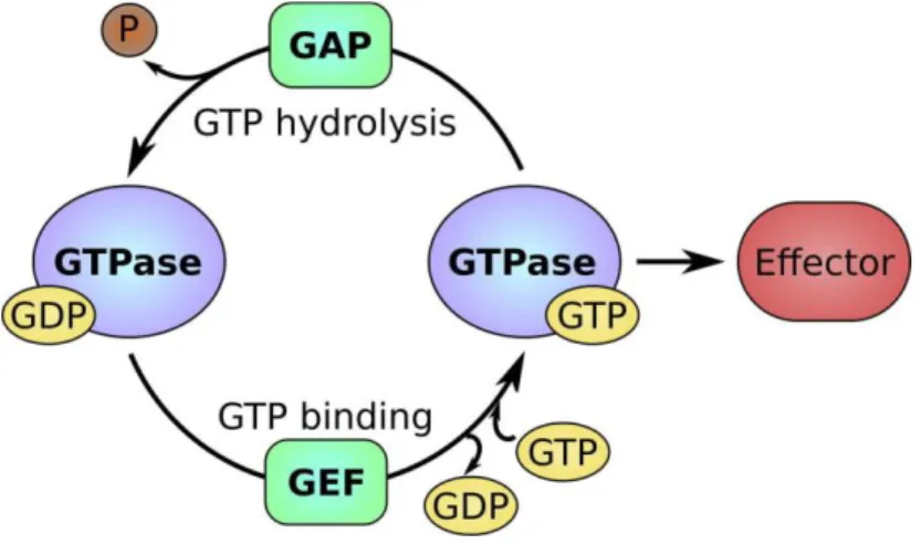
The Ras (Rat sarcoma) superfamily of GTP-binding (G) proteins is composed of low molecular weight monomeric proteins that bind GTP and are able to hydrolyze it, i.e., they have intrinsic GTPase activity (Goitre et al, 2014). In humans, this large superfamily includes over 150 members, which can be further grouped into five major families, based on sequence and functional similarities: Ras, Rho, Rab, Arf and Ran (Goitre et al, 2014). All members of this family are binary molecular switches that can be found in two conformational states: a GDP-bound inactive state and a GTP-bound active state (Figure 1.4). Binding to GTP leads to a conformational change that allows small G proteins to bind to their effectors (Herrmann, 2003). In turn, GTP hydrolysis disrupts this interaction, inactivating the small G proteins (Herrmann, 2003).
The GTP hydrolysis activity of the small G proteins is intrinsically low (Goitre et al, 2014). However, it can be stimulated by GTPase-activating proteins (GAPs), promoting the inactive conformation (Bernards and Settleman, 2004) (Figure 1.4). Moreover, guanine nucleotide exchange factors (GEFs) promote GDP dissociation, allowing the binding of GTP, which has naturally higher intracellular concentrations than GDP. This promotes the formation of the active GTP-bound state (Schmidt and Hall, 2002). Small G proteins that carry a farnesyl or geranylgeranyl group in their C-terminal domain are additionally regulated by guanine nucleotide dissociation inhibitors (GDIs), which
7 extract small G proteins from membranes by shielding the hydrophobic tail that is responsible for the membrane association (Cherfils and Zeghouf, 2013). Therefore, this precludes membrane association and interaction with regulators or effectors.
Figure 1.4 – The GTP/GDP cycle of small GTP-binding (G) proteins. Small G proteins can be found in two
different states – an active GTP-bound state and an inactive GDP-bound state. The active conformation that results from GTP binding allows small G proteins to interact with their effectors. On the other hand, the inactive conformation does not allow binding to effectors. Small G protein activity is regulated by guanine nucleotide exchange factors (GEFs) and GTPase-activating proteins (GAPs), which can stimulate GDP dissociation and GTPase activity, respectively. P, phosphate. Taken from (Carvalho et al, 2015).
Ras superfamily members are associated with several cellular processes, such as proliferation, differentiation, polarity, adhesion and migration. The different families and subfamilies of Ras G proteins regulate distinct biological functions, which is determined by a variation in their sequences and consequently in the effectors and regulators with which they interact (Goitre et al, 2014). The different subfamilies also differ in their membrane targeting domains, which determine cellular localization and spatiotemporal regulation (Goitre et al, 2014).
Given their crucial role in so many fundamental cellular processes, alterations in the expression and activity of several small G proteins are associated with human diseases (Seixas et al, 2013; Casalou
et al, 2016; Simanshu et al, 2017). The Rho (Ras homologous) family members, for example, participate
in various signaling networks which influence, among other processes, actin cytoskeleton organization, cell adhesion, polarity and cell migration (Heasman and Ridley, 2008). Thus, deregulation of Rho GTPases is frequently linked to cancer hallmarks, including oncogenic transformation, cell survival and metastasis (Porter et al, 2016). Furthermore, the Arf (ADP-ribosylation factor) family of small G proteins has also frequently been described as having a role in cancer progression (Casalou et al, 2016).
1.2.1. The Arf family
Besides six mammalian Arf proteins (Arf1-Arf6), the Arf family of small G proteins also comprises the subfamily of Arl (Arf-like), Arp (Arf-related proteins) and the more remotely related Sar
8 (secretion-associated and Ras-related) proteins (Pasqualato et al, 2002). Members of the Arf family are major regulators of intracellular membrane traffic (including vesicle budding, transport and tethering), organelle structure and cytoskeleton organization (Donaldson and Jackson, 2011; Casalou et al, 2016). Naturally, since cell migration is highly dependent on polarized vesicle trafficking, protein recycling to the leading edge of migrating cells and actin cytoskeleton dynamics, Arf proteins are also implicated in the regulation of this important cellular process.
Arf and Arl proteins and their modulators of activity (GAPs and GEFs) have emerged as candidate regulators of cancer progression, due to their key roles on various cellular processes that are relevant to the progression of the disease. Namely, they have been shown to modulate cell-cell adhesion (for example, through regulation of E-cadherin internalization and recycling); cellular trafficking of integrins, whose turnover is crucial for cell migration; and actin cytoskeleton remodeling, influencing the formation of actin-dependent structures such as lamellipodia, invadopodia and CDRs (Casalou et al, 2016).
1.2.2. Arl13b
Arl13b is an atypical member of the Arl subfamily of Arf small G proteins, which comprises 22 members of generally similar structure (Ivanova et al, 2017). While most Arf proteins have a single Arf domain of approximately 20 kDa, Arl13b has an additional 24 kDa C-terminal domain (Larkins et al, 2011). Additionally, it does not contain a highly conserved glutamine residue in the nucleotide binding G-3 motif that in other members is necessary for GTPase activity (Ivanova et al, 2017). Currently, there are no known Arl13b GAPs or GEFs. Nevertheless, it is known that Arl13b functions as a GEF for Arl3 (Gotthardt et al, 2015).
Different missense mutations in ARL13B have been identified as causative of the autosomal recessive ciliopathy Joubert syndrome, which is characterized by several developmental defects (Thomas et al, 2015). Arl13bhnn (hennin) mutant mice carry a null mutation of Arl13b, which results in
defects in cilia structure and Sonic hedgehog (Shh) signaling and is embryonic lethal (Caspary et al, 2007). Mouse embryonic fibroblasts derived from these mice have impaired cilia formation, shorter cilia with altered axoneme structure and defects in Shh signaling (Larkins et al, 2011).
Besides its known functions in primary cilia development and Shh signaling, our laboratory has demonstrated that Arl13b also regulates endocytic recycling traffic, colocalizes with the actin cytoskeleton and interacts with actin (Barral et al, 2012). More recently, it has been shown that this protein is essential for cell migration, as observed in vitro in mouse fibroblasts, HeLa and breast and gastric cancer cells and in vivo with neural crest cells in zebrafish development and breast cancer mouse models (Casalou et al, 2014, 2016; Shao et al, 2017; Casalou et al, unpublished results). The role of Arl13b in cell migration can be explained at least in part by the fact that it is needed for the formation of CDRs in response to growth factor stimulation, where it associates with the actin cytoskeleton (Casalou
9
et al, 2014). Finally, Arl13b is also necessary for in vitro and in vivo breast and gastric cancer cell
invasion (Shao et al, 2017; Casalou et al, unpublished results).
Although the molecular mechanism through which Arl13b regulates cell migration is not yet fully understood, our laboratory has identified the non-muscle myosin heavy chain IIA (NMIIA) as a bona fide Arl13b effector in fibroblasts (Casalou et al, 2014). Similarly to what was observed for Arl13b, NMIIA has also demonstrated to be necessary for CDR formation, where it colocalizes with Arl13b and actin (Casalou et al, 2014). Furthermore, NMIIA is essential for the interaction between Arl13b and actin (Casalou et al, 2014).
1.3. NMIIA and cancer cell migration
Non-muscle myosin II (NMII) is part of the myosin superfamily, which comprises a group of motor proteins with key roles in cellular processes that depend on force and translocation (Vicente-Manzanares et al, 2009). Many NMII functions are possible due to its interaction with actin and its ability to walk along actin filaments, crosslink them and slide them past each other (Vicente-Manzanares et al, 2009; Newell-Litwa et al, 2015). This sliding, in turn, allows contraction and generation of tension force on actomyosin filament bundles (Vicente-Manzanares et al, 2009; Newell-Litwa et al, 2015).
Each NMII molecule is composed of two heavy chains, two regulatory light chains (RLC) and two essential light chains (ELC) (Chen et al, 2016) (Figure 1.5). The heavy chain includes a globular head domain, which binds actin and has ATPase activity; a neck domain, which binds RLC and ELC; and a tail domain, with a region of helical homodimerization and a short non-helical tail (Winkelmann et
al, 1984; Rayment et al, 1993a, 1993b; Sandquist and Means, 2008). There are three NMII heavy chain
isoforms (A, B and C), which in mammalian cells are encoded by the MYH9, MYH10 and MYH14 genes, respectively (Newell-Litwa et al, 2015). The NMII isoform – NMIIA, NMIIB and NMIIC – is determined by the isoform of the heavy chain that it contains, and since they form homodimers, deletion of a specific heavy chain isoform leads to loss of the respective NMII isoform (Vicente-Manzanares et al, 2009).
The RLC allows regulation of NMII conformation and activity, which depends on its state of phosphorylation on Ser19 and/or Thr18 (Umemoto et al, 1989). Phosphorylation of both residues leads to a conformational change that causes increased association with actin, ATPase activity and actomyosin filament formation (Scholey et al, 1980; Umemoto et al, 1989; Vicente-Manzanares and Horwitz, 2010). Finally, the function of ELC is to stabilize the heavy chain (Vicente-Manzanares et al, 2009).
There are several known regulators of NMII activity, namely various kinases, such as RhoA-associated kinase (ROCK) and myosin light chain kinase (MLCK), which function downstream of different signaling pathways, such as those associated with small Rho G proteins and Ca2+ (Somlyo and
10
Figure 1.5 – Structure of the non-muscle myosin II (NMII). NMII comprises two heavy chains, two essential light
chains (ELC) and two regulatory light chains (RLC). The heavy chain molecules contain a globular head domain, with actin-binding regions and ATPase activity; a neck domain, to which ELC and RLC bind; and a tail domain, which has a helical region, where dimerization occurs, and a non-helical region. RLC can be phosphorylated on the Ser19 and Thr18 domains, which regulates NMII activity. Adapted from (Newell-Litwa et al, 2015).
NMII is necessary for many essential cellular processes, such as cell division, polarity, migration and adhesion. Therefore, altered NMII expression and activity contributes to several pathologies, including neuronal disorders, cancer and cardiovascular diseases (Ma and Adelstein, 2014). In fact, differential expression and/or activation of NMII isoforms have been observed in several types of cancer, either due to alterations on the genes that encode NMII or its regulatory proteins. The development of cancer phenotypes has been attributed to NMII-associated changes in cell division, differentiation, apoptosis, cell-cell and cell-matrix adhesion and motility (Newell-Litwa et al, 2015). However, NMII does not always have the same role in distinct types of cancer. Although poor prognosis is frequently associated with upregulation or increased activation of NMII, various cancer types and conditions present decreased expression and/or activity of NMII and associated regulators (Newell-Litwa et al, 2015).
Many studies have investigated the role of NMII in cell migration. In fact, there are several mechanisms through which NMII regulates this process. During cell migration, distinct NMII isoforms show different subcellular distributions and localized activities. For example, NMIIA generally localizes to the cell’s leading edge, where it regulates actomyosin contraction and adhesion maturation, whereas NMIIB usually mediates contraction at the cell’s trailing edge and has also a role on adhesion maturation, nucleus orientation and detachment from the ECM (Vicente-Manzanares et al, 2007). NMIIA has also been reported to mediate retraction of the trailing edge (Vicente-Manzanares et al, 2009). Furthermore, during collective cell migration, NMII is highly expressed in border cells and contributes to the generation of traction forces that drag the following cells (Gaggioli et al, 2007; Combedazou et al, 2016).
NMIIA does not have a relevant localization or function at the lamellipodium, but instead localizes and influences the actin retrograde flow at the lamella, as mentioned above. It has been
11 observed in several cell types that NMII induces periodic contractions of the lamellipodium during the advancement of the leading edge, which could contribute to a decrease in the protrusion rate (Vicente-Manzanares et al, 2007, 2009). Upon inhibition of NMII with blebbistatin or deletion of the corresponding gene, the periodic contractions are absent, the retrograde flow at the lamella decreases and protrusiveness increases (Vicente-Manzanares et al, 2009).
NMII is also important in the control of integrin-mediated adhesion, which is also essential for cell migration. Although NMII is dispensable for the turnover of nascent adhesions at the lamellipodium, it is required for their maturation, through actin bundling and contractile activity (Choi et al, 2008). In fact, higher levels of active NMII lead to an increased likelihood that a nascent adhesion undergoes maturation instead of disassembly (Choi et al, 2008). Moreover, NMII depletion leads to decreased numbers of large FAs, while it has less effect on small adhesions (Jorrisch et al, 2013; Chen et al, 2016).
Distinct cell types show different roles for NMII on cell migration. For example, highly migratory cells, such as leukocytes, do not usually form large adhesion structures, which can be associated to low levels of NMII activation (Vicente-Manzanares et al, 2009). On the other hand, adhesions of mildly migratory cells, such as fibroblasts, tend to undergo maturation to large and elongated structures, which can be associated to NMII activation (Vicente-Manzanares et al, 2009). In fact, in vitro two-dimensional migration assays with NMIIA-depleted cells have frequently led to contradictory results. While in some cell types, NMIIA deficiency leads to impaired cell migration (Betapudi et al, 2006; Casalou et al, 2014; Liu et al, 2015), in others it results in increased migratory capacity (Sandquist et al, 2006; Even-Ram et
al, 2007; Doyle et al, 2012; Jorrisch et al, 2013). Even in the same cell type, such as NIH/3T3 fibroblasts,
opposing phenotypes have been observed, which can be explained by differences in the employed assays, distinct experimental conditions and/or genotypic or phenotypic drifts of the cell lines (Casalou
et al., 2014; Chen et al., 2016).
It has been observed that NMIIA-deficient cells adhere to the substrate without distinct focal adhesions or actin stress fibers and migrate faster and more persistently than wildtype or NMIIB-deficient cells (Jorrisch et al, 2013). Additionally, NMIIA-deficient cells exert significantly reduced traction forces on the substrates, while NMIIB-deficient cells exert traction forces similar to wildtype cells (Jorrisch et al, 2013). This could be due to the increase in intracellular actin monomer availability deriving from the absence of actin stress fibers and promoting actin polymerization, or to the decreased requirement for contractile retraction force deriving from the absence of strong adhesion structures (Jorrisch et al, 2013).
Thus, given all the variations observed in the roles of NMII in different contexts, such as type of cancer, cell type and mode of migration, it is important to understand the specific molecular mechanisms that are employed by a given cancer or cell type. This will provide information about how fundamental/prevailing the mechanism is and, consequently, how suitable it is for therapeutic approaches.
13
2. Objectives
As described above, previous results from our laboratory have uncovered a role for Arl13b in breast cancer cell migration and invasion. Indeed, Arl13b silencing decreases the migration and invasion of highly invasive MDA-MB-231 and poorly invasive MCF7 breast cancer cells (Casalou et al, unpublished results). Accordingly, Arl13b overexpression leads to the opposite phenotype. Furthermore, it has previously been shown that Arl13b is involved in the regulation of the actin cytoskeleton dynamics in cell migration, through the interaction with its effector, the non-muscle myosin IIA (Casalou et al, 2014). Considering these results, the main goal of this work was to assess the NMIIA requirement for
the function of Arl13b in breast cancer cell migration, thus contributing to the unraveling of the
molecular mechanisms of Arl13b in cancer cell migration.
Specific aim 1: Assess Arl13b colocalization with actin and NMII isoforms and interaction
with NMIIA in breast cell lines
As described above, although cell migration depends on the formation and action of conserved structures and proteins, some variations can be observed on the molecular mechanisms of cell migration in different cell types. Therefore, we aimed to visualize the colocalization of Arl13b, actin and NMII isoforms in breast cancer cells, which had already been observed in fibroblasts (Casalou et al, 2014), and study the importance of Arl13b-NMIIA interaction in the motility of breast cells with different migratory capacities.
Specific aim 2: Determine if Arl13b regulates the migratory capacity of breast cancer cells
through its effector NMIIA
Since the silencing of Arl13b leads to a decrease in the migratory capacity of breast cancer cells, we aimed to assess if the same phenotype is obtained upon NMIIA silencing.
Specific aim 2: Identify new Arl13b-interacting partners with a direct role in breast cancer
cell migration
Since the ultimate goal of this project was to characterize the molecular mechanisms of Arl13b in cancer progression, we also aimed to find new candidate proteins that can mediate the function of Arl13b in breast cancer cell migration.
15
3. Materials and Methods
3.1. General
All reagents were purchased from Sigma-Aldrich unless otherwise stated.
3.2. Cell culture
MDA-MB-231 and MCF7 breast cancer cells were incubated at 37ºC and 5% CO2 and cultured
in Dulbecco’s Modified Eagle’s Medium (DMEM; Gibco) supplemented with 10% heat-inactivated fetal bovine serum (FBS; Gibco), 100 U/mL penicillin-streptomycin (Gibco), 2 mM GlutaMAX (Gibco) and 15 mM HEPES (Gibco) (DMEM Complete medium).
MCF10A breast cells were incubated at 37ºC and 5% CO2 and cultured with DMEM/F-12
medium (Gibco) supplemented with 7.5% heat-inactivated horse serum, 100 U/mL penicillin-streptomycin (Gibco), 10 μg/mL insulin, 0.1 μg/mL cholera toxin, 0.5 μg/mL hydrocortisone and 20 μg/mL EGF.
3.3. Cell migration
3.3.1. Stimulation of cell migration stimulation for protein extraction
To stimulate cell migration for protein extraction from migrating cells, MDA-MB-231, MCF7 or MCF10A cells were grown to a confluent monolayer in DMEM Complete medium before replacing the medium by DMEM supplemented with 0,5% BSA, 2 mM GlutaMAX and 15 mM HEPES (serum-free DMEM) and incubating the cells for 18 hours at 37ºC and 5% CO2. Multiple wound scratches were made
on the confluent monolayers with 200 μL tips and a multichannel pipette and serum-free DMEM was replaced by DMEM Complete medium. After 1 to 2 hours of migration at 37ºC and 5% CO2, cells were
collected for protein extraction.
3.3.2. Wound healing assay
After siRNA transfection, MDA-MB-231 or MCF7 cells were grown to a confluent monolayer in a 24-well plate in DMEM Complete medium. When cells were nearly confluent, growth media was replaced by serum-free DMEM and cells were incubated overnight at 37ºC and 5% CO2. After a single
scratch using a 200 μL pipette tip, cells were washed once with PBS and growth media was replaced by DMEM Complete medium. Images were taken from each well immediately (t (time) = 0h) and 4 (t = 4h) and 8 (t = 8h) hours after the scratching using an Axiovert 40 CFL inverted microscope. The area of
16 the wounds at the different time points was measured using the ImageJ software. Percentage of wound closure was determined as follows: [1 – (wound area at t = 4h or t = 8h / wound area at t = 0h) x 100].
3.4. Wound healing assay
After siRNA transfection, MDA-MB-231 or MCF7 cells were grown to a confluent monolayer in a 24-well plate in DMEM Complete medium. When cells were nearly confluent, the medium was replaced by serum-free DMEM and cells were incubated overnight at 37ºC and 5% CO2. After a single
scratch using a 200 μL pipette tip, cells were washed once with PBS and growth media was replaced by DMEM Complete medium. Images were taken from each well immediately (t = 0h) and 4 (t = 4h) and 8 (t = 8h) hours after the scratching using an Axiovert 40 CFL inverted microscope. The area of the wounds at the different time points was measured using ImageJ software. The percentage of wound closure was determined as follows: [1 – (wound area at t = 4h or t = 8h / wound area at t = 0h) x 100].
3.5. Cell transfection
3.5.1. Overexpression
MDA-MB-231 cells (1.25 x 10⁵ cells/well) were seeded 24 hours before transfection in 24-well
plates with a glass coverslip (ø 13 mm) and cultured in DMEM Complete medium. Before transfection, the medium was exchanged for Opti-MEM (Gibco). The cells were then transfected with 1.5 μg DNA and 1.5 μL Lipofectamine 2000 (Invitrogen) in 100 μL (final volume) Opti-MEM, according to the manufacturer’s instructions. The DNA plasmids used are listed in Table 3.1. Four hours after transfection, the medium was replaced by DMEM Complete medium and cells were incubated at 37ºC and 5% CO2 for 24 hours.
All overexpression plasmids were produced and purified from transformed Escherichia coli glycerol stocks, using the Plasmid Midi Kit for DNA purification (QIAGEN) according to the manufacturer’s instructions.
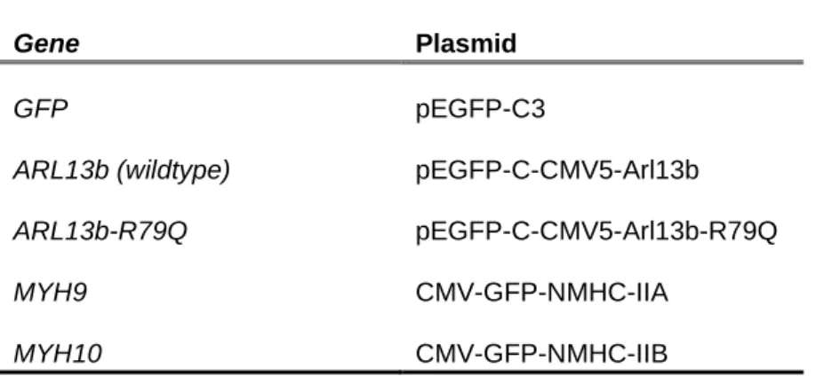
Table 3.1 – DNA overexpression plasmids used for MDA-MB-231 transfection
Gene Plasmid
GFP pEGFP-C3
ARL13b (wildtype) pEGFP-C-CMV5-Arl13b
ARL13b-R79Q pEGFP-C-CMV5-Arl13b-R79Q
MYH9 CMV-GFP-NMHC-IIA
17
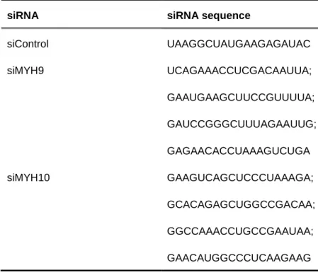
3.5.2. Silencing
NMIIA and NMIIB were silenced using siGENOME SMARTpool oligonucleotides (Dharmacon) specific for the human genes (MYH9 and MYH10, respectively). The sequences of small interfering RNAs (siRNAs) used are listed in Table 3.2. MDA-MB-231 cells (8 x 10⁴ cells/well) were seeded 24
hours before transfection in 24-well plates and cultured in DMEM Complete medium. Transfection was performed using 20 μM siRNA and 1 μL DharmaFECT 4 (Dharmacon) in 100 μL (final volume) Opti-MEM, according to the manufacturer’s instructions. Sixteen hours after transfection, the medium was replaced by DMEM Complete medium and cells were incubated at 37ºC and 5% CO2.
Table 3.2 – Sequences of siRNAs used for gene silencing
siRNA siRNA sequence
siControl UAAGGCUAUGAAGAGAUAC siMYH9 UCAGAAACCUCGACAAUUA; GAAUGAAGCUUCCGUUUUA; GAUCCGGGCUUUAGAAUUG; GAGAACACCUAAAGUCUGA siMYH10 GAAGUCAGCUCCCUAAAGA; GCACAGAGCUGGCCGACAA; GGCCAAACCUGCCGAAUAA; GAACAUGGCCCUCAAGAAG
3.6. RNA extraction, cDNA production and real-time quantitative PCR
RNA from MDA-MB-231 or MCF7 cells was extracted using the RNeasy Mini Kit (QIAGEN) according to the manufacturer’s instructions. Total RNA (500 ng) was transcribed into complementary DNA (cDNA) by incubation with 0.8 mM of dNTP mix (Thermo Scientific) and 0.25 μg/μL of random primers p(dN)6 (Roche) at 65ºC for 5 minutes. After placing the samples on ice for a few seconds, they
were incubated with 1x first-strand buffer (Invitrogen), 10 mM of DTT (Invitrogen) and 2 U/μL of RNaseOUT Recombinant Ribonuclease Inhibitor (Invitrogen) at 25ºC for 2 minutes. Finally, 2.5 U/μL of SuperScript II Reverse Transcriptase (Invitrogen) were added and samples were incubated at 25ºC for 10 minutes, at 42ºC for 50 minutes and at 70ºC for 15 minutes.
18 Real-time quantitative PCR (RT-qPCR) was performed in a qPCR LightCycler (Roche), using the FastStart Essential DNA Green Master kit (Roche) according to the manufacturer’s instructions. Amplification of the GAPDH housekeeping gene was done as an endogenous control for normalization of the expression level of each analyzed gene. The used primers are all specific for the human genes and are listed in Table 3.3. Analysis of the RT-qPCR data was done using the Roche LightCycler 96 software.
Table 3.3 – Primers used in RT-qPCR
Gene Primer sequence (5’-3’)
GAPDH Forward: CATTTCCTGGTATGACAACGA Reverse: GTCTACATGGCAACTGTGAG MYH9 Forward: AGAAGGTGAAGGTGAACAAGG Reverse: GGTGTAGATGAGCCCTGAGTAG MYH10 Forward: AGCCCAGACCAAAGAACAG Reverse: ATGAAAGATGTCCCTGACG
3.7. Immunofluorescence microscopy
MDA-MB-231 cells (1.25 x 10⁵ cells/well) were seeded on glass coverslips (ø 13 mm) in a
24-well plate and transfected as described previously. Twenty-four hours after transfection, the medium was removed and cells were fixed with 4% paraformaldehyde (Alfa Aesar) in PBS during 15 minutes at room temperature. After washing 3 times with PBS, the cells were blocked and permeabilized with 1% BSA and 0.05% saponin in PBS (blocking/permeabilization solution) for 30 minutes at room temperature. Cells were then incubated with the primary antibodies diluted in blocking/permeabilization solution for 60 minutes at room temperature in a humidified chamber. Coverslips were washed 5 times with PBS and incubated with the secondary antibodies or Alexa-Fluor-568-conjugated phalloidin diluted in the blocking/permeabilization solution for 60 minutes at room temperature in a humidified chamber. After 4 washes with PBS, cells were incubated with 1 μg/mL DAPI diluted in PBS for 5 minutes. Finally, coverslips were mounted in mounting media containing 10% Mowiol 4-88 in 100 mM Tris-HCl (pH = 8.5) and 25% glycerol. Images were acquired with a ZEISS LSM 710 confocal laser scanning microscope with a Plan-Apochromat 63x/1.4 NA oil-immersion objective and processed with the ImageJ software. The primary and secondary antibodies used are listed in Table 3.4 and Table 3.5, respectively.
19
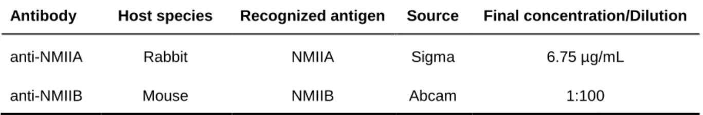
Table 3.4 – Primary antibodies used for immunofluorescence
Antibody Host species Recognized antigen Source Final concentration/Dilution
anti-NMIIA Rabbit NMIIA Sigma 6.75 µg/mL
anti-NMIIB Mouse NMIIB Abcam 1:100
Table 3.5 – Secondary antibodies used for immunofluorescence
Antibody Host species Label Source Final concentration
anti-mouse Goat Alexa Fluor 568 Invitrogen 2 µg/mL
anti-rabbit Goat Alexa Fluor 568 Invitrogen 2 µg/mL
3.8. Protein extraction
MDA-MB-231, MCF7 or MCF10A cells were lysed with ice-cold lysis buffer containing 50 mM Tris-HCl (pH = 7.5), 1 mM EDTA, 1 mM EGTA, 150 mM NaCl, 2 mM MgCl2, 1 mM DTT and
either 1% IGEPAL or 0.1% TX-100. Immediately before use, the buffer was supplemented with 1x cOmplete (Roche) protease inhibitor cocktail and 1 μM sodium orthovanadate and cells were incubated with the supplemented lysis buffer for 30 minutes on ice. After centrifugation at 13,800 x g for 30 minutes at 4ºC, the supernatants were collected. Protein concentration was determined using the DC Protein Assay kit (Bio-Rad), according to the manufacturer’s instructions.
3.9. Immunoprecipitation
For Arl13b immunoprecipitation, 800 to 1,500 μg of total protein extract were pre-cleared with Protein G-Sepharose 4 Fast Flow beads (GE Healthcare) for 1 hour at 4ºC, with rotation. Immunoprecipitation was performed overnight at 4ºC with rotation, using 3 μg of rabbit anti-Arl13b antibody (Barral et al, 2012). Protein G-Sepharose 4 Fast Flow beads were then added and incubated for 4 to 5 hours at 4ºC with rotation. The samples were centrifuged at 13,800 x g for 5 minutes at 4ºC and the supernatant was discarded. The pellet was washed once with lysis buffer containing 500 mM NaCl and three times with lysis buffer containing 150 mM NaCl. Finally, samples were solubilized in 2x sample buffer (20 mM sodium phosphate, 2% SDS, 0.001 % bromophenol blue, 0.2 M DTT and 2% glycerol) supplemented with 1,43 M β-mercaptoethanol, boiled at 95ºC for 5 minutes and centrifuged at 13,800 x g for 5 minutes at 4ºC. The supernatant containing immunoprecipitated proteins was collected and analyzed.
20 For the immunoprecipitation involving GTPɣS or GDP loading, 0.5 mM GTPɣS or 5 mM GDP were added to the pre-cleared protein extracts for 15 minutes at room temperature with agitation, before incubation with the antibody.
For GFP-tagged Arl13b immunoprecipitation performed by Paul Greiner and Cristina Casalou, GFP-Trap beads (ChromoTek) were equilibrated in lysis buffer containing 150 mM NaCl, before incubation with 500 μg of total protein extract for 2 hours at 4ºC. The samples were then centrifuged at 2,500 x g for 3 minutes at 4ºC, and the supernatants were discarded. The beads were then washed once with lysis buffer containing 500 mM NaCl and three times with lysis buffer containing 150 mM NaCl. Finally, samples were solubilized in 2x sample buffer supplemented with 1,43 M β-mercaptoethanol, boiled at 95ºC for 5 minutes and centrifuged at 13,800 x g for 5 minutes at 4ºC. The supernatant containing immunoprecipitated proteins was collected and analyzed.
3.10.
SDS-Polyacrylamide Gel Electrophoresis
Tris-glycine sodium dodecyl sulfate (SDS)-polyacrylamide gels containing 8% polyacrylamide resolving gel (pH= 8.8) and 5% polyacrylamide stacking gel (pH = 6.8) were prepared using a 1 mm spacer, mounted in the gel electrophoresis system (Amersham Biosciences) and covered with running buffer containing 25 mM Trizma base, 192 mM glycine and 0,1% SDS. Protein samples previously solubilized in 2x sample buffer supplemented with 1,43 M β-mercaptoethanol and boiled at 95ºC for 5 minutes were applied on the gel along with 10 μL Precision Plus All Blue Protein Standards (Bio-Rad). SDS-Polyacrylamide Gel Electrophoresis was performed at 20 mA per running gel during 1 to 2 hours.
3.11.
Immunoblotting
Protein samples separated on an 8% SDS-PAGE gel were transferred onto a nitrocellulose blotting membrane (0.45 μm pore; GE Healthcare) in transfer buffer containing 25 mM Trizma base, 192 mM glycine, 0.25% SDS and 20% ethanol for 75 minutes at 100V. Membranes were blocked in blocking buffer (5% non-fat dried milk and 0.1% Tween-20 in PBS) for 1 hour at room temperature, before incubation with primary antibodies in a humidified chamber for 1 hour at room temperature. Membranes were then washed 3 times for 5 minutes in 0.1% Tween-20 in PBS and incubated with HRP-conjugated secondary antibodies for 1 hour at room temperature under constant agitation. Finally, membranes were washed 3 times for 5 min in 0.1% Tween-20 in PBS and the antibodies were detected using Amersham ECL Select (GE Healthcare), according to the manufacturer’s instructions. Chemiluminescence was detected using the ChemiDoc Touch Imaging System. Band intensities were quantified using the ImageJ software and normalized using GAPDH as a loading control. The primary and secondary antibodies were diluted in blocking buffer and are listed in Table 3.6 and Table 3.7, respectively.
21
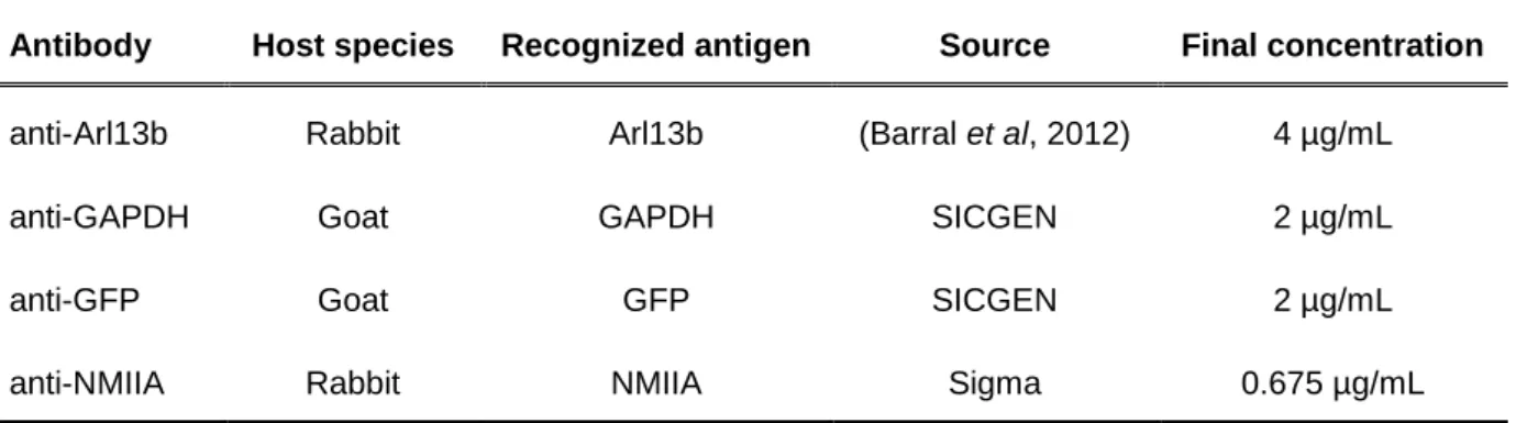
Table 3.6 – Primary antibodies used for immunoblotting
Antibody Host species Recognized antigen Source Final concentration
anti-Arl13b Rabbit Arl13b (Barral et al, 2012) 4 µg/mL
anti-GAPDH Goat GAPDH SICGEN 2 µg/mL
anti-GFP Goat GFP SICGEN 2 µg/mL
anti-NMIIA Rabbit NMIIA Sigma 0.675 µg/mL
Table 3.7 – Secondary antibodies used for immunoblotting
Antibody Host
species
Label Source Final
concentration/Dilution
Anti-rabbit Donkey HRP GE Healthcare 1:5,000
Anti-rabbit - light chain specific
Mouse HRP Jackson ImmunoResearch 0.16 µg/mL
Anti-goat Donkey HRP Jackson ImmunoResearch 0.16 µg/mL
3.12.
Gel staining
3.12.1. Coomassie gel staining
Immunoprecipitated proteins were analyzed on an 8% SDS-PAGE gel. The gel was fixed with a 40% ethanol and 10% acetic acid solution for 1 hour with agitation, before washing twice with distilled water for 10 minutes with agitation. The gel was stained in 0.12% Coomassie Brilliant Blue G-250 or Coomassie Brilliant Blue R-250, 10% orthophosphoric acid, 10% ammonium sulfate and 20% anhydrous methanol, overnight at room temperature with constant agitation. Finally, the gel was washed with a 1% acetic acid solution with agitation until the background was clear.
3.12.2. Silver nitrate gel staining
Following Coomassie staining, the gel was incubated with a 50% methanol and 5% acetic acid solution for 30 minutes with agitation, before washing twice for 2 minutes and once for 2 hours with agitation in distilled water. The gel was incubated with 0.02% sodium thiosulfate (pentahydrate) for
22 2 minutes before incubation with cold 0.1% silver nitrate for 30 minutes with agitation. After washing twice for 30 seconds with distilled water, the gel was incubated with 2% anhydrous sodium carbonate and 0.04% formaldehyde. When all bands were clearly visible, the reaction was stopped using a 1% acetic acid solution.
3.13.
Mass spectrometry
Mass spectrometry analysis was performed at the ITQB (Instituto de Tecnologia Química e Biológica António Xavier) Mass Spectrometry Unit (UniMS). Individual immunoprecipitated bands of interest excised from the silver nitrate stained gel were digested with trypsin before protein desalting and concentration in C18 micro-columns. Nanoscale liquid chromatography coupled to tandem mass spectrometry (nano LC-MS/MS) runs of 45 minutes were performed using a TripleTOF 6600 mass spectrometry system (SCIEX). Protein identification was obtained by screening in protein sequence databases.
3.14.
Statistical analysis
Numerical data are presented as mean ± standard deviation for wound healing assays and RT-qPCR. One-way ANOVA with Dunnett’s multiple comparison test was used to analyze the distinct wound healing assays data sets relatively to siControl. Statistical analysis was performed using GraphPad Prism software (version 7.03).
23
4. Results
4.1. Sub-cellular localization of Arl13b and cytoskeleton proteins
Interaction and co-localization of the actin cytoskeleton and the NMII isoforms NMIIA and NMIIB is conserved between different types of cells. Previous results from our laboratory have shown that the small G protein Arl13b co-localizes with actin and NMIIA in mouse fibroblasts (Casalou et al, 2014). In these cells, Arl13b interacts with actin and NMIIA, which proved to be required for cell migration. Since cells with different migratory capacities often show variation on the molecular mechanisms used for cell migration, co-localization of Arl13b with actin, NMIIA or NMIIB was assessed on the highly migratory MDA-MB-231 cells by confocal microscopy. Immunocytochemistry was performed using phalloidin to visualize filamentous actin and NMIIA or NMIIB antibodies to visualize the respective proteins. MDA-MB-231 cells overexpressing GFP-tagged Arl13b, NMIIA or NMIIB were also used to assess the localization of these proteins.
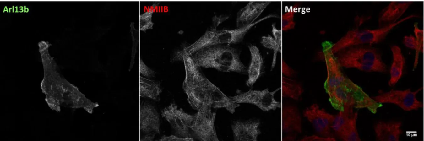
We observed that Arl13b co-localizes with actin, especially with structures localized near the cell periphery and at the plasma membrane (Figure 4.1A and B). It is also interesting to note the presence of Arl13b-positive vesicles surrounded by actin (Figure 4.1C and D). Arl13b also co-localizes with NMIIA (Figure 4.2). Namely, it is possible to observe some overlapping between Arl13b tubular networks and NMIIA (Figure 4.2C and D). Lastly, there is little, if any co-localization between Arl13b and NMIIB (Figure 4.3).
Confirmation of the co-localization of actin and NMIIA or NMIIB in MDA-MB-231 cells was also performed. Indeed, actin and NMIIA co-localize in several subcellular structures, such as stress fibers, FAs and lamellipodia (Figure S1). The typical periodic pattern of alternating actin and myosin was also possible to visualize on the stress fibers of these cells (Figure S1B and D). Similarly, NMIIB co-localizes with the same type of actin cytoskeleton structures, although not as prominently as NMIIA (Figure S1C and D).
24
Figure 4.1 – Arl13b co-localizes with actin in MDA-MB-231 cells. MDA-MB-231 cells overexpressing
Arl13b-GFP were fixed and stained with Alexa Fluor-568-conjugated phalloidin. (A and B) Arl13b co-localizes with actin mainly in structures localized at the cell periphery and at the plasma membrane. (C and D) Arl13b-positive vesicular structures are surrounded by smaller phalloidin-stained structures. B and D correspond to enlarged views of areas indicated by the boxes in A and C, respectively. All images correspond to representative Z stacks obtained by confocal microscopy. Scale bars: (A and C) 10 μm; (B) 5 μm; (D) 2 μm.
25
Figure 4.2 - Arl13b co-localizes with NMIIA in MDA-MB-231 cells. MDA-MB-231 cells overexpressing
Arl13b-GFP were fixed and stained with anti-NMIIA antibody. (A and B) Arl13b co-localizes with NMIIA in a protrusive structure that resembles a CDR. Arl13b also co-localizes with NMIIA in lamellipodia. (C and D) Arl13b and NMIIA co-localize in the leading edge of a cell with distinctive leading (right) and trailing edges (left). Arl13b-positive tubular structures are stained and co-localize with NMIIA. B and D correspond to enlarged views of areas indicated by the boxes in A and C, respectively. All images correspond to representative Z stacks obtained by confocal microscopy. Scale bars: (A and C) 10 μm; (B and D) 5 μm.