Cell migration in the postnatal
subventricular zone
1Laboratório de Neuroanatomia Celular, Departamento de Anatomia,
Instituto de Ciências Biomédicas, and 2Instituto de Biofísica Carlos Chagas Filho,
Universidade Federal do Rio de Janeiro, Rio de Janeiro, RJ, Brasil J.R.L. Menezes1, M. Marins1,
J.A.J. Alves1, M.M. Fróes1
and C. Hedin-Pereira2
Abstract
New neurons are constantly added to the olfactory bulb of rodents from birth to adulthood. This accretion is not only dependent on sustained neurogenesis, but also on the migration of neuroblasts and immature neurons from the cortical and striatal subventricular zone (SVZ) to the olfactory bulb. Migration along this long tangential pathway, known as the rostral migratory stream (RMS), is in many ways opposite to the classical radial migration of immature neurons: it is faster, spans a longer distance, does not require radial glial guidance, and is not limited to postmitotic neurons. In recent years many molecules have been found to be expressed specifically in this pathway and to directly affect this migration. Soluble factors with inhibitory, attractive and inductive roles in migration have been described, as well as molecules mediating cell-to-cell and cell-sub-strate interactions. However, it is still unclear how the various mol-ecules and cells interact to account for the special migratory behavior in the RMS. Here we will propose some candidate mechanisms for roles in initiating and stopping SVZ/RMS migration.
Correspondence
J.R.L. Menezes
Departamento de Anatomia ICB, CCS, Bloco F, UFRJ 21941-590 Rio de Janeiro, RJ Brasil
Fax: +55-21-290-0587 E-mail: [email protected]
Presented at the IV International UNESCO Course on “What the Developing Cerebral Cortex Tells About the Adult Cortex (and Vice Versa)”, Rio de Janeiro, RJ, Brazil, December 3-7, 2001.
Research supported by CNPq, TWAS, FUJB, and FAPERJ.
Received July 12, 2002 Accepted September 17, 2002
Key words
·Postnatal neurogenesis ·Chain migration ·Rostral migratory stream ·Astrocytes
·Gap junctions ·Radial glia
Introduction
Cell migration is an imperative requiment during developrequiment of most CNS re-gions. Newly generated neurons must leave the germinal layers and move, sometimes a great distance, to reach their final destina-tion. This migratory behavior can be classi-fied as radial or tangential depending on the orientation of the cell movement in relation to the pial surface (1). Alternatively, migra-tion can be viewed as gliophilic, neurono-philic, and homophilic in relation to the cell substrates of migration (1-3). The glia-de-pendent radial migration of immature neu-rons, ubiquitous in the embryonic CNS, is
precur-sors en route to the cerebral cortex in which corticofugal axons appear to function as their substrate (6). Finally, homophilic migration was thus termed because in this modality, migrating cells, neurons and neuroblasts, use each other as substrate, forming long chains of cells in situ and in vitro (3,7). This form of migration was first described for the cells that leave the anterior subventricular zone (SVZ) towards the olfactory bulb (Figure 1) and form the rostral migratory stream (RMS; 8). This tangential migratory movement of neurons and neuroblasts is directly associ-ated with the neurogenesis of olfactory bulb interneurons that occurs postnatally in the SVZ, a highly proliferative layer in the early postnatal animal that persists well into adult-hood (7-10).
Besides the unusual mode of migration, cells within the SVZ/RMS display other un-usual features, such as simultaneous
migra-tion and proliferamigra-tion (11), as well as a pre-cocious biochemical differentiation (11). Recently, there has been a resurgence of interest in the SVZ, both in the neonatal and adult brain, where this system has been used in studies ranging from cell migration to stem cell biology.
In brief, new neurons derived from the SVZ migrate long distances, invade the ma-ture nervous tissue and establish new func-tional circuits with the existing neurons (12). This is exactly the behavior one would wish from a source of new neurons that could be used to substitute neuron depletion. The un-derstanding of the mechanisms by which SVZ-derived cells migrate may be then as important as understanding how this region sustains con-tinued neurogenesis throughout adulthood. Here we will review the special characteris-tics of this tangential migration and the cel-lular interactions that drive this behavior.
Progenitors
Mitosis
Radial glia
Radially migrating neuron
Neuroblast or immature neuron
Periglomerular cell
Granular cell Cx
cc
lv
St
Tu AOB
1
3
2 MOB
Figure 1. Summary of subven-tricular zone/rostral migratory stream (SVZ/RMS) tangential mi-gration towards the olfactory bulb (OB) in a parasagittal representa-tion of the anterior forebrain in the early postnatal animal. Migra-tion in the RMS can be divided into three overlapping phases that correspond roughly to differ-ent portions of the pathway (num-bers in circles). 1) Initially cells go through a phase in which they migrate but are still able to divide. That happens mostly in regions of the SVZ close to the lateral ventricles where mitosis is more frequent. 2) At a given moment, already within the RMS, cells defi-nitely abandon the cell cycle and continue migration towards the OB. 3) Upon reaching the OB, cells switch from tangential to ra-dial migration and invade the OB parenchyma, turning on their dif-ferentiation into granular and periglomerular cells. In early post-natal life radial glia form a scaffold for this pathway as represented
Cellular composition of the postnatal SVZ
The SVZ is not a homogeneous and stable region throughout the life of an ani-mal. The cellular composition of young and adult SVZ differs little except for relative numbers, with both containing immature neu-rons, neuroblasts, undifferentiated precur-sor cells, astrocytes and microglia (13,14). A main difference is that in the young SVZ the major glial component is the radial glia, absent in the adult (15,16). The astrocytes of the adult SVZ/RMS surround the migrating cells providing an astrocytic scaffold to the SVZ that forms long cylinders denominated glial tubes (7,17). Recently, we have demon-strated that such a scaffold does in fact exist in the young SVZ formed by the cell bodies and processes of transforming radial glia (17).
Compartments in the postnatal SVZ
Proliferation in the SVZ was thought to occur homogeneously, in contrast to, for example, the embryonic ventricular zone, where a characteristic nuclear movement segregates cells in different stages of the cell cycle (18). Recently, we have demonstrated that the S-phase cells in the young SVZ/ RMS are preferentially concentrated in the periphery of the SVZ/RMS tube-like struc-ture spatially segregated from migratory cells (19). A similar periphery-to-core pattern of distribution was recently described for the expression of zebrin II (20), which rein-forces the idea that the early postnatal SVZ is organized in discrete cellular compartments. In addition, we have described a discrete spatial pattern of gap junction-mediated cell coupling involving neuroblasts and radial glia that closely resembles the S-phase dis-tribution in the young SVZ (21). It is pos-sible that some compartmentalization per-sists to adulthood since discrete islands of proliferative cells are also found in the mature
SVZ (14). Additional evidence for this comes from infusion of truncated ephrins that by disturbing proliferation cause the appearance of large islands of proliferating cells (22). The relevance of these discrete compartments is still not fully understood. They may reflect the segregated distribution of specific cells and/or growth factors with proliferation-inducing ca-pacities. Alternatively, they may arise from the preferential adhesion between homophilic migrating cells, thus excluding proliferating cells from the migratory chains.
Tangential migration within the SVZ/RMS
Cellular substrates for migration
Evidence converges to support the idea that migration within the RMS is glia inde-pendent. Most important for this conclusion was the demonstration that SVZ/RMS cells can migrate in culture without the presence of glial cells (3). In addition, in the young SVZ/RMS, migration is orthogonal to the radial glial palisade (16) and because of this arrangement RMS cell migration has been described as glia independent (10,16). How-ever, a glial sheath encircles migrating cells both in young and adult brains (7,15,17). Until now it has not been elucidated if these scaffolds have any function in guiding SVZ/ RMS migration. Apparently, astrocytes of the SVZ have the ability of inducing neuro-blast proliferation through a cell-to-cell con-tact mechanism (25). In addition, ensheath-ing astrocytes and possibly radial glial cells
may be the source of a secreted protein with a migration-inducing activity (MIA) (26). In contrast, it is possible that the interaction with glial cells may reduce migratory speed, which could account for the much lower speeds, 30 to 50 µm/h, for in situ migration within the RMS (7,10) or in SVZ explant cultures rich in glia (26) when compared to cultures with Matrigel which are poor in glia, and migration reaches speeds well over 100 µm/h (3).
Push, pull and roll: molecular substrates for migration
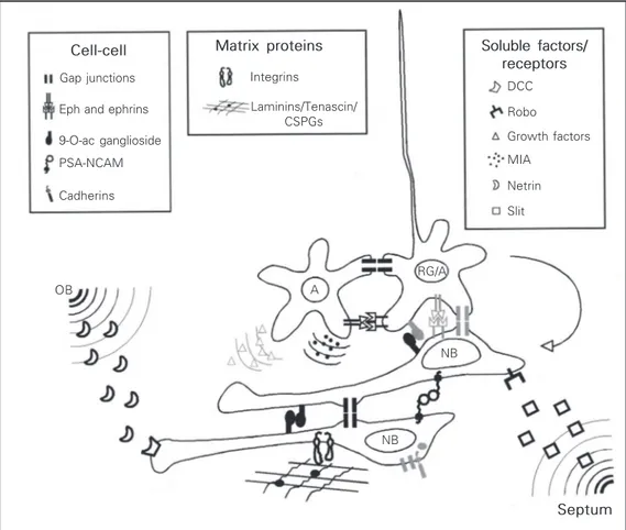
Despite the growing interest in the mechanisms underlying migration in the RMS, the factors that control this phenome-non remain largely unknown. Several mol-ecules have already been shown to regulate this type of migration (Figure 2), but we are
Cell-cell Gap junctions
Eph and ephrins
9-O-ac ganglioside
PSA-NCAM
Cadherins
Matrix proteins
Integrins
Laminins/Tenascin/ CSPGs
Soluble factors/ receptors
DCC
Robo
Growth factors
MIA
Netrin
Slit
RG/A
NB A
NB OB
Figure 2. Summary scheme of confirmed and putative mol-ecules governing cell migration in the subventricular zone/rostral migratory stream (SVZ/RMS) from birth to adulthood. Influ-ences of diffusible factors, cell-cell and cell-cell-substrate interac-tions on migration are illustrated. Only neuroblasts (NB), astrocytes (A) and radial glia (RG) are repre-sented. To simplify the scheme RG and A were considered equivalent (RG/A). Presently, we can identify many different fac-tors involved in directed chain mi-gration to the olfactory bulb (OB) through the RMS. Illustrated are known soluble factors, mem-brane-bound and extracellular molecules that affect this migra-tion, and their confirmed cellular and tissue location (in black). Mol-ecules in light gray represent pos-sible but not demonstrated inter-actions of these molecules. Al-though most molecules repre-sented have been shown to be directly involved in RMS migra-tion, little is known about how they interact with each other. See text for details. CSPGs, chon-droitin sulfate proteoglycans; DCC, deleted in colorectal cancer receptor; MIA, migration-induc-ing activity.
far from a working model comparable to the one proposed for radial migration (27). Simi-lar to radial migration one would expect to find attractant, repulsive, and permissive molecules, as well as initialization and stop signals.
Soluble factors
Attractants. Judging from the stereotypi-cally olfactory bulb-directed migration, a powerful attractant gradient originating in the olfactory bulb could be postulated; how-ever, unidirectional migration was still de-tected in the absence of the olfactory bulb (28). Sensory deprivation also did not affect cell migration in the RMS although it did alter the number of surviving interneurons (24,29). It was only recently that a putative attractant from the olfactory bulb was pro-posed. Murase and Horwitz (30) demon-strated an effect of a netrin or netrin-like molecule mediated by the receptor deleted in colorectal cancer (DCC) in the guidance of migrating cells within the RMS.
Repellents and inhibitors. The secreted proteins, Slit1 and 2, were the only mol-ecules described so far that could influence the migration direction of RMS cells via a repulsion-mediated mechanism (31). These molecules are expressed in the septum and choroid plexus, regions that could be impor-tant in driving RMS cells rostrally in the olfactory bulb direction (32). In mammals, Slit was first described to serve as an axonal guidance signal that impedes commissural axon recrossing, in association with its re-ceptor Robo present in the commissural axons (32). These molecules also appear to have a role in directing tangential migration from the ganglionic eminence, so that the migrat-ing cells avoid entermigrat-ing the Slit expressmigrat-ing striatal primordium in their pathway towards the cerebral cortex (32). Recent evidence suggested that, rather than having a repul-sive action, Slit could be inhibitory for SVZ/ RMS cells, reducing the chance of RMS
cells to end up in the septum (26). Regard-less of whether its main effect is inhibitory or repulsive, Slit alone is not sufficient to account for the directed migration to the olfactory bulb. It does not explain, for ex-ample, why cells do not abandon the RMS to invade adjacent tissue, such as the cortical white matter or the striatum. A frequent assumption is that the astrocytic sheath or alternatively the radial glial sheath acts as a barrier for this outflow (33). However, this is yet to be verified.
Growth factors. Several growth factors have been shown to promote proliferation of the SVZ population, such as EGF, FGF (34, 35), TGF-ß (36) and BDNF (37,38). Albeit no direct effect on cell migration has been demonstrated, intrathecal and intraventricu-lar infusion of growth factors increased the numbers of cells moving down the SVZ/ RMS pathway (34,35,37,38), as well as the number of cells straying from the SVZ and invading neighboring tissue (35,37). These results may be due solely to the increase in cell number; however, a role of specific growth factors augmenting SVZ/RMS cell migration cannot as yet be discarded.
Cell contact-mediating molecules
Few molecules that mediate cell-to-cell interactions have been implicated in tangen-tial cell migration within the SVZ/RMS. To date these are the polysialylated form of NCAM (PSA-NCAM; 39,40), ganglioside 9-O-acetylated GD3 (9-O-acGD3; 41-43), ephrins and eph receptors (22), and connex-ins (21,44). Despite their very diverse na-ture, perturbing any of these molecules greatly reduces or stops migration of SVZ cells. This indicates that many different intracellu-lar signaling pathways converge to elicit this behavior.
its role in olfactory bulb postnatal neurogen-esis was postulated (45). Migrating and pro-liferating cells of the SVZ typically express the polysialylated form of NCAM (10,46). The genetic deletion of the embryonic NCAM (180 kDa; 9,40,46), as well as enzymatic removal of polysialic acid (PSA; 39,40) greatly reduces or abolishes SVZ/RMS cell migration. However, given the molecular nature of PSA-NCAM, it is difficult to un-derstand how this molecule could mediate homophilic interactions, since the PSA moi-eties would, if anything, impede cell adhe-sion (39,40). It can be reasoned that PSA-NCAM favors migration by acting as a nega-tive regulator for cell-cell interaction, pre-venting cells from interacting too tightly. Indeed, NCAM knockout mice in which all isoforms were deleted revealed a speed re-duction but not abolition of chain migration of SVZ/RMS cells (46). Therefore, due to these results and the nature of the PSA-NCAM homophilic interaction, it could be expected that other molecules would partici-pate in the migration of SVZ cells.
Eph receptors and their ligands ephrins were shown to be directly involved in regu-lating migration and proliferation in the post-natal SVZ (22). This family of receptor ty-rosine kinase and their ligands mediate bidi-rectional signaling between cells and were shown to be implicated in a variety of devel-opmental events (47). In general, ephrin and eph receptor interaction lead to contact-de-pendent cell repulsion involved in guiding axons and migrating cells (47). This repul-sion effect may also play a part in boundary formation restricting cell intermingling and communication (47). Nevertheless, ephrin and eph receptor activation can trigger intra-cellular signaling and the outcome may not be just repulsive, since, for example, eph receptor activation may lead to an increase in the expression of cell adhesion molecules (47). Expression of EphB1-3, EphA4 and ephrins-B2/3 was detected in the SVZ/RMS, although cellular localization was only
pos-sible for ephrin-B, which was specifically located to astrocytes (22). Infusion of clus-tered soluble EphB2 and ephrin-B2 disrupted chain migration in the SVZ/RMS. However, this result may be due to a direct effect on proliferation (22)
intracellu-lar phosphorylation of focal adhesion ki-nases (50). It is possible that the acetylation of this molecule could regulate its interac-tion with the extracellular matrix during cell migration and axon extension.
Recently gap junction intercellular com-munication (GJIC) has been implicated in the regulation of SVZ cell migration, based on connexin 43 expression (51), ultrastruc-tural localization of gap junctions (15) and the presence of dye coupling (21) within the SVZ/RMS. Cell coupling partners within the SVZ/RMS have not yet been determined, but dye coupling studies indicate that homo-cellular and heterohomo-cellular cell coupling is present, involving both glial cells and neuro-blasts (44 and Marins M, Fróes MM and Menezes JRL, unpublished observations). It is unusual to associate gap junctions with cell migration, since these require the estab-lishment of complex and intimate adhesive contacts. Moreover, in the nervous system, cell uncoupling, as a rule, is considered to be a necessary step for initiating migration of recent postmitotic neurons (52). However, pharmacological inhibition of GJIC reduced migration and proliferation of postnatal SVZ cells in vitro (44), suggesting that GJIC is positively involved in SVZ/RMS migration. In the nervous system, the only other ex-ample in which gap junctions were shown to positively modulate migration is that of neu-ral crest cells destined to the heart. For these sympathetic nerve cells, connexin 43 ge-netic deletion abolished migration (53). Re-cently, it has been demonstrated that this effect does not require functional coupling, and may be due to the interaction of connex-ins with N-cadherin (54). In most systems where gap junctions were shown to be posi-tively linked to migration, cells do not mi-grate as individual cells but as cohorts (55), as neural crest and RMS cells, suggesting that gap junction expression favors adhesion of these migrating cells. Since GJIC inhibi-tion, besides migrainhibi-tion, also affects prolif-eration (44) we postulate that cell coupling
may be a key component for the initiation of migration, in contrast to the radial migration in the embryonic cortex, where uncoupling is a possible start signal for migration (52).
Extracellular matrix molecules. From birth to adulthood the SVZ/RMS pathway provides a diverse extracellular environment to migrating cells, in which some molecules are specifically enriched, such as tenascin-C, proteoglycans (33,56), and laminins (30). Extracellular matrix molecules can be seen as providing an instructive and/or permis-sive environment, important for tangential cell migration, in a similar fashion as seen for neuronal radial migration (27).
Extracellular molecules involved in boundary formation such as tenascin-C and chondroitin sulfate proteoglycans were shown in the RMS pathway (33,57). Since it is possible that the major source of these molecules could be the scaffolding glial cells, tenascin-C and chondroitin sulfate proteo-glycans may somehow prevent migrating cells from straying from the SVZ/RMS path-way (33). However, no obvious defects were found in the brains of tenascin-C-deficient mice (58) albeit effects on SVZ migration were not specifically addressed. Another pro-teoglycan, brevican, and a metalloprotein-ase inhibitor, TIMP-4, are also expressed in a restricted manner in the postnatal RMS, suggesting they may also act to guide migra-tion (59). Although proteoglycans and te-nascin-C are generally related to inhibitory effects in process outgrowth, a permissive role in migration cannot be excluded. How-ever, if they do function as inhibitory cues it is possible that they help elicit chain migra-tion behavior, since cells would prefer to make adhesive contacts essential for migra-tion with other migrating cells rather than with these substrates in the pathway.
integrin subunits in migrating SVZ cells. Integrins are an important group of adhesion molecules that bind to the extracellular ma-trix and have been shown to participate in the migration of progenitors and young neu-rons (27). In the SVZ/RMS integrin subunits such as a1, ß1 and ß8 were detected in early postnatal ages whereas aV and ß6 subunits appeared postnatally and persisted to adult-hood. Immunoblockade of some of these integrins inhibited the translocation and pro-trusion of the leading processes of SVZ cells (30). Immunoblockade of a6ß1 integrins also disrupts chain migration from EGF expanded neurospheres derived from postnatal fore-brain cells (60).
Long and winding road: Instruction or restriction?
While a complete picture of the mol-ecules involved in the RMS flow of cells is not available, uncertainty remains as to what is unique about the SVZ/RMS pathway.
Could the plasticity displayed by the SVZ cells in postnatal life, such as long-range migration and invasion of differentiated tis-sue be conferred to cells transplanted to the SVZ? Or is that capacity exclusive of the SVZ cells? While the SVZ/RMS could sus-tain migration of some exogenous progeni-tor cell populations (61-63), cells from the embryonic ventricular zone and external granular layer failed to migrate when trans-planted into the postnatal SVZ (57,64). In spite of their normal radial migration it could be expected that external granular layer and ventricular zone cells would respond to the local cues of the postnatal SVZ/RMS, for, as we have seen above, many factors involved in radial migration are also present in RMS mi-grating cells. Overall, these data suggest that SVZ/RMS may provide a permissive/instruc-tive environment only for responsive cells,
not being therefore a universal niche for migration.
If the SVZ/RMS cells are under a default migratory program, should the SVZ/RMS function primarily to contain these cells therein? In fact, removing SVZ cells from their normal sites in vivo and plating them as explants in vitro does not halt their outward migration (3,26). However, heterotopic trans-plantation strategies provide conflicting evi-dence. When injected into the lateral ven-tricle of the embryonic brain, SVZ cells do not invade the ventricular zone of the cere-bral cortex, but are found in few regions of the brain such as the mesencephalon (25). On the other hand, young and adult SVZ can invade and disperse in the striatum of the adult brain (65). In agreement with a con-tainment role for the SVZ/RMS, recent evi-dence indicates that neuroblasts can escape the RMS in response to cellular lesions of neighboring tissue and RMS (66-68). Pres-ently, we are far from determining the capac-ity for autonomous migration of SVZ/RMS cells. Heterotopic transplantation and post-lesion experiments may still prove to be a useful strategy to dissect the rich potentiali-ties of these cells for CNS dispersion.
Concluding remarks
References
1. Rakic P (1990). Principles of neural cell migration. Experentia, 46: 882-891. 2. Hatten ME (1999). Central nervous
sys-tem neuronal migration. Annual Review of Neuroscience, 22: 511-539.
3. Wichterle H, Garcia-Verdugo JM & Alva-rez-Buylla A (1997). Direct evidence for homotypic, glia-independent neuronal mi-gration. Neuron, 18: 779-791.
4. Komuro H & Rakic P (1998). Distinct modes of neuronal migration in different domains of developing cerebellar cortex. Journal of Neuroscience, 18: 1478-1490. 5. Nadarajah B, Brunstrom JE, Grutzendler
J, Wong RO & Pearlman AL (2001). Two modes of radial migration in early devel-opment of the cerebral cortex. Nature Neuroscience, 4: 143-150.
6. Denaxa M, Chan CH, Schachner M, Parnavelas JG & Karagogeos D (2001). The adhesion molecule TAG-1 mediates the migration of cortical interneurons from the ganglionic eminence along the corticofugal fiber system. Development, 128: 4635-4644.
7. Lois C, Garcia-Verdugo J-M & Alvarez-Buylla A (1996). Chain migration of neu-ronal precursors. Science, 271: 978-981. 8. Altman J (1969). Autoradiographic and
his-tological studies of postnatal neurogen-esis IV. Cell proliferation and migration in the anterior forebrain, with special refer-ence to persisting neurogenesis in the olfactory bulb. Journal of Comparative Neurology, 137: 36-47.
9. Smart I (1961). The subependymal layer of the mouse brain and its cell production as shown by radioautography after thymi-dine-H3 injection. Journal of Comparative
Neurology, 116: 325-348.
10. Luskin MB (1993). Restricted proliferation and migration of postnatally generated neurons derived from the forebrain sub-ventricular zone. Neuron, 11: 173-189. 11. Menezes JRL, Smith CM, Nelson KC &
Luskin MB (1995). The division of neu-ronal progenitor cells during migration in the neonatal mammalian forebrain. Mo-lecular and Cellular Neurosciences, 6: 496-508.
12. Carlén M, Cassidy RM, Brismar H, Smith GA, Enquist LW & Frisen J (2002). Func-tional integration of adult-born neurons. Current Biology, 12: 606-608.
13. Stensaas LJ & Gilson BC (1972). Ependy-mal and subependyEpendy-mal cells of the caudato-pallial junction in the lateral ven-tricle of the neonatal rabbit. Zeitschrift für
Zellforschung und Mikroskopische Anato-mie, 132: 297-322.
14. Doetsch F, Garcia-Verdugo JM & Alvarez-Buylla A (1997). Cellular composition and three dimensional organization of the sub-ventricular germinal zone in the adult mammalian brain. Journal of Neurosci-ence, 17: 5046-5061.
15. Kishi K, Peng JY, Kakuta S, Murakami K, Kuroda M, Yokota S, Hayakawa S, Kuge T & Asayama T (1990). Migration of bipolar subependymal cells, precursor of the granule cells of the rat olfactory bulb, with reference to the arrangement of the radial glial fibers. Archives of Histology and Cy-tology, 53: 219-226.
16. Alves JAJ, Barone P, Engelender S, Fróes MM & Menezes JRL (2002). Initial stages of radial glia astrocytic transformation in the early postnatal anterior subventricular zone. Journal of Neurobiology, 52: 251-265.
17. Peretto P, Merighi A, Fasolo A & Bonfanti L (1997). Glial tubes in the rostral migra-tory stream of the adult rat. Brain Re-search Bulletin, 42: 9-21.
18. Takahashi T, Nowakowski RS & Caviness VS (1992). BUdR as an S-phase marker for quantitative studies of cytokinetic behav-ior in the murine cerebral ventricular zone. Journal of Neurocytology,21: 185-197. 19. Menezes JRL, Dias F, Garson AVB & Lent
R (1998). Restricted distribution of S-phase cells in the anterior subventricular zone of the postnatal mouse forebrain. Anatomy and Embryology, 198: 205-211. 20. Staugaitis SM, Zerlin M, Hawkes R, Levine JM & Goldman JE (2001). Aldo-lase C/zebrin II expression in the neonatal rat forebrain reveals cellular heterogene-ity within the subventricular zone and early astrocyte differentiation. Journal of Neuroscience, 21: 6195-6205.
21. Menezes JR, Froes MM, Moura Neto V & Lent R (2000). Gap junction-mediated cou-pling in the postnatal anterior subventricu-lar zone. Developmental Neuroscience, 22: 34-43.
22. Conover JC, Doetsch F, Garcia-Verdugo JM, Gale NW, Yancopoulos GD & Alvarez-Buylla A (2000). A disruption of Eph/ephrin signaling affects migration and prolifera-tion in the adult subventricular zone. Na-ture Neuroscience, 3: 1091-1097. 23. Kishi K (1987). Golgi studies on the
devel-opment of granule cells of the rat olfac-tory bulb with reference to migration in the subependymal layer. Journal of
Com-parative Neurology, 258: 112-124. 24. Frazier-Cierpial L & Brunjes PC (1989).
Early postnatal cellular proliferation and survival in the olfactory bulb and rostral migratory stream of normal and unilater-ally odor-deprived rats. Journal of Com-parative Neurology, 289: 481-492. 25. Lim DA & Alvarez-Buylla A (1999).
Interac-tion between astrocytes and adult sub-ventricular zone precursors stimulates neurogenesis. Proceedings of the Na-tional Academy of Sciences, USA, 96: 7526-7531.
26. Mason HA, Ito S & Corfas G (2001). Extra-cellular signals that regulate the tangen-tial migration of olfactory bulb neuronal precursors: inducers, inhibitors, and re-pellents. Journal of Neuroscience, 21: 7654-7663.
27. Rakic P, Cameron RS & Komuro H (1994). Recognition, adhesion, transmembrane signaling and cell motility in guided neu-ronal migration. Current Opinion in Neu-robiology, 4: 63-69.
28. Kirschenbaum B, Doetsch F, Lois C & Alvarez-Buylla A (1999). Adult subven-tricular zone neuronal precursors continue to proliferate and migrate in the absence of the olfactory bulb. Journal of Neurosci-ence, 19: 2171-2180.
29. Corotto FS, Henegar JR & Maruniak JA (1994). Odor deprivation leads to reduced neurogenesis and reduced neuronal sur-vival in the olfactory bulb of the adult mouse. Neuroscience, 61: 739-744. 30. Murase S & Horwitz AF (2002). Deleted in
colorectal carcinoma and differentially ex-pressed integrins mediate the directional migration of neural precursors in the ros-tral migratory stream. Journal of Neuro-science, 22: 3568-3579.
31. Wu W, Wong K, Chen J, Jiang Z, Dupuis S, Wu JY & Rao Y (1999). Directional guid-ance of neuronal migration in the olfac-tory system by the protein Slit. Nature, 400: 331-336.
32. Brose K & Tessier-Lavigne M (2000). Slit proteins: key regulators of axon guidance, axonal branching, and cell migration. Cur-rent Opinion in Neurobiology, 10: 95-102. 33. Thomas LB, Gates MA & Steindler DA (1996). Young neurons from the adult sub-ependymal zone proliferate and migrate along an astrocyte, extracellular matrix-rich pathway. Glia, 17: 1-14.
endogenous subependymal neural pre-cursor cell populations in the adult mouse brain. Journal of Neuroscience, 16: 2649-2658.
35. Khun HG, Winkler J, Kemperman G, Thal LJ & Gage FH (1997). Epidermal growth factor and fibroblast growth factor-2 have different effects on neural progenitors in the adult rat brain. Journal of Neurosci-ence, 17: 5820-5829.
36. Tropepe V, Craig CG, Morshead CM & van der Kooy D (1997). Transforming growth factor-a null and senescent mice show decreased neural progenitor cell proliferation in the forebrain subepen-dyma. Journal of Neuroscience, 17: 7850-7859.
37. Zigova T, Pencea V, Wiegand SJ & Luskin MB (1998). Intraventricular administration of BDNF increases the number of newly generated neurons in the adult olfactory bulb. Molecular and Cellular Neuroscien-ces, 11: 234-245.
38. Pencea V, Bingaman KD, Wiegand SJ & Luskin MB (2001). Infusion of brain-de-rived neurotrophic factor into the lateral ventricle of the adult rat leads to new neurons in the parenchyma of the stria-tum, sepstria-tum, thalamus, and hypothala-mus. Journal of Neuroscience, 21: 6706-6717.
39. Hu H, Tomasiewicz H, Magnuson T & Rutishauser U (1996). The role of poly-sialic acid in migration of olfactory bulb interneuron precursors in the subventricu-lar zone. Neuron, 16: 735-743.
40. Ono K, Tomasiewicz H, Magnuson T & Rutishauser U (1994). N-CAM mutation inhibits tangential neuronal migration and is phenocopied by enzymatic removal of polysialic acid. Neuron, 13: 595-609. 41. Mendez-Otero R & Cavalcante LA (1996).
Expression of 9-O-acetylated gangliosides is correlated with tangential cell migration in the rat brain. Neuroscience Letters, 204: 97-100.
42. Miyakoshi LM, Mendez-Otero R & Hedin-Pereira C (2001). The 9-O-acetyl GD3 gan-gliosides are expressed by migrating chains of subventricular zone neurons in vitro. Brazilian Journal of Medical and Bio-logical Research, 34: 669-673.
43. Hedin-Pereira C, Miyakoshi LM & Men-dez-Otero R (2001). Ganglioside 9-O-acetyl GD3 is involved in the migration of subventricular zone neuroblasts in vitro. Society for Neuroscience Abstracts, 27: 248.6.
44. Marins M, Moura Neto V, Fróes MM & Menezes JR (2001). Gap junctions are in-volved in the migration and proliferation
of postnatal subventricular zone cells. So-ciety for Neuroscience Abstracts, 27: 248.5.
45. Miragall F, Kadmon G, Husmann M & Schachner M (1988). Expression of cell adhesion molecules in the olfactory sys-tem of the adult mouse: presence of the embryonic form of N-CAM. Developmen-tal Biology, 129: 516-531.
46. Chazal G, Durbec P, Jankovski A, Rougon G & Cremer H (2000). Consequences of neural cell adhesion molecule deficiency on cell migration in the rostral migratory stream of the mouse. Journal of Neuro-science, 20: 1446-1457.
47. Mellitzer G, Xu Q & Wilkinson DG (2000). Control of cell behaviour by signalling through Eph receptors and ephrins. Cur-rent Opinion in Neurobiology, 10: 400-408.
48. Mendez-Otero R & Santiago MF (2001). Functional role of a glycolipid in direc-tional movements of neurons. Anais da Academia Brasileira de Ciências, 73: 221-229.
49. Santiago MF, Berredo-Pinho M, Costa MR, Gandra M, Cavalcante LA & Mendez-Otero R (2001). Expression and function of ganglioside 9-O-acetyl GD3 in postmi-totic granule cell development. Molecular and Cellular Neurosciences, 17: 488-499. 50. Probstmeier R, Michels M, Franz T, Chan BM & Pesheva P (1999). Tenascin-R inter-feres with integrin-dependent oligoden-drocyte precursor cell adhesion by a gan-glioside-mediated signaling mechanism. European Journal of Neuroscience, 11: 2474-2488.
51. Miragall F, Albiez P, Bartels H, de Vries U & Dermietzel R (1997). Expression of gap junction protein connexin43 in the sub-ependymal layer and rostral migratory sys-tem of the mouse: evidence for an in-verse correlation between intensity of staining of connexin43 expression and cell proliferation activity. Cell and Tissue Re-search, 287: 243-253.
52. Bittman K, Owens DF, Kriegstein AR & LoTurco JJ (1997). Cell coupling and un-coupling in the ventricular zone of devel-oping neocortex. Journal of Neurosci-ence, 17: 7037-7044.
53. Huang GY, Cooper ES, Waldo K, Kirby ML, Gilula NB & Lo CW (1998). Gap junc-tion-mediated cell-cell communication modulates mouse neural crest migration. Journal of Cell Biology, 143: 1725-1734. 54. Xu X, Li WEI, Huang GY, Meyer R, Chen
Y, Luo Y, Thomas MP, Radice GI & Lo CW (2001). Modulation of mouse neural crest cell motility by N-cadherin and connexin
43 gap junctions. Journal of Cell Biology, 154: 217-230.
55. Froes M & Menezes JRL (2002). Coupled heterocellular arrays in the brain. Neuro-chemistry International (in press). 56. Gates MA, Thomas LB, Howard EM,
Laywell ED, Sajin B, Faissner A, Gotz B, Silver J & Steindler DA (1995). Cell and molecular analysis of the developing and adult mouse subventricular zone of the cerebral hemispheres. Journal of Com-parative Neurology, 361: 249-266. 57. Jankovski A & Sotelo C (1996).
Subven-tricular zone-olfactory bulb migratory path-way in the adult mouse: cellular composi-tion and specificity as determined by heterochronic and heterotopic transplan-tation. Journal of Comparative Neurology, 371: 376-396.
58. Saga Y, Yagi T, Ikawa Y, Sakakura T & Aizawa S (1992). Mice develop normally without tenascin. Genes and Develop-ment, 6: 1821-1831.
59. Jaworski DM & Fager N (2000). Regula-tion of tissue inhibitor of metalloprotein-ase-3 (Timp-3) mRNA expression during rat CNS development. Journal of Neuro-science Research, 61: 396-408. 60. Jacques TS, Relvas JB, Nishimura S,
Pytela R, Edwards GM, Streuli CH & French-Constant C (1998). Neural precur-sor cell chain migration and division are regulated through different ß1 integrins. Development, 125: 3167-3177.
61. Suhonen JO, Peterson DA, Ray J & Gage FH (1996). Differentiation of adult hippo-campus-derived progenitors into olfactory neurons in vivo. Nature, 383: 624-627. 62. Flax JD, Aurora S, Yang C, Simonin C,
Wills AM, Billinghurst, Jendoubi M, Sidman RL, Wolfe JH, Kim SU & Snyder EY (1998). Engraftable human neural stem cells respond to developmental cues, re-place neurons, and express foreign genes. Nature Biotechnology, 16: 1033-1039.
63. Englund U, Fricker-Gates RA, Lundberg C, Bjorklund A & Vwictorin K (2002). Transplantation of human neural progeni-tor cells into the neonatal rat brain: exten-sive migration and differentiation with long-distance axonal projections. Experi-mental Neurology, 173: 1-21.
65. Zigova T, Pencea V, Betarbet R, Wiegand SJ, Alexander C, Bakay RA & Luskin MB (1998). Neuronal progenitor cells of the neonatal subventricular zone differentiate and disperse following transplantation into the adult rat striatum. Cell Transplan-tation, 7: 137-156.
66. Magavi SS, Leavitt BR & Macklis JD (2000). Induction of neurogenesis in the
neocortex of adult mice. Nature, 405: 951-955.
67. Nait-Oumesmar B, Decker L, Lachapelle F, Avellana-Adalid V, Bachelin C & Van Evercooren AB (1999). Progenitor cells of the adult mouse subventricular zone pro-liferate, migrate and differentiate into oli-godendrocytes after demyelination. Euro-pean Journal of Neuroscience, 11:
4357-4366.