UNIVERSIDADE DE LISBOA FACULDADE DE FARMÁCIA
Ana Sofia Gregório Fernandes
M
ODULATION OF OXIDATIVE STRESS BY SUPEROXIDEDISMUTASE MIMETICS
DOUTORAMENTO EM FARMÁCIA (Toxicologia)
UNIVERSIDADE DE LISBOA FACULDADE DE FARMÁCIA
Ana Sofia Gregório Fernandes
M
ODULATION OF OXIDATIVE STRESS BY SUPEROXIDEDISMUTASE MIMETICS
DOUTORAMENTO EM FARMÁCIA (Toxicologia)
Supervisor: Professor Doutor Nuno Oliveira Co-supervisor: Professora Doutora Judite Costa
i
Preface
Toxicology, once said to be the science of poisons, is presently defined by The Society of Toxicology as “the study of the adverse effects of chemical, physical or biological agents on living organisms and the ecosystem, including the prevention and amelioration of such adverse effects”. It is therefore an inherently multidisciplinary subject that encompasses many areas, such as chemistry, biology and pharmacology.
Free radical toxicology is a very relevant and emerging field in Toxicology. During the past three decades, a remarkable rise of new information implicating reactive species in the mechanisms of action of many toxicants and pathological phenomena has driven an increasing interest of this scientific area. This thesis, by aiming at developing and studying antioxidant compounds, gathers concepts of chemistry and biology towards the discovery of novel approaches to overcome toxicity of oxidative stress-related conditions. Carrying out my PhD thesis in such a multidisciplinary area of research was very motivating, absorbing and challenging since the very beginning. The results presented herein contribute to the understanding of superoxide dismutase mimetics, representing a further step towards a potential clinical use of these promising antioxidants.
iii
Acknowledgments
I want to express my deepest gratitude to my supervisor Professor Nuno Oliveira. Thank you for your mentorship, commitment, tireless, availability and scientific rigor. Our numerous broad scientific discussions were invaluable. More than just a supervisor, you have been a present and caring friend. Thank you for your support and friendship. It has been a pleasure to work under your guidance and I am looking forward to continue this fruitful partnership in future years.
I also want to thank my co-supervisor Professor Judite Costa. Thank you for giving me the opportunity to work with this research group, by joining your project POCTI/49114/QUI/2002. I want to thank all your support during this fellowship, as well as during these four years of my PhD. Thank you so much for your friendship, help and guidance.
My deepest gratitude to Professor Matilde Castro, who invited me to join her scientific studies when I was still an undergraduate student. Thank you for believing in me. I fell deeply honored in working in your group and in being mentored by someone who I profoundly admire personally and professionally. Even in your hardest moments, you are always there, caring and encouraging. Thanks for your leadership, vision, support, and constant help. And, most of all, thank you for your friendship.
I want to thank the colleagues of the Chemical Biology and Toxicology group. A special word to Professor Fátima Cabral: thank you for your constant availability and precious help. I also want to thank Ana Francisca for her friendship and permanent help. It has been a pleasure to work next to you. I remember gladly our trips to scientific meetings, as well as the simple moments we have shared in the Lab. Also to Joana Miranda, who recently joined the group, thank you for contributing to the nice and friendly environment of the Lab.
I want to express my acknowledgments to all the colleagues from Centro de Investigação em Genética Molecular Humana. A special word to Professor José Rueff, coordinator of this institute. Thank you for receiving me in your Lab and for providing what I needed to carry out my work. Your vision and vast knowledge were precious contributions for this research. I want to highlight the very important help of Professor Jorge Gaspar. Thank you for your support, assistance, and for the many fruitful
iv
scientific discussions. I also want to acknowledge Michel Kranendonk for helping me in the DNA strand break analysis.
I would like to thank to all the other researchers that somehow were involved in this work. To Rita Guedes and Daniel dos Santos from the Medicinal Chemistry group of iMed.UL, thank you for carrying out the theoretical calculations on the copper(II) complexes. My acknowledgment to Professor Ines Batinic-Haberle, from the Department of Radiation Oncology of Duke University Medical School. Thank you for giving me the opportunity to study your ortho-substituted manganese(III) porphyrins, and thanks for the very constructive scientific discussions that we had. I also want to thank Octávia Gil and Vanda Martins from Instituto Tecnológico Nuclear who are now involved in the studies of radioprotection with the manganese(III) porphyrins.
I want to express my gratitude to Professors Teresa Chaveca and Ana Rita Conde for their support and friendship. Thank you for giving me the opportunity to integrate your pedagogical activities at Faculdade de Farmácia. I also want to thank my colleagues from Universidade Lusófona and from Escola Superior de Saúde Ribeiro Sanches for their contribution to my pedagogical development.
Being surrounded by such a group of extraordinary people made my scientific work a pleasant activity. But beyond science, I have the privilege to have very special friends, always helpful and enthusiastic… I want to thank all of you, and specially Ana, Andrea, Mafalda, Mara and Cátia, for enriching my life with happiness and great moments.
To my dear family, especially to my parents, sister and grandma… thank you for your constant support, encouragement, and most of all thank you so much for you unconditional love. I owe you everything.
A very special thank to Pedro, for being always by my side, walking with me every step of this way. Thank you for your love, attention, understanding and constant encouragement. Thank you for making our home a very special, warm and happy place. I also want to thank my little Rodrigo who, right from his in utero life, had to share his mother’s attention with this thesis. Thank you for the joy that you brought to my life.
v
Finally, I want to acknowledge Fundação para a Ciência e a Tecnologia that financially supported the work presented in this thesis, through the Project POCTI/49114/QUI/2002 and through my PhD fellowship (SFRH/BD/28773/2006).
vii
Abstract
Superoxide anion, along with other reactive species, is involved in many toxicological processes, as well as in a number of pathophysiological phenomena. Superoxide dismutase mimetics (SODm), i.e., synthetic compounds that mimic the functional properties of superoxide dismutase converting O2•– to H2O2 and O2, have thus
emerged as prospective pharmaceutical candidates to overcome toxicity in oxidative stress-related conditions. Several metal-containing compounds were previously shown to possess SOD-like activity. This thesis is focused on two promising classes of macrocyclic complexes: manganese(III) porphyrins (MnPs) and macrocyclic copper(II) complexes.
The effects of MnTM-4-PyP, a para-substituted MnP, against the cell injury induced by three different oxidants were evaluated in V79 cells. This MnP protected against the cytotoxicity and increase in intracellular O2•– levels induced by the
xanthine-xanthine oxidase system (XXO; an extracellular O2•– generator) and by
tert-butylhydroperoxide (TBHP; an analogue of lipid hydroperoxides). However, MnTM-4-PyP did not show considerable protection against the anticancer drug doxorubicin (Dox) that, among other mechanisms of cytotoxicity, generates O2•– intracellularly. The effects
of MnTM-4-PyP were not observed at the cell division level, as shown by the mitotic index analysis. The results of this well established SODm in XXO- and TBHP-treated cells permitted the validation of cell-based models of oxidative stress to be used in the evaluation of the macrocyclic copper(II) complexes developed within the scope of this thesis.
Two optimized ortho-substituted MnPs, MnTE-2-PyP and MnTnHex-2-PyP, were also studied in this thesis and their role against TBHP-induced cell injury was evaluated. The two MnPs, even at low concentrations, counteracted remarkably the effects of TBHP in cell viability and intracellular O2•– levels. While the exposure of V79
cells to TBHP resulted in a significant depletion of total and reduced glutathione and in an increase in GSSG, MnPs augmented markedly the total and reduced glutathione contents in TBHP-treated cells.
Two sets of macrocyclic copper(II) complexes, without (CuL1-CuL5) or with a pyridine ring in the macrocyclic backbone (CuL6-CuL9), were synthesized and
viii
evaluated in vitro for their O2•– scavenging ability. The complexes were chemically
characterized in order to correlate their biochemical activity with thermodynamic, structural and electrochemical features. The complexes CuL3, CuL4 and CuL8 were selected to be further evaluated in cell-based assays since they presented high thermodynamic stability and effective ability to scavenge O2•–, and were devoid of
significant cytotoxicity. These complexes did not show considerable antioxidant activities in the oxidative stress models previously validated (XXO and TBHP). CuL3, CuL4 and CuL8 were also studied as redox modulators of anticancer drugs. The three complexes were shown to undergo Fenton chemistry in vitro. However, CuL3 and CuL4 did not potentiate the cytotoxicity of Dox in breast carcinoma (MCF7) cells. Conversely, CuL8 was shown to improve the therapeutic window of Dox, and especially of oxaliplatin, by both protecting non-tumoral human mammary cells (MCF10A) and boosting the cytotoxic effects in breast carcinoma cells.
The results presented in this thesis contribute to the comprehension of SODm as promising pharmaceutical agents both for antioxidant and cancer therapies, reinforcing the high therapeutic potential of this type of compounds.
Keywords: superoxide dismutase mimetics, manganese(III) porphyrins, macrocyclic copper(II) complexes, oxidative damage, antioxidant protection, chemotherapy sensitizers.
ix
Resumo
A produção de espécies reactivas, nomeadamente de O2•–, está associada a
diversos fenómenos toxicológicos e fisiopatológicos. Entre estes, destacam-se os processos de lesão tecidular e inflamação, os quais são factores etiológicos chave num grande número de patologias, incluindo isquémia-reperfusão, aterosclerose, cancro, lesão por radiação, doenças neurodegenerativas, doenças inflamatórias pulmonares, intestinais e cardiovasculares, entre outras. A remoção de O2•– pode ser assim uma via
importante para modular um vasto número de fenómenos patológicos. As enzimas superóxido dismutases (SOD), que catalizam a reacção de dismutação do O2•– em H2O2
e O2, são metaloproteínas com um papel fulcral na defesa contra o excesso de espécies
reactivas. Estudos pré-clínicos e clínicos com SOD têm demonstrado protecção contra os efeitos nocivos de várias situações de stress oxidativo. No entanto, a utilização clínica da enzima nativa apresenta diversas limitações. Tendo em conta que a remoção de O2•– permite minorar fenómenos inflamatórios, e de modo a ultrapassar as limitações
do uso da SOD nativa, têm sido desenvolvidos nos últimos anos compostos de baixo peso molecular com a capacidade de mimetizar as propriedades funcionais da SOD. Estes compostos designam-se usualmente por SOD miméticos (SODm). Várias classes de SODm têm sido desenvolvidas, tendo demonstrado efeitos benéficos notáveis em diferentes modelos de stress oxidativo, quer em cultura de células, quer em modelos animais. A maioria destes compostos contém um ião metálico no seu centro activo, habitualmente Mn(III), Mn(II), Cu(II) ou Fe(III), o qual é estabilizado por coordenação com um ligando. A utilização de ligandos macrocíclicos tem revelado vantagens em termos de estabilidade termodinâmica e actividade catalítica. Neste âmbito, a presente tese incide sobre duas classes de complexos macrocíclicos com actividade mimética da SOD: porfirinas de Mn(III) (MnPs) e complexos macrocíclicos de Cu(II).
A classe das MnPs contém os SODm mais potentes e estáveis desenvolvidos até ao momento. Apesar de serem amplamente estudados, os efeitos ao nível celular destes compostos ainda não são totalmente conhecidos. A porfirina para-substituída MnTM-4-PyP é um SODm de eficácia bem estabelecida, disponível comercialmente, que tem vindo a ser estudado em vários modelos de situações fisiopatológicas. Os papel deste SODm face à toxicidade induzida por três oxidantes, cujos mecanismos de toxicidade diferem nas espécies reactivas envolvidas e no local de formação destas, foi avaliado em
x
células V79, uma linha celular de fibroblastos de pulmão de hamster chinês. Os sistemas oxidantes estudados foram: xantina-xantina oxidase (XXO), um gerador extracellular de O2•–; tert-butilhidroperóxido (TBHP), um análogo de cadeia curta dos
hidroperóxidos lipídicos; e o fármaco citotóxico Doxorrubicina (Dox) que, entre outros mecanismos, gera O2•– intracelularmente. A porfirina MnTM-4-PyP demonstrou efeitos
protectores muito significativos face à XXO e ao TBHP, quer em termos de viabilidade celular (avaliada através dos ensaios MTT e Violeta cristal), quer no que respeita aos níveis intracelulares de O2•– (avaliados por fluorimetria, com a sonda dihidroetídio -
DHE). No entanto, a análise do índice mitótico demonstrou que o efeito protector da MnP não foi observado ao nível da divisão celular. Relativamente à Dox, não se verificaram efeitos protectores consideráveis na presença de MnTM-4-PyP. Através dos resultados obtidos para os sistemas TBHP e XXO, estabeleceram-se protocolos para avaliar os possíveis efeitos antioxidantes dos complexos macrocíclicos de cobre(II) desenvolvidos no âmbito desta tese.
Estudos de relação estrutura-actividade anteriormente realizados na Duke University deram origem aos compostos MnTE-2-PyP e MnTnHex-2-PyP, duas MnPs optimizadas substituídas na posição orto, que apresentam uma elevada actividade catalítica. O efeito destas MnPs em células V79 expostas a TBHP foi avaliado. Ambas as MnPs, mesmo em concentrações muito baixas, revelaram uma capacidade notável para reverter o decréscimo de viabilidade celular induzido por este oxidante. Em relação aos níveis intracelulares de O2•–, o efeito protector das MnPs foi também muito
significativo. Foram ainda estudados os efeitos destas MnPs ao nível do glutationo. Em células expostas a TBHP observou-se uma depleção acentuada de glutationo total (quantificado pelo ensaio DTNB) e reduzido (avaliada por fluorimetria, usando a sonda monoclorobimano). Observou-se ainda um aumento considerável da proporção de glutationo na forma oxidada (pelo método DTNB). Em culturas simultaneamente tratadas com TBHP e MnTE-2-PyP ou MnTnHex-2-PyP, registou-se um aumento muito significativo nos níveis de glutationo total, assim como da forma reduzida, restabelecendo-se o equilíbrio do estado redox das células.
A segunda parte da presente tese é dedicada ao desenvolvimento e estudo de complexos macrocíclicos de cobre(II) com actividade mimética da SOD. Apesar de terem sido já estudados muitos complexos de Cu(II) para este fim, a descoberta de
xi
novos complexos com elevada estabilidade e actividade, juntamente com baixa toxicidade constitui ainda um desafio. Foram assim sintetizados dois tipos de complexos macrocíclicos de cobre(II), contendo ou não um grupo piridínico no anel macrocíclico. Para o primeiro grupo de complexos (CuL1-CuL5), os respectivos ligandos foram sintetizados pelo método de Richman e Atkins, seguido da remoção dos grupos protectores por cisão reductiva. No caso do segundo tipo de complexos (CuL6-CuL9), contendo um grupo piridínico no anel macrocíclico, recorreu-se a um processo de ciclização assistida por ião metálico para a obtenção dos ligandos. Os ligandos sintetizados foram caracterizados por ressonância magnética nuclear (1H-RMN e 13 C-RMN). A estabilidade termodinâmica dos respectivos complexos de cobre(II) foi avaliada por técnicas potenciométricas. Foram ainda realizados estudos estruturais (ressonância paramagnética electrónica e absorção na região do visível) e electroquímicos (por voltametria cíclica), com o objectivo de correlacionar as propriedades químicas dos complexos com a respectiva capacidade de degradar O2•–. A
avaliação desta actividade sequestradora de O2•– dos complexos foi realizada in vitro,
por dois métodos diferentes: a redução do NBT e a oxidação do DHE. Com base nestes estudos seleccionaram-se os complexos com elevada estabilidade termodinâmica, actividade sequestradora de O2•– efectiva e baixa citotoxicidade, para serem estudados
mais aprofundadamente em ensaios celulares. As capacidades antioxidantes dos complexos CuL4, CuL5 e CuL8, foram então avaliadas nos dois modelos de stress oxidativo previamente validados (XXO e TBHP). No entanto, não se observaram efeitos protectores consideráveis face a estes sistemas oxidantes.
Os compostos com actividade mimética da SOD têm-se também revelado promissores adjuvantes em quimio- e radioterapia, quer por poderem potenciar a acção antitumoral destes regimes, quer pela possibilidade de protegerem o tecido não tumoral dos seus efeitos adversos. Assim, foram realizados estudos no sentido de avaliar uma possível utilização dos complexos CuL3, CuL4 e CuL8 no contexto do cancro da mama. Muitos dos complexos de cobre(II), para além de dismutarem o O2•–, podem reagir com
o H2O2 formado produzindo HO•, o que poderá ter vantagens na supressão da
proliferação de células tumorais. Os três complexos foram assim avaliados pelo ensaio de clivagem do DNA plasmídico, tendo demonstrado capacidade de gerar HO•. No entanto, os complexos CuL3 e CuL4 não potenciaram a citotoxicidade da Dox em células humanas de carcinoma da mama (MCF7). Por outro lado, o complexo CuL8
xii
revelou a capacidade de modulação redox dos efeitos da Dox e, sobretudo da oxaliplatina, em células mamárias humanas. Este complexo exerceu um efeito citoprotector em células mamárias não tumorais (MCF10A) e potenciou o efeitos dos fármacos citotóxicos nas células tumorais MCF7. O complexo CuL8, ao melhorar a janela terapêutica dos fármacos antitumorais Dox e oxaliplatina, poderá ser útil como adjuvante em regimes quimioterápicos e deverá ser mais estudado neste sentido.
Em suma, o desenvolvimento de SODm é uma área em grande expansão, dadas as múltiplas oportunidades terapêuticas destes compostos. Os resultados descritos na presente tese contribuem para um conhecimento mais aprofundados dos efeitos dos SODm quer em terapêutica antioxidante, quer em terapêutica antitumoral, reforçando o elevado potencial farmacoterapêutico destes compostos.
Palavras-chave: miméticos da superóxido dismutase, porfirinas de manganês(III), complexos macrocíclicos de cobre(II), lesão oxidativa, protecção antioxidante, sensibilizadores em quimioterapia.
xiii
List of publications and communications
From the results presented in this thesis, the following full papers were published in international refereed journals:
1. Protective role of ortho-substituted Mn(III) N-alkylpyridylporphyrins against the oxidative injury induced by tert-butylhydroperoxide
A.S. Fernandes, J. Gaspar, M.F. Cabral, J. Rueff, M. Castro, I. Batinic-Haberle, J. Costa, N.G. Oliveira
Free Radical Research (2010) 44, 430-440.
2. Oxidative injury in V79 Chinese hamster cells: protective role of the superoxide dismutase mimetic MnTM-4-PyP
A.S. Fernandes, J. Serejo, J. Gaspar, M.F. Cabral, A.F. Bettencourt, J. Rueff, M. Castro, J. Costa, N.G. Oliveira
Cell Biology and Toxicology (2010) 26, 91–101.
3. Macrocyclic copper(II) complexes: superoxide scavenging activity, structural studies and cytotoxicity evaluation
A.S. Fernandes, J. Gaspar, M.F. Cabral, C. Caneiras, R. Guedes, J. Rueff, M. Castro, J. Costa, N.G. Oliveira
Journal of Inorganic Biochemistry (2007) 101, 849-858.
Also, the following full-length paper was submitted and is being revised:
Two macrocyclic pentaaza compounds containing pyridine evaluated as novel chelating agents in copper(II) and nickel(II) overload
A.S. Fernandes, M.F. Cabral, J. Costa, M. Castro, R. Delgado, M.G.B. Drew, V. Félix Journal of Inorganic Biochemistry (2010), under final revision.
In addition, two full papers are being prepared for submission:
1. Pyridine-containing macrocyclic copper(II) complexes with superoxide scavenging activity: CuPy[15]aneN5 as a novel chemotherapy sensitizer for breast cancer
A.S. Fernandes, J. Costa, J. Gaspar, J. Rueff, M.F. Cabral, M. Castro, N.G. Oliveira,
xiv
2. Macrocyclic copper(II) complexes as superoxide scavengers: Insights from a DFT study
J.M. Matxain, D.J.V.A. dos Santos, A.S. Fernandes, M.F. Cabral, J. Costa, M. Castro, N.G. Oliveira, R.C. Guedes, to be submitted.
This thesis also contains data and methods published in the following paper:
Cytotoxicity and chromosomal aberrations induced by acrylamide in V79 cells: Role of glutathione modulators
N.G. Oliveira, M. Pingarilho, C. Martins, A.S. Fernandes, S. Vaz, V. Martins, J. Rueff, J.F. Gaspar
Mutation Research (2009) 676, 87–92.
Within the scope of this thesis, the following oral communications were presented in scientific meetings:
1. Macrocyclic copper(II) complexes as superoxide scavengers: Insights from a DFT study
J.M. Matxain, D.J.V.A. dos Santos, A.S. Fernandes, M.F. Cabral, J. Costa, M. Castro, N.G. Oliveira, R.C. Guedes
9º Encontro Nacional de Química-Física, 2009, Portugal.
2. Modulação do stress oxidativo por compostos miméticos da superóxido dismutase A.S. Fernandes
III Conferências de Biologia Molecular da Escola Superior de Saúde Egas Moniz, 2008, Portugal.
3. Development and evaluation of novel copper(II) complexes with superoxide scavenger activity
A.S. Fernandes, M.F. Cabral, J. Serejo, A.F. Bettencourt, C. Caneiras, R. Guedes, D. dos Santos, J. Rueff, J. Gaspar, J. Costa, N.G. Oliveira, M. Castro
xv
The results obtained in this thesis were also presented in scientific meetings in the following poster communications:
1. The superoxide scavenger Cu(py[15]aneN5) is a novel oxaliplatin sensitizer for
breast cancer
A.S. Fernandes, J. Costa, J. Gaspar, J. Rueff, M.F. Cabral, M. Castro, N.G. Oliveira Society for Free Radical Research - Europe Meeting 2010, Norway; awarded with an Early Career Investigator Prize.
2. Role of the superoxide dismutase mimetic MnTMPyP on the modulation of the oxidative injury induced by tert-butylhydroperoxide
A.S. Fernandes, J. Serejo, J. Gaspar, M.F. Cabral, A.F. Bettencourt, J. Rueff, M. Castro, J. Costa, N.G. Oliveira
SFRR-E Free Radical Summer School, 2008, Greece.
3. Evaluation of the protective effect of superoxide scavengers on the cytotoxicity induced by tert-butylhydroperoxide
A.S. Fernandes, J. Serejo, J. Gaspar, M.F. Cabral, A.F. Bettencourt, J. Rueff, M. Castro, J. Costa, N.G. Oliveira
AOAC Europe Section International Workshop / II Encontro Nacional de Bromatologia, Hidrologia e Toxicologia, 2008, Portugal.
4. Evaluation of the protective effect of MnTMPyP on the cytotoxicity induced by different oxidant agents
A.S. Fernandes, J. Gaspar, M.F. Cabral, A.F. Bettencourt, C. Caneiras, J. Rueff, M. Castro, J. Costa, N.G. Oliveira
14th Annual Meeting of the Society for Free Radical Biology & Medicine, 2007, USA.
5. Pyridine-containing macrocyclic copper(II) complexes: studies on their potential role as superoxide scavengers
A.S. Fernandes, N.G. Oliveira, J. Gaspar, M.F. Cabral, C. Caneiras, J. Rueff, M. Castro, J. Costa
Society for Free Radical Research - Europe Meeting 2007, Portugal.
6. Novel copper(II) macrocyclic complexes with superoxide scavenging activity
A.S. Fernandes, M.F. Cabral, J. Rueff, C. Caneiras, J. Gaspar, M. Castro, J. Costa, N.G. Oliveira
13th Annual Meeting of the Society for Free Radical Biology and Medicine, 2006, USA.
xvi
7. 15-membered macrocyclic compounds: thermodynamic stability studies for copper(II) and cytotoxicity evaluation in mammalian cells
A.S. Fernandes, M.F. Cabral, C. Caneiras, N.G. Oliveira, M. Castro, J. Costa
9th International Symposium on Metal Ions in Biology and Medicine, 2006, Portugal.
xvii Table of contents Preface i Acknowledgments iii Abstract vii Resumo ix
List of publications and communications xiii
Table of contents xvii
List of Figures xxiii
List of Tables xxvii
List of abbreviations xxix
Chapter 1 – General Introduction 1
1.1. Oxidative stress and reactive oxygen species 2
1.1.1. Antioxidant defences 5
1.1.2. Consequences of oxidative stress 11
1.2. Superoxide dismutase mimetics 14
1.2.1. Considerations for drug design 15
1.2.2. Classes of SODm 19
1.2.3. Therapeutic opportunities of SODm 29
1.3. References 35
Chapter 2 - Aim 51
Chapter 3 - Oxidative injury in V79 Chinese hamster cells: protective role
of the superoxide dismutase mimetic MnTM-4-PyP 55
Abstract 56
xviii
3.2. Materials and Methods 61
3.2.1. Chemicals 61
3.2.2. V79 Cells culture 61
3.2.3. Cytotoxicity assays 61
3.2.3.1. MTT Reduction assay 62
3.2.3.2. Crystal Violet assay 63
3.2.4. Evaluation of the Mitotic Index 63
3.2.5. DHE fluorimetric assay 64
3.2.6. Statistical analysis 65
3.3. Results and Discussion 66
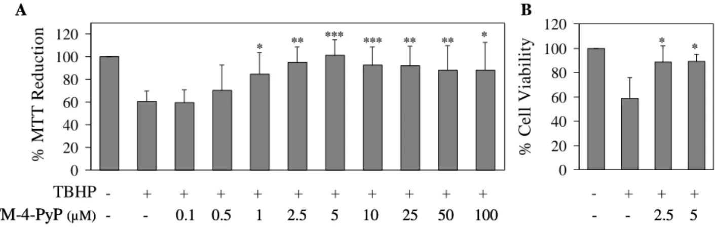
3.3.1. Effects of MnTM-4-PyP 66
3.3.2. Effects of MnTM-4-PyP in XXO-treated cells 68 3.3.3. Effects of MnTM-4-PyP in TBHP-treated cells 70 3.3.4. Effects of MnTM-4-PyP in Dox-treated cells 73
3.3.5. Conclusion 75
3.4. References 77
Chapter 4 - Protective role of ortho-substituted Mn(III)
N-alkylpyridylporphyrins against the oxidative injury induced by
tert-butylhydroperoxide 83
Abstract 84
4.1. Introduction 85
4.2. Materials and Methods 88
4.2.1. Chemicals 88
4.2.2. MTT Reduction assay 88
4.2.3. Crystal Violet assay 88
4.2.4. DHE fluorimetric assay 89
4.2.5. DTNB assay 89
xix
4.2.7. Stability of MnPs 91
4.2.8. Statistical analysis 91
4.3. Results and Discussion 92
4.3.1. Cytotoxicity profile of MnTE-2-PyP and MnTnHex-2-PyP 92 4.3.2. Effects of MnTE-2-PyP and MnTnHex-2-PyP against
TBHP-induced cytotoxicity 92
4.3.3. Effects of MnTE-2-PyP and MnTnHex-2-PyP against
TBHP-induced ROS generation 97
4.3.4. Effects of MnTE-2-PyP and MnTnHex-2-PyP on the changes in
glutathione status induced by TBHP 98
4.3.5. Conclusion 101
4.4. References 102
Chapter 5 - Development of macrocyclic copper(II) complexes: synthesis,
superoxide scavenging activity, structural studies and biological
evaluation 109
Abstract 110
5.1. Introduction 111
5.2. Materials and Methods 113
5.2.1. Chemicals 113
5.2.2. Synthetic procedures 113
5.2.2.1. Synthesis of the macrocycles 113
5.2.2.2. Synthesis of the macrocyclic copper(II) complexes 116
5.2.3. Superoxide scavenging activity 117
5.2.3.1 NBT assay 117
5.2.3.2 DHE assay 118
5.2.3.3. Xanthine oxidase inhibition assay 119
5.2.4. Structural studies 119
xx
5.2.4.2. Electrochemistry 120
5.2.5. Evaluation of the cytotoxicity profile of the macrocyclic
copper(II) complexes in V79 cells 120
5.2.6. Evaluation of potential protective effect of CuL3 and CuL4
against the oxidative injury induced by XXO and by TBHP 121
5.2.6.1. MTT Reduction assay 121
5.2.6.1. Crystal Violet assay 121
5.2.6.3. DHE fluorimetric assay 122
5.2.7. DNA strand break analysis 122
5.2.8. Evaluation of the cytotoxicity profile of the macrocyclic
copper(II) complexes CuL3 and CuL4 in MCF7 cells 123
5.2.8.1. MCF7 Cells culture 123
5.2.8.2. MTT Reduction assay 124
5.2.9. Evaluation of possible role of the macrocyclic copper(II) complexes CuL3 and CuL4 on the potentiation of Dox
cytotoxicity in MCF7 cells 124
5.2.10. Statistical analysis 124
5.3. Results and Discussion 125
5.3.1. Chemical characterization and superoxide scavenging activity of
the complexes 125
5.3.2. Cytotoxicity profile of the complexes 135
5.3.3. Studies on CuL3 and CuL4 as potential antioxidants 136 5.3.4. HO• radical generation by CuL3 and CuL4 139 5.3.5. Studies on CuL3 and CuL4 as potential anticancer agents 142
5.3.6. Conclusion 144
xxi Chapter 6 - Development of pyridine-containing macrocyclic copper(II)
complexes with superoxide scavenging activity. Studies on CuL8 as a
novel chemotherapy sensitizer for breast cancer 153
Abstract 154
6.1. Introduction 155
6.2. Materials and Methods 159
6.2.1. Chemicals 159
6.2.2. Synthesis of the macrocycles 159
6.2.3. Synthesis of the macrocyclic copper(II) complexes 163
6.2.4. Species distribution curves 163
6.2.5. Structural studies 164
6.2.5.1 Spectroscopic studies 164
6.2.5.2. Electrochemistry 164
6.2.6. Superoxide scavenging activity 165
6.2.6.1 NBT assay 165
6.2.6.2 DHE assay 165
6.2.6.3. Xanthine oxidase inhibition assay 165
6.2.7. Cytotoxicity profile of the complexes 165
6.2.7.1. Cell culture 165
6.2.7.2. MTT reduction assay 166
6.2.8. Evaluation of potential protective effect of CuL8 against the
oxidative injury induced by XXO and by TBHP 166
6.2.9. DNA strand break analysis 167
6.2.10. Evaluation of possible role of CuL8 on the modulation of the
cytotoxicity of the anticancer drugs Dox and oxaliplatin 167
6.2.11. Statistical analysis 167
6.3. Results and Discussion 168
6.3.1. Chemical characterization and superoxide scavenging activity of
the complexes 168
xxii
6.3.3. Studies on CuL8 as a potential antioxidant 177 6.3.4. Studies on CuL8 as a potential anticancer agent 178
6.3.5. Conclusion 182
6.4. References 183
xxiii
List of Figures
Page Fig. 1.1 Generation of the most relevant reactive oxygen species. 3 Fig. 1.2 Basic reaction sequence of lipid peroxidation. 4
Fig. 1.3 Enzymatic antioxidant defenses. 6
Fig. 1.4 Catalytic cycle of glutathione peroxidase. 9
Fig. 1.5 Cellular responses to oxidative stress. 11
Fig. 1.6 Different roles of O2•– in inflammation. 12
Fig. 1.7 Redox diagram for O2•– oxidation and reduction and the placement of
some SODm on it. 16
Fig. 1.8 Chemical structures of some representative MnPs. 20 Fig. 1.9 Chemical structures of four representative salen-manganese complexes. 21 Fig. 1.10 Chemical structure of four representative Mn(II) cyclic polyamines. 23
Fig. 1.11 Chemical structures of Tempol and PBN. 27
Fig. 1.12 Schematic representation of the relationship between intracellular H2O2
levels and cell proliferation. 33
Fig. 3.1 Generation of O2 •–
by the xanthine – xanthine oxidase system. 57 Fig. 3.2 Chemical structure of tert-butylhydroperoxide. 58
Fig. 3.3 Doxorubicin redox-cycling. 58
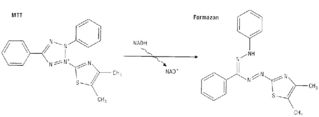
Fig. 3.4 Chemical structure of the Mn(III) porphyrin MnTM-4-PyP. 59 Fig. 3.5 MTT reduction in live cells by mitochondrial reductase results in the
formation of a formazan derivative. 62
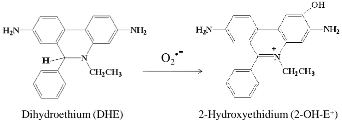
Fig. 3.6 Reaction of dihydroethidium with O2•–, originating the fluorescent
product 2-hydroxyethidium. 64
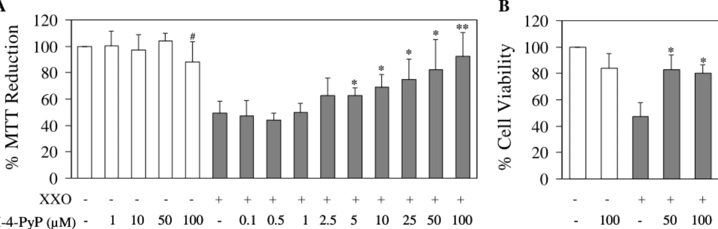
Fig. 3.7 Effect of MnTM-4-PyP on the cytotoxicity induced by xanthine (240 M) plus xanthine oxidase (20 U/L) in V79 cells. 66 Fig. 3.8 Effect of MnTM-4-PyP on the intracellular superoxide anion levels, in
the absence or presence of xanthine (240 M) plus xanthine oxidase (100 or 200 U/L), as evaluated by the oxidation of DHE. 68 Fig. 3.9 Effect of MnTM-4-PyP on the cytotoxicity induced by TBHP (100 M)
xxiv
Fig. 3.10 Effect of MnTM-4-PyP on the cytotoxicity induced by TBHP in V79
cells, as evaluated by the MTT assay. 71
Fig. 3.11 Effect of MnTM-4-PyP on the intracellular superoxide anion levels in cells treated with TBHP (1.0 or 2.0 mM), as evaluated by the oxidation
of DHE. 72
Fig. 3.12 Effect of MnTM-4-PyP on the cytotoxicity induced by Dox in V79 cells. 73 Fig. 3.13 Effect of MnTM-4-PyP on the cytotoxicity induced by Dox in V79 cells,
as evaluated by the MTT assay. 74
Fig. 3.14 Effect of MnTM-4-PyP on the superoxide anion levels in cells treated with Dox (2.5, 5 or 10 M), as evaluated by the oxidation of DHE. 75 Fig. 4.1 Chemical structures of the Mn(III) porphyrins MnTE-2-PyP and
MnTnHex-2-PyP. 86
Fig. 4.2 Fundamentals of the DTNB assay. 89
Fig. 4.3 Fundamentals of the mCB assay. 91
Fig. 4.4 Cytotoxicity evaluation of MnTE-2-PyP (A) and MnTnHex-2-PyP (B). 92 Fig. 4.5 Effect of MnTE-2-PyP on the cytotoxicity induced by TBHP (100 M)
in V79 cells. 93
Fig. 4.6 Effect of MnTE-2-PyP on the cytotoxicity induced by TBHP in V79
cells, as evaluated by the MTT assay. 93
Fig. 4.7 Effect of MnTnHex-2-PyP on the cytotoxicity induced by TBHP (100
M) in V79 cells. 95
Fig. 4.8 Effect of MnTnHex-2-PyP on the cytotoxicity induced by TBHP in V79
cells, as evaluated by the MTT assay. 95
Fig. 4.9 Effect of MnTE-2-PyP (A) and MnTnHex-2-PyP (B) on the DHE oxidation in V79 cells treated with TBHP (100 µM). 97 Fig. 4.10 Effect of MnTE-2-PyP and MnTnHex-2-PyP (5 µM) on the glutathione
depletion induced by TBHP (100 µM) in V79 cells, as evaluated by the
mCB assay. 100
Fig. 5.1 Potential activity of Cu(II) complexes (CuL) with SOD-like activity as
boosters of Doxorubicin anticancer properties. 112
Fig. 5.2 Schematic representation of the synthesis of the ligands L1 and L2. 114 Fig. 5.3 Schematic representation of the synthesis of the ligands L3 and L4. 115 Fig. 5.4 Schematic representation of the synthesis of the ligand L5. 116
xxv
Fig. 5.5 Fundamentals of the DNA strand break analysis. 122 Fig. 5.6 Effect of the macrocyclic copper(II) complexes CuL1- CuL5 on the
inhibition of the NBT reduction (A) and DHE oxidation (B) by the XXO
generated superoxide. 128
Fig. 5.7 EPR X-band spectra of the Cu(II) complexes of L2-L5. 131
Fig. 5.8 B3LYP Optimized geometry of CuL2 complex. 132
Fig. 5.9 B3LYP Optimized geometry of CuL3 complex. 133
Fig. 5.10 B3LYP Optimized geometry of CuL4 complex. 133
Fig. 5.11 B3LYP Optimized geometry of CuL5 complex. 134
Fig. 5.12 Cell viability of V79 cells treated with different concentrations of the
macrocyclic copper(II) complexes CuL1-CuL5 and Cu(II), for 24 h. 135 Fig. 5.13 Effect of CuL3 and CuL4 on the cytotoxicity induced by xanthine (240
M) plus xanthine oxidase (20 U/L) in V79 cells. 136 Fig. 5.14 Effect of CuL3 on the cytotoxicity induced by TBHP (100 M) in V79
cells. 137
Fig. 5.15 Effect of CuL3 on the DHE oxidation in V79 cells treated with TBHP
(100 µM). 138
Fig. 5.16 Effect of CuL4 on the cytotoxicity induced by TBHP (100 M) in V79
cells. 139
Fig. 5.17 Validation of the DNA strand break analysis protocol. 140 Fig. 5.18 Evaluation of HO• generation by CuL3 (A) and CuL4 (B), by the DNA
strand break analysis. 141
Fig. 5.19 Cell viability of MCF7 cells treated with different concentrations of
CuL3 and CuL4, for 24 h. 143
Fig. 5.20 Effect of CuL3 and CuL4 on the cell viability of MCF7 cells treated
with doxorubicin (Dox), as evaluated by the MTT assay. 143 Fig. 6.1 Potential activity of Cu(II) complexes (CuL) with SOD-like activity as
redox modulators of oxaliplatin cytotoxicity. 157
Fig. 6.2 Schematic representation of the synthesis of the ligand L6. 160 Fig. 6.3 Schematic representation of the synthesis of the ligand L7. 161 Fig. 6.4 Schematic representation of the synthesis of the ligands L8 and L9. 162
xxvi
Fig. 6.5 Effect of the macrocyclic copper(II) complexes CuL6-CuL9 on the inhibition of NBT reduction (A) and DHE oxidation (B) by the XXO
generated superoxide. 172
Fig. 6.6 Cell viability of V79 cells treated with different concentrations of the
complexes CuL6-CuL9, as evaluated by the MTT assay. 175 Fig. 6.7 Cell viability of MCF10A cells treated with different concentrations of
the complexes CuL6-CuL9, as evaluated by the MTT assay. 176 Fig. 6.8 Cell viability of MCF7 cells treated with different concentrations of the
complexes CuL6-CuL9, as evaluated by the MTT assay. 176 Fig. 6.9 Effect of CuL8 on the cytotoxicity induced by xanthine (240 M) plus
xanthine oxidase (20 U/L) (A) or by TBHP (100 M) (B), in V79 cells. 178 Fig. 6.10 Evaluation of HO• generation by CuL8, by the DNA strand break
analysis. 179
Fig. 6.11 Effect of CuL8 on the cell viability of human mammary cells treated
with doxorubicin (Dox), as evaluated by the MTT assay. 180 Fig. 6.12 Effect of CuL8 on the cell viability of human mammary cells treated
xxvii
List of Tables
Page
Table I.1 Types of SOD enzymes in mammalians 7
Table I.2 Clinical conditions in which the involvement of superoxide anion has
been suggested 13
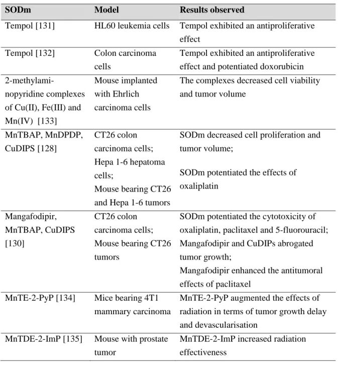
Table I.3 Examples of protective effects reported using SODm in cell-based
models 29
Table I.4 Examples of protective effects reported using SODm in animal models 30 Table I.5 Examples of protective effects shown by SODm against adverse effects
of chemotherapy and radiotherapy 32
Table I.6 Examples of studies focusing on the potential of SODm as anticancer
agents 34
Table III.1 Mitotic indices presented by V79 cells treated with the oxidants under study, in the absence or presence of MnTM-4-PyP 67 Table IV.1 Effect of MnTE-2-PyP and MnTnHex-2-PyP on the changes in
glutathione status induced by TBHP in V79 cells, as evaluated by the
DTNB assay 99
Table V.1 Stepwise stability constants (log units) for the copper(II) complexes of
L1-L5 126
Table V.2 Species distribution and pCu values calculated for an aqueous solution containing Cu(II) (10 µM) and each ligand (10.5 µM) at a molar ratio
of 1:1 127
Table V.3 Superoxide scavenging activity for the copper(II) compounds and
native Cu,Zn-SOD 128
Table V.4 Spectroscopic X-band EPR data for the Cu(II) complexes of L2-L5 and
Cu,Zn-SOD 131
Table VI.1 Stepwise stability constants (log units) and pCu values for copper(II)
complexes 169
Table VI.2 Spectroscopic data for the Cu2+ complexes of L6-L9 170 Table VI.3 Cyclic voltammetric data for the copper(II) complexes of L8 and L9 171 Table VI.4 Superoxide scavenging activity for the copper(II) compounds and
xxix
Abbreviations
AP-1 activator protein 1
B3LYP Becke 3-Parameter, Lee, Yang and Parr (hybrid functional)
CAT catalase
CuDIPs copper(II) 3',5'-diisopropylsalicylate
CuL1 copper(II) complex of 1-oxa-4,7-diazacyclononane (Cu[9]aneN2O)
CuL2 copper(II) complex of 1-oxa-4,7-diazacyclononane-4,7-diacetic acid; Cu(N-ac2[9]aneN2O)
CuL3 copper(II) complex of 1-oxa-4,7, 10,
13-tetraazacyclopentadecane; Cu([15]aneN4O)
CuL4 copper(II) complex of 1-oxa-4,7,10,13-tetraazacyclopentadecane-4,7,10,13-tetraacetic acid ; Cu(N-ac4[15]aneN4O)
CuL5 copper(II) complex of 1,4-dioxa-7, 10, 13-triazacyclopentadecane; Cu([15]aneN3O2)
CuL6 copper(II) complex of 7-methyl-3,7,11,17-tetraazabi- cyclo[11.3.l]heptadeca-1(17),13,15-triene; Cu(N-Mepy[14]aneN4)
CuL7 copper(II) complex of 3,7,11-tris(carboxymethyl)-3,7,11,17-tetraazabicyclo[11.3.l]heptadeca-1(17),13,15-triene;
Cu(N-ac3py[14]aneN4)
CuL8 copper(II) complex of 3,6,9,12,18-pentaaza-
bicyclo[12.3.1]octadeca-1(18),14,16-triene; Cu(py[15]aneN5)
CuL9 copper(II) complex of 3,6,10,13,19-pentaaza-
bicyclo[13.3.1]nonadeca-1(19),15,17-triene; Cu(py[16]aneN5)
CuPs copper(II) porphyrins
Cu(py[14]aneN4) copper(II) complex of
3,7,11,17-tetraazabicyclo[11.3.l]heptadeca-1(17),13,15-triene
Cu,Zn-SOD copper, zinc-superoxide dismutase (SOD1)
CV crystal violet
DHE dihydroethidium
DMEM Dulbecco’s Modified Eagle’s Medium DMEM/F12 DMEM/Nutrient Mixture F-12 Ham
xxx
DMSO dimethylsulfoxide
Dox doxorubicin
DTNB 5,5’-dithiobis(2-nitrobenzoic acid)
E1/2 half wave potential
EC-SOD extracellular superoxide dismutase (SOD3) edta ethylenediaminetetraacetic acid
EPR electronic paramagnetic resonance
FePs iron(III) porphyrins
FOLFOX folinate, oxaliplatin, 5-fluorouracil regimen γ-GCS γ-glutamylcysteine synthetase
G-6-PDH glucose-6-phosphate dehydrogenase
GPx glutathione peroxidase
GR glutathione reductase
GSH reduced glutathione
GSHt total glutathione content GSSG oxidised glutathione GST glutathione-S-transferase
4-HNE 4-hydroxynonenal
HIF-1α hypoxia-inducible factor 1α
ICD NADP+-dependent isocitrate dehydrogenase
IL-1β interleukin-1β
IL-6 interleukin-6
LPO lipid peroxidation
mCB monochlorobimane
MDA malondialdehyde
MI mitotic index
MnSOD manganese-superoxide dismutase (SOD2)
MnPs manganese (Mn) porphyrins
MnTDE-2-ImP Mn(III) 5,10,15,20-tetrakis(N,N'-diethylimidazolium-2-yl)- porphyrin
MnTBAP Mn(III) meso-tetrakis(4-carboxyphenyl)porphyrin
MnTE-2-PyP5+ Mn(III) 5,10,15,20-tetrakis(N-ethylpyridinium-2-yl)porphyrin MnTM-4-PyP5+ Mn(III) 5,10,15,20-tetrakis(N-methylpyridinium-4-yl)porphyrin
xxxi
MnTnHex-2-PyP5+ Mn(III) 5,10,15,20-tetrakis(N-n-hexylpyridinium-2-yl)porphyrin MTT thiazolyl blue tetrazolium bromide
NADH nicotinamide adenine dinucleotide
NADPH nicotinamide adenine dinucleotide phosphate NBT nitro blue tetrazolium
NF-kB nuclear factor kB
NHE normal hydrogen electrode NMR nuclear magnetic resonance
PARP poly-ADP-ribose polymerase
PBS phosphate buffered saline
PBN α-phenyl-tert-butylnitrone
pCu -log [Cu2+]
PHGPx phospholipid hydroperoxide glutathione peroxidase PLED dipyridoxyl ethylenediamine diacetate
RFU relative fluorescence units RNS reactive nitrogen species
ROS reactive oxygen species
RS reactive species
salen N,N’-bis-(salicylideneamino)ethane
SOD superoxide dismutase
SODm superoxide dismutase mimetics TBHP tert-butylhydroperoxide
TNB 5-thio-2-nitrobenzoic acid TNFα tumor necrosis factor α
XO xanthine oxidase
XXO xanthine-xanthine oxidase system
1
Chapter 1
2
1.1. Oxidative stress and reactive oxygen species
The term “oxidative stress” can be defined as an imbalance between oxidants and antioxidants in favor of the oxidants, potentially leading to damage [1]. Oxidants are formed as normal products of aerobic metabolism and their production is balanced by the antioxidant defenses [1]. Under pathophysiological conditions, due to an increased production of oxidants or to a failure of antioxidant defenses, oxidative damage may occur [2]. Oxidative damage seems to be implicated in several pathological processes including tissue injury, inflammatory disorders, cardiovascular diseases, pulmonary diseases, neurodegenerative diseases, and cancer [3, 4].
The collective term reactive species (RS) includes oxygen, nitrogen, chlorine, bromine and sulphur transient species with high chemical reactivity [4, 5]. These include free radicals, i.e., species containing one or more unpaired electrons, and non-radical derivatives [4]. Reactive oxygen species (ROS) encompass a variety of diverse chemical species including superoxide anion (O2•–), hydroxyl radical (HO•), hydrogen
peroxide (H2O2), singlet oxygen (1O2), alkoxyl radicals (RO •
), and peroxyl radicals (ROO•) [4-6]. Some ROS, such as O2
•–
or HO•, are extremely unstable and reactive. In contrast, other ROS like H2O2 or ROO• are relatively stable, with half-lives in the range
of seconds. These species may diffuse away from their site of generation, transporting the radical or oxidant function to other target sites [1, 6]. Reactive nitrogen species (RNS), such as peroxynitrite (ONOO-), are also relevant in numerous pathophysiological phenomena [7, 8]. Fig. 1.1 shows the generation pathways of ROS. Superoxide anion is formed in biological systems by the partial reduction of molecular oxygen. Reduction of O2•– with a second electron, as well as a two-electron reduction of
O2, generates O22-, which leads to H2O2. The one-electron reduction of H2O2, that
occurs in the presence of reduced transition metals like Cu(I) and Fe(II), originates HO• [9, 10]. Other reactive species can be generated by the reaction of ROS with biological molecules (e.g. polyunsaturated lipids, thiols and nitric oxide (NO)) [3]. For example, the reaction between O2•– and NO originates ONOO-, which is unstable at physiological
pH and rapidly decomposes to form potent nitrating and oxidizing species [3, 4]. Other species that result from the reaction of RS with biomolecules are the radicals alkoxyl and peroxyl [11]. These can be generated by a variety of routes, including the reaction
3
of lipid peroxides with HO2•, the breakdown of organic peroxides, and the reaction of
RS with lipids or amino acid radicals [4, 11].
Fig. 1.1 – Generation of the most relevant reactive oxygen species (adapted from [3]).
ROS can be generated as a result of normal intracellular metabolism. The majority of intracellular ROS production is derived from the mitochondria [6]. In fact, O2•– is formed from the uncoupling of the mitochondrial electron transport chain during
oxidative phosphorylation [3, 10]. Also, the catalytic action of a variety of intracellular and extracellular oxidases like NADPH oxidase, xanthine oxidase or lipooxygenase, as well as the metabolism of arachidonic acid, gives rise to O2•– [4, 7]. Superoxide can also
be produced by the phagocytic NADPH oxidase, during host defense responses, where O2•– is thought to act in cell-signaling and in the killing of foreign bacteria [3, 8, 10].
Other ROS are also generated in peroxisomes, as well as from a variety of cytosolic enzyme systems [6, 10]. Other process closely related with ROS formation is the lipid peroxidation (LPO), i.e., the oxidative deterioration of polynsaturated lipids (Fig. 1.2) [4]. LPO is initiated by addition of a RS (e.g. HO•, HO2
•
, RO•, ROO•) to an unsaturated lipid or by hydrogen abstraction from a methylene group by a RS, forming a carbon radical. Carbon radicals often stabilize by molecular rearrangement generating conjugated dienes. Carbon radicals can also react with O2, giving lipid peroxyl radicals
O
2O
2•–H
2O
2HO
•ONOO
–NO
ROS + Biomolecules1e
–1e
–2 e
–1e
–RO
•ROO
•2 H
+2 H
+2
4
(LOO•). These radicals can abstract H• from an adjacent lipid, propagating the process. The combination of LOO• with H• generates a lipid hydroperoxide (LOOH). The decomposition of LOOH in the presence of transition metal ions produces lipid alkoxyl radicals (LO•) that may also abstract H• from unsaturated lipids, continuing the propagation of LPO. These phenomena may occur in biological membranes, leading to oxidative damage. In fact, the continued oxidation of fatty-acid side chains and their fragmentation to produce aldehydes and hydrocarbons may lead to loss of membrane integrity. In addition, products of lipid peroxidation (e.g. isoprostanes, malondialdehyde, 4-hydroxynonenal) may exert themselves deleterious effects [4], namely DNA damage and inhibition of proteins, leading to cytotoxicity, allergy, mutagenicity, and carcinogenicity phenomena [12].
Fig. 1.2 – Basic reaction sequence of lipid peroxidation (adapted from [4, 13, 14]). (LH, lipid; RS, reactive species; L•, carbon-centered radical; LOO•, lipid peroxyl radical;
LOOH, lipid
hydroperoxide; LO•, lipid alkoxyl radical; LOH,
alcohol; MDA, malondialdehyde; 4-HNE, 4-hydroxynonenal). LH H.abstraction or RS addition L. Molecular rearrangement L. O2 LOO. LOOH LO. LOH L. LH Transition metals MDA, 4-HNE L. LH
In
iti
ati
on
P
r
op
agati
on
5
Despite the endogeneous sources of ROS, a number of external agents can trigger ROS production [6]. Different types of radiation, like X-rays, ultraviolet, ultrasound, microwave, and ionizing radiation are known to generate ROS [1, 6]. Also, chemotherapeutic agents, hyperthermia, inflammatory cytokines and environmental toxins can shift cells into a state of oxidative stress [6].
Although high concentrations of ROS trigger oxidative damage, low concentrations of ROS are needed to regulate several key physiological processes [9, 10, 15]. These include cell differentiation, cell proliferation and apoptosis that are regulated by redox-sensitive signal transduction pathways [9, 10, 15].
1.1.1. Antioxidant defences
The production of ROS is counteracted by an intricate antioxidant defense system [6, 9]. An antioxidant has been defined as a substance that, when present at low concentrations compared with those of an oxidizable substrate, significantly delays or prevents oxidation of that substrate [2, 4, 11]. More recently, Halliwell and Gutteridge [4] altered this definition in order to account chaperones, repair systems and inhibitors of RS generation. An antioxidant is presently defined as any substance that delays, prevents or removes oxidative damage to a target molecule [4]. This concept includes non-enzymatic compounds, as well as antioxidant enzymes [1].
Non-enzymatic antioxidant defenses comprise a number of low molecular weight molecules with the ability to scavenge ROS [6, 10]. These include compounds synthesized in vivo (e.g. glutathione, bilirubin, pyruvate, melatonin, coenzyme Q, uric acid), as well as agents obtained from the diet (e.g. ascorbate, tocopherol, carotenoids, flavonoids) [4].
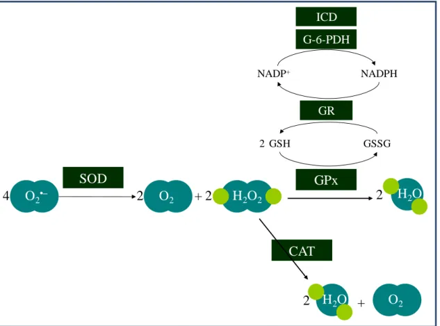
Eukaryotic cells possess an efficient antioxidant enzymatic network, as depicted in Fig. 1.3. The three major classes of antioxidant enzymes are the superoxide dismutases (SOD), catalases (CAT) and glutathione peroxidases (GPx) [1].
6
Fig. 1.3 – Enzymatic antioxidant defenses: role of superoxide dismutases (SOD), catalases (CAT), glutathione peroxidases (GPx), glutathione reductase (GR), glucose-6-phosphate dehydrogenase (G-6-PDH), and NADP+-dependent isocitrate dehydrogenase (ICD) on ROS detoxification (adapted from [9, 15]).
Superoxide dismutase (SOD)
Superoxide dismutases, which enzymatic activity was discovered in 1969 by McCord and Fridovich, are metalloproteins with oxido-reductase capacity that catalyze the dismutation of O2•– [16]. A dismutation reaction is defined as a reaction in which
two like-molecules react to produce two different products (i.e. A + A → B + C) [3]. In the case of SOD, oxygen and H2O2 are formed from two O2
•– [3], as shown in the following equations: O2•– + M(n+1)+ → O2 + Mn + (Eq 1) O2•– + Mn+ + 2H+ → H2O2 + M(n+1)+ (Eq 2) Net reaction: 2O2•– → O2 + H2O2 O2•– O2 H2O2 H2O O2 SOD CAT GPx GR G-6-PDH GSH GSSG NADPH NADP+ H2O + ICD 4 2 + 2 2 2 2
7
The oxidized form of the metalloenzyme SOD (M(n+1)+) reacts with one O2•– to
form O2 and generate the reduced form of the enzyme (Mn+) (Eq 1). In the second step
of the dismutation reaction, the reduced enzyme reacts with another O2•– and two
protons to form H2O2, regenerating the oxidized form of the enzyme (Eq 2).
Three different isoforms of SOD have been characterized in mammals. The SOD enzymes have a distinct genomic structure and are well compartmentalized [8]. Table I.1 summarizes the differences between the three SOD isoforms.
Table I.1 – Types of SOD enzymes in mammals [4, 5, 8].
SOD1 SOD2 SOD3 (EC-SOD)
Active center Cu(II)/(I) and Zn(II) Mn(III)/(II) Cu(II)/(I) and Zn(II) Protein
Structure Homodimer Homotetramer Tetrameric glycoprotein
Genetic locus 21q22.1 6q25.3 4p15.3-p15.1 Location Cytosol, nuclear compartments and mitochondrial inter-membrane space Mitochondria matrix Extracellular compartments (plasma, lymph, synovial fluid)
The involvement of the three SOD isoforms in several pathological conditions has been unraveled by modulating the expression of the enzymes using knockout and transgenic models [8]. Down-regulation of SOD1 has been associated with neuronal death, reduced fertility and increased susceptibility to paraquat toxicity [5, 8]. On the other hand, the overexpression of this enzyme in transgenic mice was shown to protect the cerebral tissue in pathological conditions such as ischemia or Parkinson’s disease [8]. Loss or reduction of the SOD2 activity has been associated with neurodegeneration, heart failure, and dilated cardiomyopathy [5, 8, 12]. The importance of SOD2 is also highlighted by the fact that, in contrast to SOD1 and SOD3, the SOD2 knock-out mice do not survive past 3 weeks of age [5, 12]. Furthermore, SOD2 gene has several polymorphisms which result in a reduction of the enzyme activity and were shown to be
8
associated with an increased risk of sporadic motor neuron disease, nonfamilial idiopathic cardiomyopathy, breast cancer and reduction of the tumour-suppressive effect of SOD2 [8, 17]. Studies with SOD3 knockout mice have shown that the loss of this enzyme activity is related to an impaired spatial learning and an increased sensitivity to hyperoxia exposure [8]
Further classes of SOD have been identified, namely nickel-containing SODs in
Streptomyces sp. and some cyanobacteria, and iron-containing SODs in bacteria, algae,
trypanosomes and higher plants [4, 5].
Catalase (CAT)
CAT enzymes are present in the peroxisomes of most aerobes and catalyze the direct decomposition of H2O2 to ground-state O2 [4]. In animals, CAT is present in all
organs and it is most concentrated in liver, kidney and erythrocytes [4, 18]. Human CAT is a tetrameric haemin-enzyme consisting of four identical subunits of 60 kDa, each containing Fe(III)-haem at its active site [4, 5, 18]. Its gene is located in chromosome 11, band p13 [18].
CAT enzymes catalyze the dismutation of H2O2. In fact, H2O2 oxidizes the heme
iron of the resting enzyme to form an oxyferryl group with a π-cationic porphyrin radical, termed Compound I (Eq 3). This step is followed by oxidation of a second molecule of H2O2 by Compound I (Eq 4) [19]. The catalatic rate of mammalian CAT is
among the highest of known enzymatic rates, and it is simply proportional to H2O2
concentration over a wide range of concentrations [19].
CAT (Por-FeIII) + H2O2 → Compound I (Por•+-FeIV=O) + H2O (Eq 3)
Compound I (Por•+-FeIV=O) + H2O2 → CAT (Por-FeIII) + H2O + O2 (Eq 4)
9
Glutathione peroxidases (GPx)
Peroxidases remove H2O2 by using it to oxidize another substrate [4]. GPx
are selenium-containing peroxidases that degrade a variety of peroxides, namely H2O2
(Eq 5) and ROOH (Eq 6), by coupling their reduction with the oxidation of reduced glutathione (GSH) [4, 5].
2 H2O2 + 2 GSH → GSSG + 2 H2O (Eq 5)
ROOH + 2 GSH → GSSG + H2O + ROH (Eq 6)
GPx enzymes are widely distributed in animal tissues [4]. Four isoenzymes have been identified in humans, being the level of each isoform dependent of the tissue type [4, 5]. The classical GPx (GPx1) is cytosolic. GPx2 is an isoform present in the gastro-intestinal tract. In plasma and other extracellular fluids, a different isoenzyme (GPx3) is found. The fourth type is the phospholipid hydroperoxide glutathione peroxidase (PHGPx or GPx4), which has the ability to act upon peroxidized fatty acid residues within membranes and lipoproteins [4]. GPx4 is a monomer, while GPx1, GPx2 and GPx3 are tetramers. Each protein unit contains a selenium atom in the active site, in the form of selenocysteine. The catalytic activity of GPx is schematized in Fig. 1.4. During GPx catalysis, the selenol form of the enzyme is oxidized to selenenic acid by peroxides. This form reacts with reduced glutathione (GSH) to give H2O and a
selenosulphide adduct. A second GSH molecule then regenerates the active form of the enzyme by attacking the selenosulphide to form oxidized glutathione (GSSG) [4, 20].
Fig. 1.4 – Catalytic cycle of glutathione peroxidase (adapted from [20]). Selenosulphide adduct Enzyme-Se-SG Selenol Enzyme-SeH Selenenic acid Enzyme-SeOH GSH GSSG GSH H2O ROH ROOH
10
The catalytic activity of GPx involves the oxidation of GSH in GSSG. However, in physiological conditions, the cellular ratio GSH/GSSG is high, because GSSG is reduced back to GSH by glutathione reductase (GR) [1, 4]. The catalytic activity of this enzyme requires NADPH, according to the following reaction (Eq 7):
GSSG + NADPH + H+ → 2 GSH + NADP+ (Eq 7)
Cellular NADPH can be provided by different sources, including Glucose-6-phosphate dehydrogenase (G-6-PDH) and NADP+-dependent isocitrate dehydrogenase (ICD) (Fig. 1.3) [4].
In addition to being a cofactor for GPx enzymes, GSH plays other important antioxidant roles. This ubiquitous thiol-containing tripeptide can directly scavenge RS, such as HO• and 1O2 [10, 21]. Moreover, it is used by glutathione S-transferases (GSTs)
to conjugate and eliminate reactive compounds, including products formed in vivo during oxidative stress [1, 4, 10, 21, 22]. Glutathione is also able to regenerate important antioxidants (e.g. Vitamins C and E) back to their active forms, being this capacity linked with the redox state of the glutathione disulphide-glutathione couple (GSSG/2GSH) [10]. This redox couple is a major contributor to the redox state of the cellular milieu. In a non-stressed cell, glutathione is present in the cytosol at a concentration between 1 and 10 mM, being ~99% in the form of GSH and ~1% as GSSG [4, 21].
11
1.1.2. Consequences of oxidative stress
A situation of oxidative stress, due to diminished antioxidant defenses or to an increase in the production of RS, may induce different cellular responses. These responses depend on the cell type and the severity of oxidative stress. As summarized in Fig. 1.5 mild oxidative stress may induce cell proliferation, while intense oxidation will result in cell injury and can even trigger cell death [4].
Fig. 1.5 – Cellular responses to oxidative stress. Many cells respond to mild oxidative stress by proliferating. As oxidative stress increases, cells may up-regulate their defense systems as an adaptive response. Greater oxidative stress will lead to damage in biomolecules such as DNA, proteins, lipids, or carbohydrates, resulting in oxidative cell injury. Cells may recover from this damage by repairing it or displacing the damaged molecules, or they can survive with persistent lesions. In case of intense oxidative stress, cells may become senescent, i.e., they survive but are no longer able to divide. Under highly oxidizing conditions, mechanisms of cell death (apoptosis and necrosis) may be activated [4]. Resting cell Increased proliferation Adaptation Cell injury Senescence Cell death Repair Survive with persistent oxidative damage
Inc re a si ng oxi da ti ve s tre ss
Activation of transcription factors Upregulation of defense systems
12
Pro-inflammatory action of superoxide
Under physiological circumstances, the levels of O2•– are kept under tight control by
endogenous SOD [7]. However, in acute and chronic inflammation, the production of O2•– is increased at a rate that may overwhelm the capacity of the endogenous SOD to
remove it. This imbalance results in superoxide-mediated damage [7, 8]. Some important pro-inflammatory roles for O2•– have been described [5, 7, 8] (Fig. 1.6),
namely:
endothelial cell damage and increased microvascular permeability; formation of chemotactic factors;
recruitment of neutrophils at sites of inflammation;
auto-catalytic destruction of neurotransmitters and hormones; lipid peroxidation and oxidation;
DNA single-strand damage and activation of poly-ADP-ribose polymerase (PARP); formation of peroxynitrite, a potent cytotoxic RS that can nitrate and deactivate SOD
and that causes the inactivation of nitric oxide;
reduction of Fe(III) to Fe(II) with its consequent release from storage sites, so that it can react with H2O2 producing HO• radicals.
Fig. 1.6 – Different roles of O2•– in inflammation. Excessive production of O2•– can lead
to inflammation through various pathways, including the depletion of beneficial NO and the generation of deleterious ONOO- (adapted from [7, 8]).
O2•– + NO ONOO -Formation of chemotactic factors Citokines release Neurotransmitter and hormone inactivation ↑ Adhesion Molecules Lipid peroxidation DNA damage Citotoxicity Receptor inactivation (e.g. steroids) Enzyme inactivation (e.g. MnSOD) PARP activation
13
These phenomena lead to tissue injury and inflammation that are involved in many diseases. Table I.2 presents some pathological conditions in which the involvement of superoxide anion has been suggested.
Table I.2 – Clinical conditions in which the involvement of superoxide anion has been suggested [7, 8, 23].
Category Examples
Inflammatory conditions Pain, Crohn’s disease, osteoarthritis, dermatitis, psoriasis Cardiovascular diseases Ischemia-reperfusion injury, shock, atherosclerosis,
stroke
Neurodegenerative diseases Parkinson’s, Alzheimer, amiotrophic lateral sclerosis Pulmonary diseases Asthma, hyperoxic lung damage, chronic obstructive
pulmonary disease
Oncology Side effects of chemotherapy and radiotherapy Reproductive system Erectile dysfunction, infertility
Additionally to the phenomena described above, O2•– also seems to be involved in
the processes of cell transformation, metastasis and angiogenesis [16].
The exposure to a number of xenobiotics may also result in the release and/or generation of O2•–, being their toxic effects related with oxidative stress. This O2•–
generation from xenobiotics can occur directly or upon metabolization, redox reactions, redox cycling processes, via lipid peroxidation or by stimulation of endogenous production of O2•–. Examples of xenobiotics whose effects seem to be somehow related
with O2•– are the pesticides paraquat and pentachlorophenol, and the transition metal
vanadium, pointing out a role for O2•– in occupational toxicology. There are also several
drugs (e.g. gentamicin, anthracyclines and rifamycin) whose action and toxic effects are, at least partially, related to O2•– generation [4, 24].
The removal of O2•– provides thus a unique strategy to manipulate numerous
pathological processes, being a very promising approach to the treatment of a variety of diseases and intoxications in which O2•– plays a deleterious role.
14
1.2. Superoxide dismutase mimetics
Some therapeutic approaches have emerged based on the fact that the removal of O2•– modulates the course of numerous pathological processes. Efforts have been made
towards a clinical use of SOD. A different strategy, that has shown very promising results, relies on the development of synthetic compounds with the capacity to mimic the native enzyme - SOD mimetics (SODm).
Protective and beneficial roles of SOD have been demonstrated in a broad range of diseases, both in preclinical and in clinical studies [8, 16]. Preclinical studies have revealed a protective role of SOD in animal models of a variety of pathological conditions, including: ischemia-reperfusion injury, transplant-induced reperfusion injury, inflammation, cancer, AIDS and pulmonary disorders [7]. Moreover, the overexpression of SOD in animal models has provided protection against the deleterious effects of a wide range of oxidative stress paradigms, such as stroke or Parkinson's disease [3, 7].
In terms of clinical data, a Cu,Zn-SOD prepared from bovine tissues – Orgotein, used to be applied as a human therapy in inflammatory conditions, namely in rheumatoid arthritis, osteoarthritis, and against the side effects associated with chemotherapy and radiotherapy [7]. Despite the encouraging anti-inflammatory properties demonstrated, Orgotein was responsible for immunological problems and was withdrawn from the market [7]. Besides this problem of antigenicity, the use of native SOD as a therapeutic agent presents high-manufacturing costs and limitations related with the large size of these proteins that limit their cell permeability and circulating half-life [3]. Moreover, SOD presents a bell-shaped dose-response curve, making difficult the precise restoration of optimal balance between O2•– and SOD [16].
Various attempts at modifying the SOD enzymes have been performed in order to improve their properties. These include the delivery of SOD by liposomes, the development of SOD conjugates, and the genetic engineering of the human proteins [4, 5, 16].
SOD mimetics constitute a different and promising strategy to remove of O2•–,
overcoming the limitations of the clinical use of native SOD. A SODm can be defined as a small synthetic molecule that achieve the destruction of O2•– at a rate of tens of
![Fig. 1.1 – Generation of the most relevant reactive oxygen species (adapted from [3])](https://thumb-eu.123doks.com/thumbv2/123dok_br/15134841.1011240/36.892.173.725.209.575/fig-generation-relevant-reactive-oxygen-species-adapted.webp)
![Fig. 1.2 – Basic reaction sequence of lipid peroxidation (adapted from [4, 13, 14])](https://thumb-eu.123doks.com/thumbv2/123dok_br/15134841.1011240/37.892.124.523.482.1117/fig-basic-reaction-sequence-lipid-peroxidation-adapted.webp)
![Fig. 1.4 – Catalytic cycle of glutathione peroxidase (adapted from [20]). Selenosulphide adduct Enzyme-Se-SGSelenolEnzyme-SeH Selenenic acidEnzyme-SeOHGSHGSSG GSHH2OROHROOH](https://thumb-eu.123doks.com/thumbv2/123dok_br/15134841.1011240/42.892.138.489.869.1144/catalytic-glutathione-peroxidase-selenosulphide-sgselenolenzyme-selenenic-acidenzyme-seohgshgssg.webp)