Modulation of the immune response by
Fonsecaea pedrosoi
morphotypes in the
course of experimental chromoblastomycosis
and their role on inflammatory response
chronicity
Isaque Medeiros Siqueira1, Raffael Ju´nio Arau´jo de Castro1, Luiza Chaves de
Miranda Leonhardt2, Ma´rcio Sousa Jeroˆnimo2, Aluı´zio Carlos Soares3, Taina´ Raiol4, Christiane Nishibe5, Nalvo Almeida5, Aldo Henrique Tavares2, Christian Hoffmann2,6,
Anamelia Lorenzetti Bocca2*
1Molecular Pathology Post-Graduate Program, School of Medicine; University of Brası´lia, Brası´lia, Brazil, 2Department of Cell Biology, Institute of Biological Sciences; University of Brası´lia, Brası´lia, Brazil, 3University Hospital, University of Brası´lia, Brası´lia, Brazil,4Institute Leoˆnidas and Maria Deane, Oswaldo Cruz Foundation, Manaus, Brazil,5School of Computing Sciences, Federal University of Mato Grosso do Sul, Campo Grande, Brazil,6Department of Food Sciences and Experimental Nutrition, School of Pharmaceutical Sciences, University of São Paulo, São Paulo, Brazil
Abstract
A common theme across multiple fungal pathogens is their ability to impair the establish-ment of a protective immune response. Although early inflammation is beneficial in contain-ing the infection, an uncontrolled inflammatory response is detrimental and may eventually oppose disease eradication. Chromoblastomycosis (CBM), a cutaneous and subcutaneous mycosis, caused by dematiaceous fungi, is capable of inducing a chronic inflammatory response. Muriform cells, the parasitic form ofFonsecaea pedrosoi, are highly prevalent in infected tissues, especially in long-standing lesions. In this study we show that hyphae and muriform cells are able to establish a murine CBM with skin lesions and histopathological aspects similar to that found in humans, with muriform cells being the most persistent fungal form, whereas mice infected with conidia do not reach the chronic phase of the disease. Moreover, in injured tissue the presence of hyphae and especially muriform cells, but not conidia, is correlated with intense production of pro-inflammatory cytokinesin vivo. High-throughput RNA sequencing analysis (RNA-Seq) performed at early time points showed a strong up-regulation of genes related to fungal recognition, cell migration, inflammation, apo-ptosis and phagocytosis in macrophages exposedin vitroto muriform cells, but not conidia. We also demonstrate that only muriform cells required FcγR and Dectin-1 recognition to be internalizedin vitro, and this is the main fungal form responsible for the intense inflammatory pattern observed in CBM, clarifying the chronic inflammatory reaction observed in most patients. Furthermore, our findings reveal two different fungal-host interaction strategies according to fungal morphotype, highlighting fungal dimorphism as an important key in under-standing the bipolar nature of inflammatory response in fungal infections.
a1111111111 a1111111111 a1111111111 a1111111111 a1111111111 OPEN ACCESS
Citation:Siqueira IM, de Castro RJA, Leonhardt LCdM, Jeroˆnimo MS, Soares AC, Raiol T, et al. (2017) Modulation of the immune response by
Fonsecaea pedrosoimorphotypes in the course of experimental chromoblastomycosis and their role on inflammatory response chronicity. PLoS Negl Trop Dis 11(3): e0005461.https://doi.org/10.1371/ journal.pntd.0005461
Editor:Todd B. Reynolds, University of Tennessee, UNITED STATES
Received:September 9, 2016
Accepted:March 6, 2017
Published:March 29, 2017
Copyright:©2017 Siqueira et al. This is an open access article distributed under the terms of the
Creative Commons Attribution License, which permits unrestricted use, distribution, and reproduction in any medium, provided the original author and source are credited.
Data Availability Statement:All sequencing data are deposited at NCBI’s GEO database (GSE 84257).
Author summary
Pathogenic fungi often present two distinct forms, which are correlated to the host’s inflammatory response and eventual disease outcome. Chromoblastomycosis is a fungal disease occurring especially in tropical areas, and it is most often caused by the dimorphic
fungusFonsecaea pedrosoi. Although it is not a notifiable disease, it is estimated that this
disease affects several million people worldwide. Treatment includes long-term antifungal chemotherapy and it is often combined with physical and surgical treatment, even so the relapse ratio is high. We were able to demonstrate the existence of two distinct fungal-host interactions. Muriform cells were responsible for inflammatory response and conidia were related to fungus persistence in the tissue, highlighting fungal dimorphism as an important element in understanding the bipolar nature of the inflammatory response in some fungal infections. These data open avenues to rethinking a better treatment schedule that takes into consideration the host’s immune response to fungal infections.
Introduction
A common theme observed across multiple fungal pathogens is their ability to impair the establishment of a protective immune response. Although early inflammation is beneficial in containing the infection, an uncontrolled inflammatory response is detrimental and may
even-tually oppose disease eradication [1,2]. In some clinical settings, disease chronicity may be the
result of an exaggerated inflammatory response that probably compromises the host’s ability
to cope with infective fungi, as opposed to an ‘intrinsic’ susceptibility to infection [2]. For
instance, the host’s inability to control infections byAspergillus fumigatus,Candida albicans
andParacoccidioides brasiliensishas been linked to a failure in controlling inflammation
relat-ing to specific fungal components [1,3,4].
Chromoblastomycosis (CBM) is a cutaneous and subcutaneous mycosis, caused by dema-tiaceous fungi, which is capable of inducing a chronic inflammatory response, making it a suit-able model to study chronic inflammation caused by dimorphic fungal infection. Despite occurring worldwide, it has a high prevalence in humid areas of tropical and subtropical
cli-mate [5,6].Fonsecaea pedrosoiis the predominant causative agent of CBM, being found as a
saprophyte in soil and plant tissues. Thorns and wood splinters are thought to promote trau-matic inoculation of fungal propagules, consisting of fungal conidia and hyphal fragments into
host skin, more frequently into lower limbs [6–8]. The incubation period can take several
years post-infection, after which time patients slowly develop polymorphic skin lesions
includ-ing nodules, verrucas and plaques, scar tissues and tumors [8]. CBM patients usually present
low disease resolution and high relapse rates after treatment, which includes long-term
anti-fungal chemotherapy and is often combined with physical and surgical treatment [9].
CBM histological examination reveals pseudo-epitheliomatous hyperplasia and highly organized suppurative granulomatous inflammation accompanied by necrosis. It presents dense inflammatory infiltrates, enclosed within a thick fibrous edge, which are rich in
granulo-cytes, especially activated neutrophils [10,11]. Macrophages are regularly observed within the
lesions, in different degrees of maturation and activation, and also form multinucleated giant
cells, sometimes containing fungal parasitic forms within [10].
Different fungal morphotypes have been associated with different immune response patterns, and the successful establishment of fungal infection in mammalian hosts usually
requires the pathogen’s ability to switch between different forms [12]. Furthermore, the ability
to exist in different forms and to reversibly switch from one to the other during infection is an
funders had no role in study design, data collection and analysis, decision to publish, or preparation of the manuscript.
important virulence factor, which allows fungal survival and persistence in the host [1,2]. CBM etiologic agents exhibit morphological and biochemical composition changes from sap-rophytic (hyphal and conidia cells) to parasitic forms denominated muriform cells (also
known as sclerotic cells or medlar bodies). These cells are credited as a key contributor toF.
pedrosoivirulence and they are highly prevalent in infected tissues, especially in long-standing
lesions [8,13–16]. Muriform cells (MCs) main features include meristematic growth of
swol-len, thick-walled cells with increased melanin deposition that provides resistance to fungal
elimination by phagocytosis [13,17].
In mice infected with fugal conidia, recognition ofF.pedrosoiby cells of the innate immune
system occurs mainly through the engagement of the C-type lectin receptors (CLRs) Dectin-1
and Dectin-2 [18].F.pedrosoiconidia (FC) are unable to promote the release of
pro-inflamma-tory cytokines, such as TNF-α, which is only re-instated after Toll like receptor (TLR)
co-stim-ulation [19]. Studies ofF.pedrosoi, mostly making use of its conidial form, have thus far been
unable to completely elucidate the pathogenesis of CBM, leaving a gap in the understanding of the disease progression and the markedly chronic inflammatory response observed in CBM [9,20–22].
In this study we show thatF.pedrosoihyphae (FH) and muriform cells are able to establish
a murine CBM displaying skin lesions and histopathological features similar to that found in humans, with muriform cells being the most persistent fungal morphotype, whereas mice
infected withF.pedrosoiconidia do not reach the chronic phase of the disease. Furthermore,
the presence of hyphae and especially muriform cells, but not conidia, is correlated with
intense production of pro-inflammatory cytokinesin vivo. High-throughput RNA sequencing
analysis showed distinct patterns of gene expression dependent on the fungal form interaction used: macrophages exposed to muriform cells, but not conidia, exhibited a strong up-regula-tion of genes related to fungal recogniup-regula-tion, cell migraup-regula-tion, inflammaup-regula-tion and apoptosis. Alto-gether, our findings reveal two different fungal-host interaction strategies according to fungal form, highlighting fungal dimorphism as an important key in understanding the bipolar nature of the inflammatory response in fungal infections.
Materials and methods
Fungal strain and infection
F.pedrosoi(ATCC 46428) was cultivated in Sabouraud Dextrose Agar medium (SDA,
Hime-dia) supplemented with 100 mg.l-1chloramphenicol at 37˚C, as described previously [23].
Fungal virulence and strain adaptation to an animal host was acquired by sequentially
inocu-latingF.pedrosoipropagules three times into experimental animal footpads at 2x10⁷cells per
ml (50μl per foot), followed by strain recovery 15 days later in SDA medium.
Purified conidia and hyphae were obtained by growing virulentF.pedrosoipropagules in
potato dextrose medium in a rotary shaker (120 rpm) at 37˚C for 7–14 days. At that time, cul-ture suspensions containing conidia and hyphal fragments were first filtered in sterile fiber-glass to remove large hyphae clumps. The filtrate was subjected to successive filtrations on
70μm and 40μm cell strainers (BD). Retained hyphae in the 40μm cell strainer (measuring 40
to 70μm) were re-suspended in phosphate buffered saline (PBS) and centrifuged twice at 1000 g,
providing more than 98% of purified hyphae (Fig 1A, FH). Filtrate containing conidia and small
hyphal fragments from the 40μm cell strainer was further filtered using a 14μm filter paper (J.
Prolab, Brazil), and centrifuged twice at 3000 g, yielding a cell suspension containing at least 98%
purified conidia (Fig 1A, FC). Fungal propagules (Fig 1A, FP) were obtained by mixing the
puri-fied hyphae and conidia at a 3:1 rate. Muriform cells were obtained from virulentF.pedrosoi
37˚C at pH 2.7 under 120 rpm, as previously described [24]. The suspension containingF.
pedro-soimuriform cells was then filtered through a 40μm cell strainer, yielding more than 90% of
puri-fied muriform cells (Fig 1A, MC). Live and purified fungal cells were finally counted in a
hemocytometer using cotton blue dye and then inoculated into experimental animal footpad at
2x10⁷conidia, hyphal fragments or muriform cells per ml (50μl per foot).
Animals and experimental design
BALB/c mice (male, 6–8 weeks old) were purchased from the University of São Paulo (USP,
SP, Brazil) and maintained under standard laboratory conditions. In addition to the healthy
group, animals were divided into four experimental groups:i.Mice infected withF.pedrosoi
conidia (FC);ii.Mice infected withF.pedrosoihyphae (FH);iii.Mice infected withF.pedrosoi
muriform cell (MC);iv.Mice infected withF.pedrosoipropagules (FP). Eight animals per
group were euthanized in CO2chamber, 3, 15, 30 and 45 days after infections. Injured tissue
was measured with a caliper and collected for downstream analysis. Zymosan-induced
inflam-mation was performed as described elsewhere [25,26]. After 15 days of infection with FP, 16
animals were treated every three days in the footpad with 20μl of a suspension containing 5
mg/ml of zymosan (Sigma-Aldrich) or PBS. Cell migration to peritoneal cavity was assessed
through peritoneal inoculation of 106cells of each fungal form. After 4 and 72 hours of
inocu-lation, leukocytes were harvested from 8 animals per group and quantified with the aid of Automated Cell Counter.
Ethics statement
All experimental procedures involving animals were approved by the Ethics Committee for Scientific Studies of the University of Brasilia, and conducted in accordance with the Brazilian Fig 1. Progression of murine chromoblastomycosis induced by differentF.pedrosoifungal forms.BALB/c mice were infected in the footpad with 1x10⁶conidia (FC), hyphae (FH), muriform cells (MC) or a combination of hyphal fragments and conidia (FP) in the ratio of 3:1 (A). Morphometric (B) and CFU data (C) showed a fast clearance of inoculated conidia, while infection with MCs was reflected in the persistence of the fungus in the tissue up to 45 days after infection. 400x magnification (A).*P<0.05 and***P<0.001 compared to FH group.
Council for the Control of Animal Experiments (CONCEA) guidelines on the use and care of
laboratory animals (UnBDoc no. 135976/2014).
Histopathology, differential fungal quantification and fungal burden
Small fragments of infected tissue were fixed dehydrated and embedded in paraffin to evaluate lesion progression. Serial sections were made and stained with hematoxylin and eosin (HE) or Masson’s trichrome stain. Differential fungal quantification was performed using three
histo-logical slides from the same tissue fragment.F.pedrosoicells were counted on twenty fields
chosen at random in these slides with the aid of a counting reticle. Cell counts were expressed
as cells per mm2of injured tissue. Other samples from each experimental animal were also
homogenized in PBS (pH 7.2) and then plated onto SDA medium, supplemented with 100 mg.
l-1chloramphenicol and cultivated at 37˚C for seven days. Fungal burden was then measured
by quantitative counts of colony-forming units (CFU) ofF.pedrosoi. Results were expressed as
number of CFU±standard deviation (SD) per gram of tissue.
Peritoneal macrophages (PMs), bone marrow-derived macrophages
(BMDMs) and dendritic cells (BMDCs)
Peritoneal macrophages (PMs) were obtained as described previously [27]. The cell suspension
acquired was re-suspended in RPMI medium (with 2% Fetal Bovine Serum (Gibco–Thermo
Fisher Scientific)) and 80μg/ml of gentamicin. Mouse bone marrow-derived macrophages
(BMDMs) and dendritic cells (BMDCs) were generated using GM-CSF, as described
previ-ously [28], and re-suspended in RPMI medium (with 10% FBS) for subsequent use. 5x10⁵cells
were plated in a 24-well plate and incubated at 37˚C, in 5% CO2, for 24 hours to allow
macro-phage adhesion and used in the subsequent fungal-phagocyte co-cultures.
Fungal–phagocyte co-cultures
Phagocytes were plated and infected withF.pedrosoiconidia or muriform cells at MOI 1 and
co-cultured for 6, 12, 24 and 48h. A positive control culture was prepared using 1μg/ml LPS (
Escher-ichia coliserotype 0111:B, Aldrich) and 100 U/ml interferon-gamma (IFN-γ,
Sigma-Aldrich). For IL-1βinduction, 500 ng/ml LPS and 5 mM ATP (InvivoGen) were used,
individu-ally or associated with fungal cells. Cell culture supernatants were collected and subjected to cyto-kines, chemokine and nitric oxide quantification. Phagocytosis index assessment was made as
previously described [29] with or without Fcblock (BD biosciences) or laminarin (InvivoGen).
Cells were rinsed to remove non-phagocytosed fungal cells, fixed with absolute methanol and stained with 20% buffered Giemsa solution. The number of attached and/or ingested fungal cells per 200 macrophages was evaluated microscopically in triplicate preparations.
Measurement of nitric oxide (NO) concentration and cytokine production
assay
NO2concentration in culture supernatants was used as an indicator of NO generation and it
was measured using Griess reagent as described previously [30].
Cytokine production was measured from homogenized tissue obtained from infected ani-mals as well as cell culture supernatants by Elisa, following manufactory instructions:
interleu-kin-1β(IL-1β), interleukin-6 (IL-6), tumor necrosis factor (TNF), monocyte chemoattractant
High-throughput RNA sequencing
Peritoneal macrophages (PMs) were infected for 6h withF.pedrosoiconidia or muriform
cells. Washed macrophages were then lysed, and total RNA was extracted with the RNeasy kit (Qiagen) according to the manufacturer’s instructions. Extracted RNA was quantified in a fluorometer (Qubit) and submitted to Bioanalyzer 2100 (Agilent) to determine its integrity. Paired-end cDNA reads (100bp) were generated using the HiSeq 2000 Sequencing system (Illumina) located at the Scripps DNA Sequencing Facility (California, USA) according to the manufacturer’s standard protocol. Quality check of the paired-end reads was performed using
FASTQC [31], and clipping and trimming was done using CUTADAPT [32] and PRINSEQ
[33] software, respectively. The filtered reads were aligned to the mouse genome downloaded
from the Ensembl database [34] using open source TopHat 2.0.9 [35]. The aligned files were
ordered and indexed using Samtools [36] followed by read count using HTSeq-count [33].
Statistical analysis was done using the R environment for statistical computing [37]. Gene
model quantifications were performed using the Bioconductor package EdgeR [38]. Genes
were considered as differentially expressed when FDR and corrected p-values were<0.05 and
Fold-change>1.4. Heatmaps of differentially expressed genes were generated using the
pack-age gplots. Genes considered differentially expressed were annotated with their Biological
Pro-cess gene ontology using the package org.Mm.eg.db [39], placed within pathways using the
package pathview [40]. Genes detected to be significantly modulated by the EdgeR analysis
were used as input on a Biological Process Gene Ontology enrichment analysis using the R
package topGO [41]. Only nodes containing at least 10 genes were considered in the analysis
and the classic algorithm was used with the Fisher’s exact test. The p-values obtained were FDR adjusted.
Accession numbers
All sequencing data are deposited at NCBI’s GEO database (GSE 84257).
Results
Different
F
.
pedrosoi
fungal forms trigger distinct infection patterns
We infected Balb/c mice in the footpad with 106cells of each of the fungal morphotypes
(conidia, hyphae and muriform cells) (Fig 1A) to determine how differentF.pedrosoifungal
forms trigger CBM establishment and development. Footpad infection withF.pedrosoi
propa-gules is already known to establish experimental CBM with skin lesions and histopathological
features similar to that found in humans [18,23,42,43] and it is used here as a positive control.
Ulcerative lesions similar to those found in humans arose after 15 days in all infected ani-mals, except for those infected with conidia which presented local edema in the first 6 days of
infection followed by reduction in the injured area size (Fig 1B). Histopathological analysis of
skin lesions of animals infected with hyphae and muriform cells showed ulceration of exuda-tive areas, with the presence of necrotic material and fungal cells, as well as a multifocal lym-phocytic infiltrate outlining a granulomatous aspect which is similar to that observed in
humans with the disease (S1A Fig). After 30 days of infection, progressive healing in the
injured area was evident in all groups. However, after 45 days only animals infected with
muri-form cells still showed significant edema (Fig 1B).
Fungal recovery from infected footpads revealed a gradual fungal elimination from the infected site. The CFU number was reduced over time in all animals, reaching undetectable levels after 45 days of infection, except for those infected with muriform cells, which still
and histopathological analysis, these data indicate that differentF.pedrosoifungal forms trig-ger distinct infection patterns so that conidia alone was not able to develop CBM characteristic skin lesions in contrast to muriform cells infection which develops a longer-lasting
experimen-tal CBM when compared to otherF.pedrosoimorphotypes.
Presence of muriform cells and hyphae in injured tissue, but not conidia,
is correlated with intense proinflammatory cytokines production and cell
migration during CBM establishment
Quantification of fungal morphotypes in the tissue lesions showed that both conidia and
hyphae were able to turn into muriform cells (Fig 2A, 2B and 2D, red arrows). Moreover, it
was possible to observe muriform cell germination generating hyphae in the tissue (Fig 2C,
brown arrow). Conidia infection induced significantly lower levels of pro-inflammatory cyto-kines when compared to infections using hyphae and muriform cells, even though some
conidia turned into muriform cells (Fig 2A, red arrow). Indeed, hyphae and muriform cells
infection induced high levels of TNF-α, IL-1βand IL-6 during the disease establishment stage
(Fig 2E–2G). A reduction in proinflammatory cytokines was observed only after 30 days of
infection, considered the resolution stage for murine CBM (Fig 2E–2H), and only muriform
cells infected animals showed significantly higher levels of IL-1βat this time point (Fig 2F).
High levels of MCP-1 were also observed after 15 days of infection in animals inoculated with
hyphae or muriform cells (Fig 2H), suggesting that those fungal cells are able to induce strong
cell migration in the tissue. Taken together, these results demonstrate that the presence of muriform cells and hyphae in injured tissue, but not conidia, is correlated with an intense pro-duction of proinflammatory cytokines and cell migration during establishment of the experi-mental CBM.
Muriform cells promote large upregulation of inflammatory response
genes
As differentF.pedrosoifungal morphotypes were able to activate distinct inflammatory
response patternsin vivo, we further evaluated the host-parasite relationships in CBM by
per-forming anin vitromacrophage infection. Peritoneal macrophages were co-cultured with
fugal conidia or muriform cells so that macrophage gene expression was assessed after 6 hours of incubation using RNA-Seq. We chose 6 hours as most cytokine and chemokine genes are
expressed a few hours after incubation [44]. A larger number of differentially expressed genes
(3672 genes) were observed when macrophages were co-cultured with muriform cells, while
only 47 genes were differentially expressed in the co-culture with conidia (Figs3B,S2A and
S2B), most of which were downregulated (Fig 3C). Among the 47 genes, 30 of them are
com-mon to the 3672 differentially expressed genes listed to the co-culture of macrophages with
muriform cells (Fig 3B). Only 3, out of the 30 shared genes, present opposite patterns
modula-tion:Gm12250,Cxcl10andDusp2(S3 Fig).
Gene Ontology (GO) enrichment analysis of macrophages stimulated with conidia showed
few differentially expressed genes correlated to immune response processes (Fig 3C).
Dif-ferentially expressed (DE) genes detected in co-culture of macrophages with muriform cells included a large amount of upregulated genes related to immune system GO categories,
espe-cially those concerning inflammatory response (Fig 3D). The majority of downregulated genes
detected in the interaction with muriform cells were annotated as belonging to GO categories
related to general metabolic processes (Fig 3D). A variety of processes related to the disease
pathogenesis are modulated by fungus so that the full list of ontologies detected to be enriched
Fig 2. Quantification ofF.pedrosoifungal cells in the tissue and production of pro-inflammatory cytokines in the course of murine CBM. Fungal cells were counted on twenty fields chosen at random in histopathological slides with the aid of a counting reticle (A-D). In all groups muriform cells (red arrows) could be identified after 15 days after infection. At the same time, hyphal fragments (brown arrow) were also present in animals infected either with hyphae (FH) (B) or muriform cells (MC) (C). Few conidia (black arrow) are observed after 15 days of infection in animals infected withF.
pedrosoiconidia (FC) (A). Cell counts were expressed as cells per mm2of injured tissue. 1000x magnification (A-D). After 15 days of infection, high
levels of TNF-α(E), IL-1β(F), IL-6 (G) and MCP-1 (H) were observed in groups with higher numbers of hyphae and muriform cells in the tissue. Cytokine production was measured by ELISA from homogenized footpad tissue.*P<0.05,**P<0.01 and***P<0.001 compared to FC group.
Global analysis of DE genes in conidia co-culture showed similar patterns to those found in
unstimulated cells (Fig 3A). Conversely, co-culture with muriform cells triggered profound
changes in macrophage gene expression profile, featuring gene expression able to promote the
establishment of inflammatory response, with intense cell migration and proliferation (S4
Fig). DE genes observed in macrophages stimulated with muriform cells included several
genes annotated as involved in inflammatory response (GO:0006954) (S4A Fig).Il1a,Il1b,
Il1f9,Il6,Tnf,Ptgs2andBdkrb1were all up regulated in muriform cell co-culture, as well as
genes coding to chemokines such asCxcl1,Ccl2,Cxcl2,Ccl3,Ccl7,Cxcl9andCxcl10, which
ac-tively participate in inflammatory processes (S4A Fig). Similarly, most differentially expressed
genes related to cell proliferation (S4C Fig), cell migration (S4D Fig) were upregulated when
macrophages were cultured with muriform cells in contrast to those stimulated with
co-nidia. Muriform cells induced down-regulation ofCCl8,Bcl6,Tlr1,Tlr8,Tlr9andIl18(S4A
Fig). High cell migration rates are indeed observed after 72h of intraperitoneal inoculation
with MC when compared with FC inoculation, confirming that the higher expression of cell migration genes observed in PM-MC interaction is actually related to increased rates of cell
migration in the presence muriform cells (S5I Fig).
Other noteworthy genes up-regulated in the interaction with muriform cells included genes coding for important receptors in fungal recognition, such as TLR2, as well as genes coding
for co-stimulatory molecules, such as CD40 (S4A Fig). Furthermore, several genes related to
angiogenesis and epidermal growth, such asEreg,Ptgs2andWars(S4B and S4E Fig) were also
up-regulated, which is consistent with common histopathological features observed in human and murine CBM, where great neovascularization is observed. Finally, several pro-apoptotic
genes, includingBcl3,Bcl10,Malt1andDedd2, were up-regulated in response to muriform
cells infection whereasBcl2l12, coding to anti-apoptotic factor, was down-regulated (S4F Fig),
suggesting that infection of macrophages with muriform cells could induce apoptosis.
Co-culture of macrophages with muriform cells strongly induces
proinflammatory cytokines and chemokines, while inhibiting nitric oxide
production
Gene expression profiling showed the modulation of several genes encoding proinflammatory cytokines and chemokines involved in the Toll-like receptor signaling pathway, which were
increased upon interaction with muriform cells (Fig 4B). Interaction with conidia did not
show significant changes in gene expression in this pathway except forTirapandIl1bgene,
which were down-regulated (Fig 4A). The production of cytokines and chemokines involved
in the activation of the innate immune response, as well as in the establishment of the inflam-matory process, were confirmed by Elisa of co-culture supernatants. Only muriform cells were
able to induce TNF-α(Fig 5A) and IL-6 secretion (Fig 5C), with intense MCP-1 production
being observed mainly after 48h of co-culture (Fig 5E). Even a higher concentration of conidia
(multiplicity of infection of conidia and macrophages, MOI 5:1) was not sufficient to raise the
levels of TNF-αand IL-12 (S5A and S5B Fig).
In our in vitro assay model, no fungal morphotype was able to induce the release of IL-1β in its active form without further stimulation. However, both conidia and muriform cells were
able to induce IL-1βrelease in the presence of LPS, with a more intense release observed in
macrophages infected with muriform cells (Fig 5B).
differentially expressed in conidia and macrophage co-culture. GO enrichment analysis in FC-PM interaction showed few differentially expressed genes correlated to immune response (C). On the other hand, MC-PM interaction promoted the up-regulation of a large number of genes related to the immune system, especially those concerning the inflammatory response (D).
Bone marrow-derived macrophages (BMDM) and dendritic cell-derived macrophages (BMDC) were used to confirm that the cytokine profile observed was not restricted to perito-neal macrophages. Similarly, BMDC and BMDM co-cultures with conidia and muriform cells
resulted in a higher secretion of proinflammatory cytokines such as TNF-αand IL-1β, without
any additional stimulus (S5E–S5H Fig). Nitric oxide (NO) production was inhibited in
perito-neal macrophage by fungal forms, especially muriform cells (Fig 5FandS5D Fig). RNA-seq
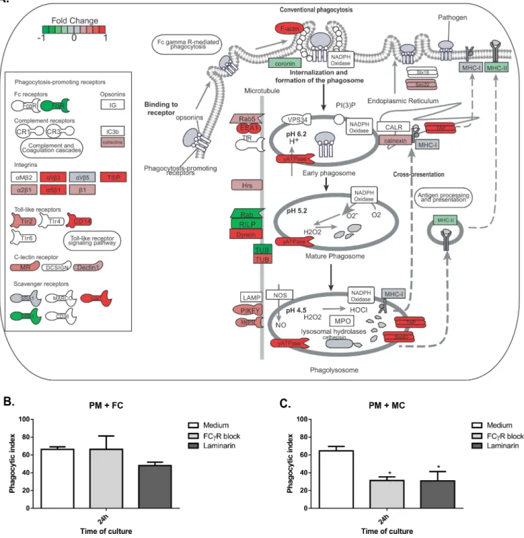
Fig 4. Toll-like receptor signaling pathway overview.Analysis of genes related to the Toll-like receptor signaling pathway showed they were mostly up-regulated in peritoneal macrophage (PM) co-culture with muriform cells (B), but not conidia (A), leading to the expression of pro-inflammatory cytokines and chemokines genes.
data showed that muriform cells, but not conidia, inducedIl12atranscript, which encodes for
IL12-p35 that forms, with IL-12p40, bioactive IL-12p70 protein (Fig 4A and 4B). Muriform
cells and conidia failed to induce the production of IL12p70 in infected macrophages (Fig 5D
andS5C Fig).
F
.
pedrosoi
conidia phagocytosis does not require Fc
γ
R and Dectin-1
recognition
F.pedrosoiconidia are not able to increase the expression ofFcyRor pattern recognition
recep-tor genes such asDectin1(Fig 6A), even though conidia were internalized after 24h of
co-culti-vation with peritoneal macrophages (Fig 7B). Macrophages stimulated by the co-culturing
with muriform cells showed an elevated expression of Dectin-1 gene while reducing the
expression of the FcγR gene (Fig 7A). Only muriform cells phagocytosis was impaired when
these membrane receptors were blocked by laminarin and Fcblock, respectively (Fig 7C).
Intense inflammatory response during murine CBM is correlated with
fungus persistence in the host
Unlike human CBM, which keeps fungal burden and chronic inflammatory processes for long periods of time, all available CBM murine models described to date tend to spontaneously heal
after a short period of infection [45]. After 30 days of infection withF.pedrosoipropagules, it
was possible to observe a significant decrease in the inflammatory response followed by a reduction in fungal load. However, it is not clear whether the reduction in the inflammatory response observed in the murine model is the result of the disease’s gradual resolution or the Fig 5.In vitrocytokine and chemokine production.The production of cytokines and chemokines by macrophage in conidia (FC) or muriform cell (MC) co-culture supernatants was assessed by ELISA (A-E). NO2concentration in culture supernatants was used as an indicator of NO generation
and measured using Griess reagent (F). High levels of TNF-α(A), IL-1β(B) and IL-6 (C) were observed in co-culture with muriform cells, but not with conidia. To assess IL-1βproduction after 24 hours, peritoneal macrophages required LPS co-stimulation (B). Muriform cells also induced higher levels of MCP-1 after 48h when compared to macrophages infected with conidia (E). Production of IL-12 (D) and NO2(F) was strongly inhibited by
muriform cells.**P<0.01 and***P<0.001.
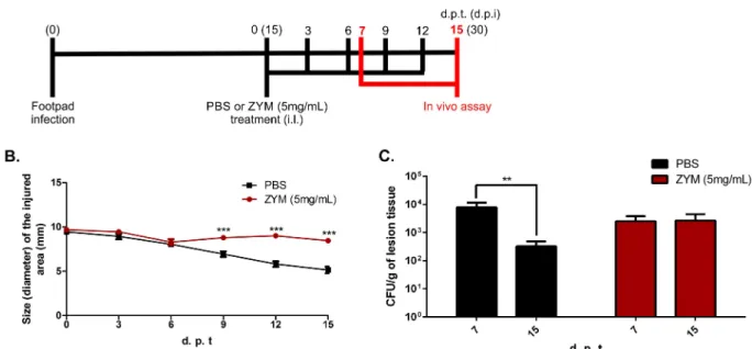
necessary environment for the healing process observed in those animals. We tested the latter
hypothesis using a model of chronic inflammation induced by zymosan (ZYM) [25,26]. After
15 days of infection with fungal propagules, animals were treated every three days with 20μl of
a suspension containing 5 mg/ml of ZYM (Fig 8A). As expected, those treated with ZYM
showed impaired edema reduction in the infected footpad, displaying intense inflammatory
response up to 30 days post infection (15 days of treatment) (Fig 8BandS6 Fig). The increase
in the inflammatory response impaired the reduction of fungal burden over time (Fig 8C).
Discussion
Chromoblastomycosis (CBM), together with sporotrichosis and mycetoma, is considered a
subcutaneous mycosis of higher incidence in the world [46]. The development of CBM starts
as a macular lesion at site of inoculation progressing to a granulomatous inflammatory reac-tion of cutaneous and subcutaneous tissues. As the initial lesion is asymptomatic, most patients only look for diagnosis during the chronic phase of the disease, with some cases showing edema and bacterial secondary infections that affect the patient’s health and their immune
response modulation [47].
Fig 6. Phagocytosis gene expression in peritoneal macrophages co-culturing with conidia.Peritoneal macrophages (PM) co-culturing with conidia (FC) did not induce much gene expression related to phagocytosis.
In nature,Fonsecaea pedrosoiis found in its saprophytic form, and it is not clear whether conidia or hyphae are responsible for initiating the disease. While hyphae and conidia have Fig 7. Phagocytosis gene expression in peritoneal macrophages co-culturing with muriform cells and phagocytosis index.Peritoneal macrophages (PM) co-culturing with muriform cells (MC) (A) induced the expression of several genes related to phagocytosis. Muriform cells displayed an elevated expression of Dectin-1 gene while reducing the expression of FcγR gene (A). By blocking Dectin-1 and FcγR with laminarin and FcγR Block, respectively, only muriform cells phagocytosis was impaired (C), while conidia (FC) phagocytosis was not affected (B).*P<0.05, compared to medium control.
been described as being present on the surface of the thorns fromMimosa pudica[7], muri-form cells, similar to those observed in the tissues of patients, are rarely observed in plants
[7,48]. Many works were carried out using only condia as the infective form [18,19,49]. Here
we demonstrate that hyphae and muriform cells are infective, with both morphotypes being able to establish murine CBM with skin lesions similar to that found in humans, whereas infec-tion with conidia did not reach the chronic phase of the disease. In this model, using immuno-competent mice, inoculated conidia were quickly eliminated from the tissue and few of them turned into muriform cells. Conidia also induced lower levels of inflammatory cytokines
secre-tion in tissue and low inducsecre-tion of transcripsecre-tionin vitro.
This low inflammatory response pattern has also been observed afterA.fumigatusspores
inhalation, with little neutrophil recruitment and macrophage activation, while the
germinat-ing conidia reverse this absent response throughβ-glucan exposure [50,51]. The absence of
β-glucan recognition by dectin-1 is also important forC.albicansescape from the host’s innate
immune response, while dendritic cells discriminate between yeast and hyphae according to
β-glucan expression [52]. An alternative explanation for this low innate immune response after
infection by conidia is that the small-sized cells can bypass structural tissue defenses, which in
turn may go unnoticed by the host immune cells [12]. Nevertheless, despite the lower immune
response observed at the inoculation site,F.pedrosoiconidia has been previously reported to
be able to reside inside phagocytes [21,53], which may constitute a survival strategy and could
be related to the slow progression of the disease in humans.
Using a different experimental model, Sousa et al [19] have shown that intraperitoneal
inoc-ulation ofF.pedrosoiconidia increased IL-10 and decreased TNF-αproduction by spleen cells.
They have also demonstrated that conidia in vitro failed to stimulate macrophages to secrete inflammatory cytokines, such as TNF-α, suggesting that failure in innate recognition can result
in chronic infection [19]. However, most CBM lesions in patients present muriform cells in a
Fig 8. CBM progression in a Zymosan-induced inflammation model.After 15 days post infection with fungal propagules (FP), animals were treated intra lesionally (i.l.) in the footpad with 20μl of a suspension containing 5 mg/ml of zymosan (ZYM) or PBS, until 15 days post treatment start (d.p.t) (A). Animals treated with ZYM displayed intense inflammatory response up to 30 days post infection (d.p.i) (B). Animals facing prolonged inflammation showed no reduction in fungal load over time, as observed for those animals treated with PBS (C).**P<0.01 and***P<0.001.
chronic granulomatous reaction, which is associated with neutrophil-rich, purulent abscesses and a chronic and highly organized inflammatory response coupled with extensive fibrosis
[9,20–22]. Our histopathological analysis did not observe conidia in granulomatous reaction
during the chronic stage of the infection. Finally, in CBM infected tissue, macrophages (some-times identified as epithelioid or giant cells) are described as highly activated with prominent
expression of proinflammatory cytokines, such as TNF-α[10], corroborating the expression of
TNF-a we observed in Figs2Eand5A. Thus, our data largely suggest a different route for the
disease progression according to fungal morphotype, so that the establishment of inflamma-tion is required for successful infecinflamma-tion chronicity.
DistinctA.fumigatusmorphotypes were shown to have a complex and distinct response
after leukocyte-fungal form interaction, with genes modulated by hyphae being implicated in
establishing or prolonging the infection [54]. Thus, understanding howF.pedrosoifungal
forms are able to modulate the macrophage response is extremely relevant to comprehend the result of the constant interaction of fungal antigens with macrophages at the site of infection and its contribution to disease chronification. Chronic granulomatous infectious diseases are usually characterized by numerous macrophages on lesional tissue so that Sotto et al. have
shown the presence of high numbers of macrophages in the skin biopsies of CBM patients [55]
and it is common to find muriform cells, sometimes within giant multinucleated cells [6]. In
addition, due to high rate of cell migration and proliferation near the infection site, the initial contact of fungus antigens with macrophages and dendritic cells occurs continuously in the course of the disease, even in the later stages.
We observed that macrophages infected in vitro with conidia or muriform cells showed dis-tinct gene expression profiles. Inflammatory response genes, as well as cytokine secretion by macrophages and dendritic cells infected with muriform cells, showed a Th17-inducing differ-entiation pattern (i.e. enhanced production of IL-6 and IL-1β) concomitant with Th1 suppres-sion (i.e. inhibition of IL-12 production). Muriform cells and conidia failed to induce the
production of IL12p70 in infected macrophages (Fig 5DandS4C Fig), suggesting a
post-tran-scriptional and tranpost-tran-scriptional negative regulation of il12a by these fungal forms, respectively.
In fact, conidia of several species ofFonsecaedisrupt nuclear IRF1 activity, which is crucial for
il12a transcription via nucleosome remodeling [56]. Furthermore, both fungal forms inhibit
the production of IL-12p70 by LPS-treated macrophages (Fig 5D). In this context, the low
pro-duction or the absence of functional IL-12 may restrict the generation of Th1 immunity lead-ing to the suppression of macrophage fungicidal activity.
Microorganism size and shape has been correlated with virulence and morphogenesis [57],
and cell size can interfere in multiple host immune responses, such as phagocytosis. Small
structures can disperse easily through the tissue, as described forCryptococcusspores [58].F.
pedrosoiconidia have 1.5 x 3μm in diameter [7], which can be easily internalized by
macro-phages. AlthoughF.pedrosoiphagocytosis mechanisms are not completely understood, it is
known that neutrophils, macrophages, dendritic cells and Langherhan cells are able to
inter-nalize the fungus [53,59,60]. The interaction between Langherhan cells and conidia, but not
muriform cells, yields a decrease in the expression of CD40 and B7-2, impairing antigen
pre-sentation and adaptive immune response [59]. Also, the fungal clearance by phagocytes is
dependent on the interaction between fungal pathogen-associated molecular patterns
(PAMPs) and phagocytic receptors [61].
Our results show that recognition by Dectin-1 or FCγR is important only to muriform cells internalization, showing that conidia and muriform cells internalization processes occur
through distinct recognition receptors. This discriminated recognition was described forS.
schenckiimorphotypes, in which phagocytosis of conidia failed to induce a pro-inflammatory
the phagocytic receptors-PAMP interaction is not only important for fungal engulfment and
killing, but also throughout the phagosome maturation [61]. Therefore, internalized conidia
without phagocytic receptor interaction can remain silent inside macrophages without activat-ing the cell, as well as the host immune system, through cytokines production and inflamma-some activation. Alternatively, other receptor-independent mechanisms for microorganism uptake, such as clathrin-mediated endocytosis, macropinocytosis and
lipidraft/caveolae-dependent endocytosis [63], can be used in the response to certain fungi, such asC.
neofor-mans[64].
This scenario may contribute to the induction and establishment of the chronic inflamma-tory response observed in CBM characterized by dense inflammainflamma-tory infiltrates that are rich in granulocytes, especially neutrophils, along with macrophages showing poor fungicidal activ-ity against muriform cells.
We also observed several genes linked to NFκB activation, including those associated with
apoptosis process. Apoptosis is an important component of protective immunity against
Histo-plasma capsulatumandP.brasiliensis, and they have been shown to be associated with
out-comes of infection [65,66]. In our model, apoptotic genes were up-regulated by muriform cells
and our group has reported similar results in an analysis of the transcriptional response of
peritoneal macrophages [67] and dendritic cells [68] toP.brasiliensisinfection in vitro. It is
reasonable to presume that despite the induction of apoptosis, phagocytes do not promptly
remove apoptotic cells and necrosis can start, increasing the inflammation [69].
Early inflammation is beneficial in containing the infection, but an uncontrolled inflamma-tory response is detrimental and may eventually oppose disease eradication, being evident in
mice with chronic granulomatous disease [2]. Animals infected with fungal propagules and
later treated with high doses of Zymozan showed no reduction in fungal burden over time, confirming the detrimental nature of an intense inflammatory response in CBM, which may contribute to a chronic infection state. Besides, previous work showed that modulation of the inflammatory response mediated by DNA-hsp65 vaccine was able to accelerate the healing
process of experimental CBM [23].
Altogether, our results show that muriform cells are able to induce inflammatory response in the course of murine CBM, allowing the fungus to persist in the host and involving inflam-mation as an important factor in CBM chronicity. In this way, CBM chronicity may not be due to a failure in inducing an inflammatory response, but rather be related to the host’s inabil-ity to regulate the exacerbated inflammatory response induced by muriform cells. With this in mind, new therapeutic approaches concerning the modulation of inflammatory response can be developed, allowing reduced chemotherapy periods and lowering relapse rates.
Supporting information
aspect was observed after 15 days of infection with all fungal forms except conidia (B). After 30 days of infection, an intense tissue repair was already observed in animals infected with conidia, while for those infected with hyphae and fungal propagules (FP), an intense healing process was only seen at 45 days post-infection, with the presence of fibroblasts and collagen deposition. At that time, only animals infected with muriform cells still exhibited exudative areas (B).
(TIF)
S2 Fig. Identification of differentially expressed genes.Funnel chart (A) and VennEuler
dia-gram (B) displaying differentially expressed genes when False Discovery Rates (FDR)<0.05
and Fold Change cutoff (FC cutoff)>1.4, respectively.
(TIF)
S3 Fig. Common differentially expressed genes to PM-FC and PM-MC interaction. Heat-map of 30 differentially expressed genes in peritoneal macrophages (PM) infected with conidia (FC) or muriform cells (MC). Heatmap was build based on fold-change values.
(TIF)
S4 Fig. Cell differentiation GO enrichment analysis.Heatmap of differentially expressed genes in peritoneal macrophages (PM) infected with conidia (FC) or muriform cells (MC) cor-related to inflammatory response (GO: 0006954), angiogenesis (GO:0001525), cell prolifera-tion (GO:0008283), cell migraprolifera-tion (GO:0016477), regulaprolifera-tion of angiogenesis (GO: 0045765) and regulation of apoptotic process (GO:0042981).
(TIF)
S5 Fig. Cell migration and cytokine production after incubation with conidia or muriform cells using distinct phagocytes and MOI.TNF-α(A) and IL-6 (B) production are not in-creased in higher concentration of conidia (MOI 5:1 of conidia and peritoneal macrophage, respectively). IL-12 (C) and NO2 (D) were not detected after 6, 12, 24 or 48 hours of PM incu-bation with FC or MCs. Fungal cells co-culture with mouse bone marrow-derived
macro-phages (BMDMs) and dendritic cells (BMDCs) showed similar patterns of TNF-α(E-F) and
IL-1β(G-H) production compared to PM cells after 24 hours. Further stimulation was not
required for IL-1βproduction in BMDM-MC (G) or BMDC-MC (H) co-culture. Peritoneal
inoculation with 106cells of each fungal form revealed intense cell migration to peritoneal
cavity induced by muriform cells compared to conidia (FC) inoculation (I).P<0.001,
P<0.01.
(TIF)
S6 Fig. Skin lesions analysis in mice infected with F. pedrosoi and treated with zymosan. After 15 days post infection with FP, animals were treated intra lesionally (i.l.) in the footpad
with 20μl of a suspension containing 5 mg/ml of zymosan (ZYM) or PBS, until 15 days post
treatment start (d.p.t). DPI and HE or Masson’s trichrome stain are indicated in the figure. (TIF)
S1 Table. Gene ontology enrichment results for biological process categories in macro-phage co-culture with F. pedrosoi conidia.
(PDF)
S2 Table. Gene ontology enrichment results for biological process categories in macro-phage co-culture with muriform cells.
Acknowledgments
The authors would like to thank Viviane Montanaro Leal, for technical assistance in the histo-chemical assay, and Maria Emı´lia Telles Walter and Marcelo de Macedo Brı´gido, for prelimi-nary discussion about bioinformatics analysis.
Author Contributions
Conceptualization:IMS ALB AHT.
Data curation:TR CH.
Formal analysis:IMS CH AHT.
Funding acquisition:ALB.
Investigation:IMS RJAdC ALB AHT.
Methodology:IMS RJAdC MSJ ACS LCdML.
Project administration:CH ALB AHT.
Resources:ALB.
Software:TR CN NA CH.
Supervision:ALB AHT.
Validation:IMS RJAdC ALB.
Visualization:IMS CH ALB AHT.
Writing – original draft:IMS CH ALB AHT.
Writing – review & editing:IMS CH ALB AHT.
References
1. Pina A, Bernardino S, Calich VLG. Alveolar macrophages from susceptible mice are more competent than those of resistant mice to control initial Paracoccidioides brasiliensis infection. J Leukoc Biol. 2008; 83(5):1088–99.https://doi.org/10.1189/jlb.1107738PMID:18281437
2. Romani L. Immunity to fungal infections. Nat Rev Immunol [Internet]. Nature Publishing Group; 2011 Apr [cited 2013 Aug 5]; 11(4):275–88. Available from:http://www.ncbi.nlm.nih.gov/pubmed/21394104 https://doi.org/10.1038/nri2939PMID:21394104
3. Romani L, Fallarino F, De Luca A, Montagnoli C, D’Angelo C, Zelante T, et al. Defective tryptophan catabolism underlies inflammation in mouse chronic granulomatous disease. Nature [Internet]. 2008 Jan 10; 451(7175):211–5. Available from:http://www.ncbi.nlm.nih.gov/pubmed/18185592 https://doi. org/10.1038/nature06471PMID:18185592
4. Zelante T, De Luca A, Bonifazi P, Montagnoli C, Bozza S, Moretti S, et al. IL-23 and the Th17 pathway promote inflammation and impair antifungal immune resistance. Eur J Immunol [Internet]. 2007 Oct; 37 (10):2695–706. Available from:http://www.ncbi.nlm.nih.gov/pubmed/17899546 https://doi.org/10.1002/ eji.200737409PMID:17899546
5. Ameen M. Chromoblastomycosis: Clinical presentation and management. Clin Exp Dermatol. 2009; 34 (8):849–54.https://doi.org/10.1111/j.1365-2230.2009.03415.xPMID:19575735
6. Santos ALS, Palmeira VF, Rozental S, Kneipp LF, Nimrichter L, Alviano DS, et al. Biology and patho-genesis of Fonsecaea pedrosoi, the major etiologic agent of chromoblastomycosis. FEMS Microbiol Rev [Internet]. 2007 Sep [cited 2012 Jul 19]; 31(5):570–91. Available from:http://www.ncbi.nlm.nih.gov/ pubmed/17645522 https://doi.org/10.1111/j.1574-6976.2007.00077.xPMID:17645522
8. Bonifaz A, Carrasco-Gerard E, Sau´l A. Chromoblastomycosis: clinical and mycologic experience of 51 cases. Mycoses [Internet]. 2001; 44(1–2):1–7. Available from:http://www.ncbi.nlm.nih.gov/pubmed/ 11398635PMID:11398635
9. Queiroz-Telles F, Esterre P, Perez-Blanco M, Vitale RG, Salgado CG, Bonifaz A. Chromoblastomyco-sis: an overview of clinical manifestations, diagnosis and treatment. Med Mycol [Internet]. 2009 Feb [cited 2012 Jul 19]; 47(1):3–15. Available from:http://www.ncbi.nlm.nih.gov/pubmed/19085206 https:// doi.org/10.1080/13693780802538001PMID:19085206
10. Esterre P, Peyrol S, Sainte-Marie D, Pradinaud R, Grimaud JA. Granulomatous reaction and tissue remodelling in the cutaneous lesion of chromomycosis. Virchows Arch A Pathol Anat Histopathol [Inter-net]. 1993 Jan [cited 2012 Sep 17]; 422(4):285–91. Available from:http://www.ncbi.nlm.nih.gov/ pubmed/8506621PMID:8506621
11. Silva AADL, Criado PR, Nunes RS, da Silva WLF, Kanashiro-Galo L, Duarte MIS, et al. In Situ Immune Response in Human Chromoblastomycosis–A Possible Role for Regulatory and Th17 T Cells. PLoS Negl Trop Dis [Internet]. 2014; 8(9):e3162. Available from:http://dx.plos.org/10.1371/journal.pntd. 0003162 https://doi.org/10.1371/journal.pntd.0003162PMID:25233082
12. Gauthier GM. Dimorphism in Fungal Pathogens of Mammals, Plants, and Insects. PLOS Pathog [Inter-net]. 2015; 11(2):e1004608. Available from:http://dx.plos.org/10.1371/journal.ppat.1004608 https://doi. org/10.1371/journal.ppat.1004608PMID:25675433
13. da Silva JP, Alviano DS, Alviano CS, de Souza W, Travassos LR, Diniz J a P, et al. Comparison of Fon-secaea pedrosoi sclerotic cells obtained in vivo and in vitro: ultrastructure and antigenicity. FEMS Immunol Med Microbiol [Internet]. 2002 Mar 25; 33(1):63–9. Available from:http://www.ncbi.nlm.nih. gov/pubmed/11985971PMID:11985971
14. Alviano DS, Kneipp LF, Lopes AH, Travassos LR, Meyer-Fernandes JR, Rodrigues ML, et al. Differenti-ation of Fonsecaea pedrosoi mycelial forms into sclerotic cells is induced by platelet-activating factor. Res Microbiol [Internet]. 2003 Dec [cited 2012 Jul 19]; 154(10):689–95. Available from:http://www.ncbi. nlm.nih.gov/pubmed/14643407 https://doi.org/10.1016/j.resmic.2003.09.002PMID:14643407 15. Chavan SS, Kulkarni MH, Makannavar JH. “Unstained” and “de stained” sections in the diagnosis of
chromoblastomycosis: a clinico-pathological study. Indian J Pathol Microbiol [Internet]. 53(4):666–71. Available from:http://www.ncbi.nlm.nih.gov/pubmed/21045389 https://doi.org/10.4103/0377-4929. 72021PMID:21045389
16. Roilides E, Xi L, Pedrozo DM, Batista M, Pana D, Colombo L, et al. Chromoblastomycosis. 2017; 30 (1):233–76.
17. Bocca AL, Brito PPMS, Figueiredo F, Tosta CE. Inhibition of nitric oxide production by macrophages in chromoblastomycosis: a role for Fonsecaea pedrosoi melanin. Mycopathologia [Internet]. 2006 Apr [cited 2012 Sep 17]; 161(4):195–203. Available from:http://www.ncbi.nlm.nih.gov/pubmed/16552481 https://doi.org/10.1007/s11046-005-0228-6PMID:16552481
18. Wu¨thrich M, Wang H, Li M, Lerksuthirat T. F. pedrosoi -induced Th17-cell differentiation in mice is fos-tered by Dectin-2 and suppressed by Mincle recognition. 2015;1–22.
19. Da Glo´ria Sousa M, Reid DM, Schweighoffer E, Tybulewicz V, Ruland J, Langhorne J, et al. Restoration of pattern recognition receptor costimulation to treat chromoblastomycosis, a chronic fungal infection of the skin. Cell Host Microbe. 2011; 9(5):436–43.https://doi.org/10.1016/j.chom.2011.04.005PMID: 21575914
20. Esterre P, Queiroz-Telles F. Management of chromoblastomycosis: novel perspectives. Curr Opin Infect Dis [Internet]. 2006 Apr; 19(2):148–52. Available from:http://www.ncbi.nlm.nih.gov/pubmed/ 16514339 https://doi.org/10.1097/01.qco.0000216625.28692.67PMID:16514339
21. Lo´pez Martı´nez R, Me´ndez Tovar LJ. Chromoblastomycosis. Clin Dermatol [Internet]. 2007 [cited 2012 Jul 19]; 25(2):188–94. Available from:http://www.ncbi.nlm.nih.gov/pubmed/17350498 https://doi.org/ 10.1016/j.clindermatol.2006.05.007PMID:17350498
22. Avelar-Pires C, Simoes-Quaresma JA, Moraes-de Macedo GM, Brasil-Xavier M, Cardoso-de Brito A. Revisiting the clinical and histopathological aspects of patients with chromoblastomycosis from the bra-zilian amazon region. Arch Med Res [Internet]. Elsevier Inc; 2013; 44(4):302–6.https://doi.org/10.1016/ j.arcmed.2013.04.008PMID:23684532
23. Siqueira IM, Ribeiro AM, de Medeiros No´brega YK, Simon KS, Souza ACO, Jeroˆnimo MS, et al. DNA-hsp65 Vaccine as Therapeutic Strategy to Treat Experimental Chromoblastomycosis Caused by Fon-secaea Pedrosoi. Mycopathologia. 2013; 175(5–6):463–75. https://doi.org/10.1007/s11046-012-9599-7PMID:23179449
25. Kim HS, Ryu HS, Kim JS, Kim YG, Lee HK, Jung JK, et al. Validation of cyclooxygenase-2 as a direct anti-inflammatory target of 4-O-methylhonokiol in zymosan-induced animal models. Arch Pharm Res [Internet]. 2015; 38(5):813–25. Available from:http://www.ncbi.nlm.nih.gov/pubmed/25074039 https:// doi.org/10.1007/s12272-014-0456-8PMID:25074039
26. Gapeyev a B, Gapeyev a B, Mikhailik EN, Mikhailik EN, Chemeris NK, Chemeris NK. Anti-inflammatory effects of low-intensity extremely high-frequency electromagnetic radiation: frequency and power dependence. Bioelectromagnetics [Internet]. 2008; 29(3):197–206. Available from:http://www.ncbi. nlm.nih.gov/pubmed/18044738 https://doi.org/10.1002/bem.20381PMID:18044738
27. Lutz MB, Kukutsch N, Ogilvie AL, Ro¨ssner S, Koch F, Romani N, et al. An advanced culture method for generating large quantities of highly pure dendritic cells from mouse bone marrow. J Immunol Methods [Internet]. 1999 Feb 1; 223(1):77–92. Available from:http://www.ncbi.nlm.nih.gov/pubmed/10037236 PMID:10037236
28. Lutz MB, Kukutsch N, Ogilvie ALJ, Ro¨ßner S, Koch F, Romani N, et al. An advanced culture method for generating large quantities of highly pure dendritic cells from mouse bone marrow. J Immunol Methods. 1999; 223(1):77–92. PMID:10037236
29. Hayakawa M, Ghosn EEB, da Gloria Teixeria de Sousa M, Ferreira KS, Almeida SR. Phagocytosis, pro-duction of nitric oxide and pro-inflammatory cytokines by macrophages in the presence of dematiac-eous [correction of dematiaceus] fungi that cause chromoblastomycosis. Scand J Immunol [Internet]. 2006 Oct [cited 2012 Jul 19]; 64(4):382–7. Available from:http://www.ncbi.nlm.nih.gov/pubmed/ 16970678 https://doi.org/10.1111/j.1365-3083.2006.01804.xPMID:16970678
30. Green LC, Ruiz de Luzuriaga K, Wagner DA, Rand W, Istfan N, Young VR, et al. Nitrate biosynthesis in man. Proc Natl Acad Sci U S A [Internet]. 1981 Dec; 78(12):7764–8. Available from:http://www.ncbi. nlm.nih.gov/pubmed/6950416PMID:6950416
31. Fastqc [Internet]. Available from:http://www.bioinformatics.babraham.ac.uk/projects/fastqc/
32. Martin M. Cutadapt removes adapter sequences from high-throughput sequencing reads. EMBnet.jour-nal [Internet]. 2011 May 2; 17(1):10. Available from:http://journal.embnet.org/index.php/embnetjournal/ article/view/200
33. Anders S, Pyl PT, Huber W. HTSeq—a Python framework to work with high-throughput sequencing data. Bioinformatics [Internet]. 2015 Jan 15; 31(2):166–9. Available from:http://www.ncbi.nlm.nih.gov/ pubmed/25260700 https://doi.org/10.1093/bioinformatics/btu638PMID:25260700
34. No Title [Internet]. Available from:http://www.ensembl.org/Mus_musculus/
35. Trapnell C, Pachter L, Salzberg SL. TopHat: discovering splice junctions with RNA-Seq. Bioinformatics [Internet]. 2009 May 1; 25(9):1105–11. Available from:http://www.ncbi.nlm.nih.gov/pubmed/19289445 https://doi.org/10.1093/bioinformatics/btp120PMID:19289445
36. Li H, Handsaker B, Wysoker A, Fennell T, Ruan J, Homer N, et al. The Sequence Alignment/Map format and SAMtools. Bioinformatics [Internet]. 2009 Aug 15; 25(16):2078–9. Available from:http://www.ncbi. nlm.nih.gov/pubmed/19505943 https://doi.org/10.1093/bioinformatics/btp352PMID:19505943 37. (R Core Team). A Language and Environment for Statistical Computing [Internet]. Vienna, Austria: R
Foundation for Statistical Computing; 2015. Available from:http://www.r-project.org
38. Robinson MD, McCarthy DJ, Smyth GK. edgeR: a Bioconductor package for differential expression analysis of digital gene expression data. Bioinformatics [Internet]. 2010 Jan 1; 26(1):139–40. Available from:http://www.ncbi.nlm.nih.gov/pubmed/19910308 https://doi.org/10.1093/bioinformatics/btp616 PMID:19910308
39. Carlson M. org.Mm.eg.db: Genome wide annotation for Mouse.
40. Luo W, Brouwer C. Pathview: an R/Bioconductor package for pathway-based data integration and visu-alization. Bioinformatics [Internet]. 2013 Jul 15; 29(14):1830–1.https://doi.org/10.1093/bioinformatics/ btt285PMID:23740750
41. Alexa A, Rahnenfu¨hrer J, Lengauer T. Improved scoring of functional groups from gene expression data by decorrelating GO graph structure. Bioinformatics [Internet]. 2006 Jul 1; 22(13):1600–7. Avail-able from:http://www.ncbi.nlm.nih.gov/pubmed/16606683 https://doi.org/10.1093/bioinformatics/btl140 PMID:16606683
42. Wang H, Mu W, Ja Q, Zhang M, Chen R, Lv G, et al. Cytokine Profile of a Self-Healing Fonsecaea ped-rosoi Infection in Murine Model. Cell Biochem Biophys [Internet]. 2013 Nov [cited 2013 Nov 3]; 67 (2):599–605. Available from:http://www.ncbi.nlm.nih.gov/pubmed/23479333 https://doi.org/10.1007/ s12013-013-9547-2PMID:23479333
44. Porksakorn C, Nuchprayoon S, Park K, Scott AL. Proinflammatory cytokine gene expression by murine macrophages in response to Brugia malayi Wolbachia surface protein. Mediators Inflamm [Internet]. 2007; 2007:84318. Available from:http://www.ncbi.nlm.nih.gov/pubmed/17641731 https://doi.org/10. 1155/2007/84318PMID:17641731
45. Salgado CG. Fungal x host interactions in Chromoblastomycosis: what we have learned from animal models and what is yet to be solved. Virulence. 2010; 1(1):3–5.https://doi.org/10.4161/viru.1.1.10169 PMID:21178406
46. Bonifaz A, Va´zquez-Gonza´lez D, Perusquı´a-Ortiz AM. Subcutaneous mycoses: chromoblastomycosis, sporotrichosis and mycetoma. J Dtsch Dermatol Ges [Internet]. 2010 Aug; 8(8):619–27; quiz 628. Avail-able from:http://www.ncbi.nlm.nih.gov/pubmed/20529168 https://doi.org/10.1111/j.1610-0387.2010. 07453.xPMID:20529168
47. Queiroz-Telles F. CHROMOBLASTOMYCOSIS: A NEGLECTED TROPICAL DISEASE. Rev do Inst Med Trop São Paulo [Internet]. 2015 Sep; 57 Suppl 1:46–50. Available from:http://www.ncbi.nlm.nih. gov/pubmed/26465369
48. Krzyściak PM, Pindycka-Piaszczyńska M, Piaszczyński M. Chromoblastomycosis. Poste¸py dermatolo-gii i Alergol [Internet]. 2014; 31(5):310–21. Available from:http://www.ncbi.nlm.nih.gov/pubmed/ 25395928
49. Teixeira de Sousa MDG, Ghosn EEB, Almeida SR. Absence of CD4+ T cells impairs host defence of mice infected with Fonsecaea pedrosoi. Scand J Immunol [Internet]. 2006 Dec [cited 2012 Jul 19]; 64 (6):595–600. Available from:http://www.ncbi.nlm.nih.gov/pubmed/17083615 https://doi.org/10.1111/j. 1365-3083.2006.01846.xPMID:17083615
50. Hohl TM, Van Epps HL, Rivera A, Morgan LA, Chen PL, Feldmesser M, et al. Aspergillus fumigatus trig-gers inflammatory responses by stage-specific beta-glucan display. PLoS Pathog [Internet]. 2005 Nov; 1(3):e30. Available from:http://www.ncbi.nlm.nih.gov/pubmed/16304610 https://doi.org/10.1371/ journal.ppat.0010030PMID:16304610
51. Gersuk GM, Underhill DM, Zhu L, Marr KA. Dectin-1 and TLRs permit macrophages to distinguish between different Aspergillus fumigatus cellular states. J Immunol [Internet]. 2006 Mar 15; 176 (6):3717–24. Available from:http://www.ncbi.nlm.nih.gov/pubmed/16517740PMID:16517740 52. d’Ostiani CF, Del Sero G, Bacci A, Montagnoli C, Spreca A, Mencacci A, et al. Dendritic cells
discrimi-nate between yeasts and hyphae of the fungus Candida albicans. Implications for initiation of T helper cell immunity in vitro and in vivo. J Exp Med [Internet]. 2000 May 15; 191(10):1661–74. Available from: http://www.ncbi.nlm.nih.gov/pubmed/10811860PMID:10811860
53. Rozental S, Alviano CS, de Souza W. The in vitro susceptibility of Fonsecaea pedrosoi to activated macrophages. Mycopathologia [Internet]. 1994 May [cited 2012 Sep 17]; 126(2):85–91. Available from: http://www.ncbi.nlm.nih.gov/pubmed/8065435PMID:8065435
54. Sugui JA, Kim HS, Zarember KA, Chang YC, Gallin JI, Nierman WC, et al. Genes differentially expressed in conidia and hyphae of Aspergillus fumigatus upon exposure to human neutrophils. PLoS One [Internet]. 2008; 3(7):e2655. Available from:http://www.ncbi.nlm.nih.gov/pubmed/18648542 https://doi.org/10.1371/journal.pone.0002655PMID:18648542
55. Sotto MN, De Brito T, Silva AMG, Vidal M, Castro LGM. Antigen distribution and antigen-presenting cells in skin biopsies of human chromoblastomycosis. J Cutan Pathol [Internet]. 2004 Jan; 31(1):14–8. Available from:http://www.ncbi.nlm.nih.gov/pubmed/14675280PMID:14675280
56. Wevers B a., Kaptein TM, Zijlstra-Willems EM, Theelen B, Boekhout T, Geijtenbeek TBH, et al. Fungal engagement of the C-type lectin mincle suppresses dectin-1-induced antifungal immunity. Cell Host Microbe [Internet]. Elsevier; 2014; 15(4):494–505.https://doi.org/10.1016/j.chom.2014.03.008PMID: 24721577
57. Wang L, Lin X. Morphogenesis in fungal pathogenicity: shape, size, and surface. PLoS Pathog [Inter-net]. 2012 Jan [cited 2015 Jan 5]; 8(12):e1003027. Available from:http://www.pubmedcentral.nih.gov/ articlerender.fcgi?artid=3516537&tool=pmcentrez&rendertype=abstract https://doi.org/10.1371/ journal.ppat.1003027PMID:23236274
58. Velagapudi R, Hsueh Y-P, Geunes-Boyer S, Wright JR, Heitman J. Spores as infectious propagules of Cryptococcus neoformans. Infect Immun [Internet]. 2009 Oct; 77(10):4345–55. Available from:http:// www.ncbi.nlm.nih.gov/pubmed/19620339 https://doi.org/10.1128/IAI.00542-09PMID:19620339 59. da Silva JP, da Silva MB, Salgado UI, Diniz JAP, Rozental S, Salgado CG. Phagocytosis of Fonsecaea
60. Rozental S, Alviano CS, de Souza W. Fine structure and cytochemical study of the interaction between Fonsecaea pedrosoi and rat polymorphonuclear leukocyte. J Med Vet Mycol [Internet]. 34(5):323–30. Available from:http://www.ncbi.nlm.nih.gov/pubmed/8912165PMID:8912165
61. Erwig LP, Gow NAR. Interactions of fungal pathogens with phagocytes. Nat Rev Microbiol [Internet]. 2016 Mar; 14(3):163–76. Available from:http://www.ncbi.nlm.nih.gov/pubmed/26853116 https://doi. org/10.1038/nrmicro.2015.21PMID:26853116
62. Guzman-Beltran S, Perez-Torres A, Coronel-Cruz C, Torres-Guerrero H. Phagocytic receptors on mac-rophages distinguish between different Sporothrix schenckii morphotypes. Microbes Infect [Internet]. 2012 Oct; 14(12):1093–101. Available from:http://www.ncbi.nlm.nih.gov/pubmed/22771955 https://doi. org/10.1016/j.micinf.2012.06.001PMID:22771955
63. Gruenberg J, van der Goot FG. Mechanisms of pathogen entry through the endosomal compartments. Nat Rev Mol Cell Biol [Internet]. 2006 Jul; 7(7):495–504. Available from:http://www.ncbi.nlm.nih.gov/ pubmed/16773132 https://doi.org/10.1038/nrm1959PMID:16773132
64. Guerra CR, Seabra SH, De Souza W, Rozental S. Cryptococcus neoformans is internalized by recep-tor-mediated or “triggered” phagocytosis, dependent on actin recruitment. PLoS One. 2014; 9(2):1–10. 65. Bo¨hme L, Rudel T. Host cell death machinery as a target for bacterial pathogens. Microbes Infect
[Inter-net]. 2009 Nov; 11(13):1063–70. Available from:http://www.ncbi.nlm.nih.gov/pubmed/19733679 https://doi.org/10.1016/j.micinf.2009.08.014PMID:19733679
66. Verı´cimo MA, Franc¸a KM, Arnholdt AC V, Kipnis TL. Increased apoptosis during the early phase of experimental paracoccidioidomycosis as a phenotypic marker of resistance. Microbes Infect [Internet]. 2006 Oct; 8(12–13):2811–20. Available from:http://www.ncbi.nlm.nih.gov/pubmed/17045508 https:// doi.org/10.1016/j.micinf.2006.08.012PMID:17045508
67. Silva SS, Tavares AHFP, Passos-Silva DG, Fachin AL, Teixeira SMR, Soares CMA, et al. Transcrip-tional response of murine macrophages upon infection with opsonized Paracoccidioides brasiliensis yeast cells. Microbes Infect [Internet]. 2008 Jan; 10(1):12–20. Available from:http://www.ncbi.nlm.nih. gov/pubmed/18096424 https://doi.org/10.1016/j.micinf.2007.09.018PMID:18096424
68. Tavares AH, Derengowski LS, Ferreira KS, Silva SS, Macedo C, Bocca AL, et al. Murine dendritic cells transcriptional modulation upon Paracoccidioides brasiliensis infection. PLoS Negl Trop Dis. 2012; 6 (1).