International
Journal
of
Paleopathology
jo u r n al h om ep ag e :w w w . e l s e v i e r . c o m / l o c a t e / i j p pCase
study
A
possible
Madura
foot
from
medieval
Estremoz,
southern
Portugal
Ana
Curto
a,c,∗,
Teresa
Fernandes
b,caSkeletalBiologyResearchCentre(SBRC),SchoolofAnthropologyandConservation,TheUniversityofKent,CanterburyCT27NR,UK bDepartmentofBiology,SchoolofScienceandTechnology,UniversityofÉvora,7002-554Évora,Portugal
cCIAS(ResearchCentreforAnthropologyandHealth),DepartmentofLifeSciences,UniversityofCoimbra,3000-456Coimbra,Portugal
a
r
t
i
c
l
e
i
n
f
o
Articlehistory:
Received18September2015
Receivedinrevisedform6January2016 Accepted29January2016 Keywords: Infectiousdisease Mycetoma Maduromycosis Trepanation
a
b
s
t
r
a
c
t
Maduromycosis,commonlycalledMadurafoot,isaninfectiouspathologycausedbyfungiorbacteria, anditisnativeofthetropical,subtropicalandequatorialareas.Thispaperpresentsawellpreservedmale skeleton,between23and57yearsoldfromamedievalnecropolis(13th–15thcenturies)inEstremoz, Portugal.Theleftfootofthisindividualshowedmarkedalterationsonthemorphologyofthecalcaneus andcuboidthatareankylosed,whichledtoarthrosisofthecalcaneousandtalus.Thefivemetatarsalshave bonedestructionandirregularsubperiostealnewboneformationwithmultiplelyticfociandprogressive osteoporosiswithverylittlereactiveboneformation.Afteracarefuldifferentialdiagnosis,takinginto considerationvariouspathologicalconditions,itwasconcludedthatthisisapossiblecaseof maduromy-cosis.ThisstudysuggeststhatinthepastthispathologycouldhavebeenpresentinEurope,particularly intheMediterraneanregion,andespeciallywhentheclimaticconditionswereconducive.Thiscouldbe oneofthefirstcasesofmaduromycosisinfectionpublishedinanarchaeologicalcontext.
©2016ElsevierInc.Allrightsreserved.
1. Introduction
Maduromycosisis a chronic, progressive and granulomatous inflammatory diseasethatmay affectsubcutaneoustissues and sometimesbones,but rarelyviscera(Arenas and Lavalle,2001; Fahal,2011).Thispathologyistheconsequenceoftraumatic inoc-ulation of the skin by the causative pathogen, which can be actinomycetes(filamentousbacteria)oreumicycetes(truefungi) (Fahal, 2011).While fungal grains grow in drier areas, actino-mycetesaremorecommoninwetareas(Diengetal.,2003;Fahal, 2004).Theactinomycetesexpandfaster,beingmoreinvasiveand having more, smaller sinuses than eumycotic variants (Davies, 1958;Magana,1984).
Maduromycosishasalong,painlessandasymptomatic incuba-tionperiod,whichvariesfromseveralweekstomonthsoreven years(ArenasandLavalle,2001;Magana,1984).Thispathology involvesthesubcutaneoustissuesandprobablyoriginateswitha traumaticinoculationofthepathogenthatlaterspreadsthrough theskinanddeepstructures,suchasbone,resultingindestruction, deformityand lossoffunction(ArenasandLavalle,2001;Fahal, 2011).Difficultyinaccuratelyidentifyingthelimitsandspreadof
∗ Correspondingauthorat.SkeletalBiologyResearchCentre(SBRC),Schoolof AnthropologyandConservation,TheUniversityofKent,CanterburyCT27NR,UK
E-mailaddresses:a.q.curto@gmail.com,arqc3@kent.ac.uk(A.Curto).
thediseaseduringsurgerymayleadtorecurrenceandamputation (ElBagi,2003).Complicationsincludesecondarybacterial infec-tionsthatcanprogresstobacteremiaorsepticaemiaresultingin death(Venkatswamietal.,2012).
Thecommonnamefor pedalmaduromycosisis Madurafoot, derivingfromthecityofMadurai,inIndia,whereitwasfirst med-icallyreportedinthemiddleofthe1800’s(Bonifazetal.,2014), althoughitseemstohavebeenfirstdescribedintheIndian San-skrittextAtharvaVeda(Hospenthal,2009).Madurafootisnative tothetropical,subtropicalandequatorialareasbetweenlatitudes 30◦Nand15◦Sinthecommonlycalled“mycetomabelt”(Bakshiand Mathur,2008),especiallyamongbarefootpopulations(Tomczyk etal.,2014)andagriculturalworkers(Bonifazetal.,2014). Envi-ronmentalfactorssuchasrainfallplay animportantroleinthe distributionofthisdisease,aswellasthenatureofthesoiland presence of abundant, sharp, thorny vegetation(Mathur et al., 1979).Forthesereasons,itismostcommoninthefoot,although itcanalsooccurinthethigh,kneeandlowerleg,thehand, fore-arm,arm, shoulder,back, abdominal walland chest, aswellas occasionallyintheheadorface(ArenasandLavalle,2001).This pathologyismorecommoninmales(BakshiandMathur,2008; Bonifazetal.,2014)duetoactivitybutalsotopossibleinhibition ofthegrowthofsomeofthesepathogenscausedbyprogesterone (Hernández-Hernández etal.,1995;Hospenthal,2009).Thefirst threeclinicalcasesreportedforEuropecamefromGreece, south-ern Italy and Sardinia (Plehn, 1928).In Portugal, two cases of
http://dx.doi.org/10.1016/j.ijpp.2016.01.006 1879-9817/©2016ElsevierInc.Allrightsreserved.
Fig.1. Ankylosisoftheleftcalcaneousandcuboid(3Dimagingavailableathttps://sketchfab.com/models/7338e70695b34439b8421d014b593610).(A)Photographyshowing externalfoci(blackarrow).(B)Radiographshowingmultiplefoci(blackarrows)andthelines(whitearrow)betweenthecalcaneusandcuboid.
Fig.2.Ankylosisoftheleftcalcaneusandcuboid,aswellasarthrosisatthetalo-calcaneusarticulationandtheirregularsubperiostealnewboneformations(circles)and lyticexternalfoci(arrows)intheleftmetatarsals.
maduromycosis caused by Nocardia asteroides were recorded in 1963 (Brandão and Figueiredo, 1963 in Cabrita, 1974) and 1970 (Nobre,1970 in Cabrita, 1974), and one infection caused byMadurellamycetomi in1971(Cabritaand Figueiredo,1971in Cabrita, 1974). Thesethree individuals had never been outside ContinentalPortugalsotheymusthavebeeninfectedinEurope, althoughitisnotclearifthepresenceofthesepathogensisrelated withpeoples’movementtoandfromAfrica,especiallywithinthe thenPortugueseOverseasProvinces,suchasMozambique,Angola andGuinea.Recently morecaseshave beenreported,especially among migrant populations due to increased mobility of peo-ple(ElMuttardietal.,2010;Buonfrateetal.,2014;Iniestaetal., 2015).Oneofthese caseswasreportedin Portugal:a 43-year-oldfemalebornin CapeVerdefromwhose infectionmolecular diagnosisrevealedthepresenceofMadurellamycetomatis(Mestre
etal.,2015).Recentlya46-year-oldPortuguesemalefarmerwith a swelling onhisrightfoot, witheightyears of evolution, was diagnosedasanactinomycetomacase(Ramosetal.,2013).This individualwasinfectedintheMadeiraIsland,followingatrauma ontheinstep,whileworkingonthefields.
Mycoticdiseaseisuncommononpaleopathology,inpartdue tothelowskeletalinvolvementforthisgroupofpathologies.Only a fewcasesofMadurafoothave beendescribed fromthe pale-opathologicalrecord,and noneofthemfromEurope.In Mexico a case ofMadura footwasreportedin a malebetween25 and 30yearsoldfromtheTlatilcocollection(1300–100B.C.)with459 skeletons(Mansilla-LoryandConteras-López,2009).Thereisalso acasereportedfromtheByzantineperiod(A.D.300–600)inIsrael (Hershkovitzetal.,1992),butthiswaslaterdiagnosedasan exam-pleofleprosy(SpigelmanandDonoghue,2001),asfirstsuggested
2. Materialsandmethods
Thestudysubjectwasanalysedfollowingvarious methodolo-gies.Sexwasaccessedbasedonpelvisfeatures(Bruzek,2002).Age atdeathwasestimatedfromthedegenerationofthepubic sym-physis(BrooksandSuchey,1990).Maximumlengthofthesecond rightmetatarsalwasusedtoestablishstature(Santos,2002).The methodologiessuggestedthattheskeletonanalysedwasamale, between23and57yearsoldandapproximately159cminheight.It wasrecoveredfromanecropolisinEstremoz,insouthernPortugal. Duringexcavation,115individualswererecovered,ofwhich 72 wereadults:32males,22females.Itwasnotpossibletoestimate biologicalsexinanadditional18skeletonsduetopoorpreservation andincompletenessoftheremains.Inthiscemeteryallskeletons wereinsupinedecubituspositionandorientedfromWesttoEast, withoutarchaeologicalartefacts,whichistypicalofChristian buri-als.Radiocarbondatingoftwootherindividuals,indicatedatime periodbetweenthe13thandthe15thcentury(BP680to530and BP670to530for2Sigmacalibration,with95%probability).This relativelylargetimeframemakesmoreprecisecharacterisation of thenecropolisimpossible since this was a period of signifi-canthistoricalandenvironmentalupheavalinPortugal.Exhaustive macroscopicandradiographicobservations(performedwitha dig-italsystemMammoDiagnostUCPhilips,at28kVand25mA,coarse focus,usingKodakMin-Rscreenfilm)wereperformedonthe skele-toninorder todescribethelesionsas wellas toidentifyother anomalieshelpfultothedifferentialdiagnosis.A3Dimageofthe leftcalcaneusandcuboidwascreatedwiththesoftwareAgisoft PhotoScan,using360◦photography.
3. Resultsanddiscussion
MostofthebonesfromskeletonRMPE-121arewellpreserved, withtheexceptionofthefacialbones,forearmbonesandrighttibia andfibula.Theleftfoot(Fig.1)ofthisindividualshowedankylosis ofthecalcaneusandcuboidindicatingareactiveprocessthatled toarthrosisofthecalcaneusandtalus.
Thefivemetatarsalshaveantemortembone destructionand irregularsubperiostealnewboneformation(Fig.2).TheRadiograph (Fig.1B)showednosignsofanyfracturelinesbut,inatleastfour metatarsals(Fig.3)andthecalcaneus,multipleroundlyticfociof similarsizewereobserved(approximately7.8mmindiameter), andprogressiveosteoporosiswithverylittlereactivebone forma-tion.Sincetherightfootisfragmented,wemayonlyobservethat therearenolesionssimilartothosedescribedfortheleftfoot.
Theleftdistaltibiaandfibulaalsodisplayperiostitis,aswellas ossificationoftheinterosseousligamentandpatellarligamentat thetibia.Therightfibularinterosseousligamentossificationiseven moremarked,buttherighttibiaistoofragmentedforcomparison. Incontrasttotheleftfoot,therightfoothasnovisiblelesions;the restoftheskeletondoesnotshowanysignofsimilarlesionsorany otherinfectiouspathology.Thereisalsonoasymmetryatthearms orfemoralrobustnessthatcouldberelatedtolimitedmobility.It wouldbeexpectedthatsuchaseverecasewouldbeincapacitating fortheleftlowerlimb,butthisabsenceofasymmetrycouldbe duetoarapidspreadoftheinfection(Aufderheideand Rodríguez-Martín,1998;Ortner,2003).
Fig.3.Radiographofleftmetatarsalsshowingmultipleroundlyticfoci(arrows)of similarsize.
3.1. Differentialdiagnosis: 3.1.1. Leprosy
Althoughthefacialbonesarepoorlypreserved,thenasalspine isintact.Thereisalsonobonelossatthephalanges(includingdistal phalanges)ofeitherhandsorfeet.This,alongsidewiththe asym-metricdistributionofthelesions,doesnotcorrespondtoaclassic patternofleprosyinfection(AufderheideandRodríguez-Martín, 1998;Ortner,2003;Waldron,2009).
3.1.2. Malignanttumours
Multiplemyelomais characterizedbysharplylocalizedlytic dissolution of bone without reactive new bone formation (AufderheideandRodríguez-Martín,1998).Thelesionsobserved here are not widely distributed, being only present in the left foot and not affecting the commonly anatomic regions such asmandible,acromion,glenoidand olecranon,scapula,clavicle, radiusandulna(AufderheideandRodríguez-Martín,1998). Chon-drosarcomacausesscallopinginthemedullasimilartotheones observedhereintheleftfoot.However,chondrosarcomaismore commoninthepelvis,femur,tibia,ribs,sternum,scapula,spine andhumerusmedulla(AufderheideandRodríguez-Martín,1998). Metastaticneoplasmscanalsocausesimilarlesionsbutareusually multipleandmorefrequentinthevertebrae,pelvis,skull,ribsand proximalfemurandhumerus(Brothwell,2008).
3.1.3. Congenitalankylosis
Theankylosisofthecalcaneusandcuboidcouldbea congen-italanomalybuttheX-rayshowsfinelinesbetweenthelimitsof
thebonesandthefoci.Thesedonotappeartobecompatiblewith congenitaltarsalblocks.
3.1.4. Tuberculosisoftheankleandsubacuteosteomyelitis
Itmaybeimpossibletoachieveadifferentialdiagnosisbetween thesetwoconditionsduetotheirsimilarities.Althoughthesetwo pathologiesareusuallyasymmetricandmaycauseacavitywitha centralspongiosasequestrumandperifocalosteosclerosis(Ortner, 2003),thesimilarsizeandnumberoffociarenotexpectedinany oftheinfectionsabove,aswellasthelittleassociatedproliferative newboneformation.Fracturelinesarenotvisibleaswell,although itdoesnotmaketheoccurrenceofatraumaticlesionfallowedby infectionimpossible.Forthesereasonsitdoesnotseemprobable thatthiscasecouldresultfromoneofthesetwoinfectionsalthough theycannotdefinitivelybeexclude.
3.1.5. Maduromycosis
A chronic disease that involves mainly the foot originating multiple foci on the bones and little reactive bone forma-tion(AufderheideandRodríguez-Martín,1998).Ankylosisofthe affectedbones,duetodestructionofarticularcartilage,isalso com-mon,andsequestraarerare(AufderheideandRodríguez-Martín, 1998).These lesionsare similar tothose observed in this case studyandalthoughactinomycetescanalsocauseMadurafoot,itis morecommononthehands,causinglyticlesionsofasmallersize andgreaternumberwhilemaduromycosistendstocreatelarger andfewerfoci(Davies,1958;Magana,1984).Similarlywiththe metropolitancasesobservedinPortugal(Nobre,1970andCabrita andFigueiredo,1971in Cabrita,1974),as wellassuggestedby Manchester(1993),onlyonefootisaffected.Fungalgrainsgrow indrierareasthanactinomycetes(Diengetal.,2003;Fahal,2004), whichrelatestotheclimateinsouthernPortugal.Indeed,between 1000and 1400A.D.(Diaz et al.,2011;Cooket al.,2015), com-monlycalledtheMedievalClimateAnomaly(Stine,1994),Europe wasdrierandwarmerthanduringtheLittleIceAge.Inmedieval times,Estremozwasasmalltown,andmostofitspopulationwere predominantlyengagedinagrarianoccupationsandthereforeat elevatedriskforsuchinfections.ArecentcasefromMadeiraIsland (Ramosetal.,2013)aswellasthreemaduromycosisinfectioncases wereobservedin Portugal,affectingindividuals whohad never beenoutsideContinentalPortugal((BrandãoandFigueiredo,1963; Nobre,1970;CabritaandFigueiredo,1971)inCabrita,1974), so theinfectionmusthaveoriginatedinEurope.However,itisnot clearifthepresenceofthesepathogensisrelatedwithmovement toandfromAfrica,especiallywithinthethenPortugueseoverseas provinces.Nowadays,Madurafootisnativetotropical, subtrop-icaland equatorialareas (Bakshiand Mathur,2008),but inthe pastthesediseasepatternscouldhavebeendifferent,especially beforefootwearandmodernmedicinebecamecommon. Assum-ingthatclinicalcasescanbeusedasreferences,itissuggestedthat inthepastMadurafootcouldhavebeenrelativelycommoninthe Mediterraneanregion,especiallywhentheclimatewasconductive. Itcanalsobetheresultofpeople´ısmovementfromandtoAfrica andMiddleEastaswellasaroundtheMediterranean.Thus,the pos-sibilitythattheindividualinthestudybecameinfectedoutsideof PortugalorevenbesidetheEuropeanterritorycannotbeexcluded. Davies(1958)identifiedfourradiologicalchangesin Madura foot:theearlychange,diseasedevelopment,thelatestage,andthe terminalstage.Thiscasestudyreflectsthelatestagewiththe for-mationofmultiplecavitiespunchedoutthroughoutthebone,with largecavitiesthatarefewinnumber,withwell-definedmargins. Thissuggeststhatthiscaseisprobablyaninfectionby eumyce-toma(Davies,1958; Magana,1984).In 2003,ElBagi suggested a newradiographicclassification of bone involvement in pedal maduromycosiswithsevenstages(0–VI),andthechangesonthe footinthestudyarecompatiblewiththosedescribedonstageV
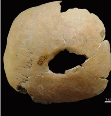
Fig.4.Ovalshapedtrauma(approximately31×21mm)attherightparietal.
andVI—amultidirectionalspreadofthelesionsbutstillwithout destructionofthefootbones.Thisskeletonistheonlyonewiththis kindoffootlesionsoftheskeletonsrecoveredfromthenecropolis inEstremoz.
3.1.6. Othermycoticinfections
Blastomycosis, paracoccidiomycosis, cryptococcosis, coccid-ioidomycosis, histoplasmosis and sporotrichosis can affect the foot bones (Aufderheide and Rodríguez-Martín, 1998; Ortner, 2003). Blastomycosis, paracoccidiomycosis, coccidioidomycosis andhistoplasmosis morecommonlyinfecttherespiratory tract, andthelesionstendtofocusonbonesinthat anatomicregion, but can spread through the blood stream (Aufderheide and Rodríguez-Martín, 1998; Ortner, 2003). Cryptococcosis tend to infectindividualswithaninadequateimmuneresponseandthe lesionscommonlyfocusatbonyprominences,cranialbonesand vertebrae(Ortner,2003).Sporotrichosisoftenaffectshands and feetbutthelesionsareperiostealratherthanlytic(Ortner,2003).
In the present skeleton, besides generalized vertebral osteoarthrosis,evidenceofculturalinterventionthatmayrelate totheMadurafootpathologyistheapparenttrepanationinthe cranial vault. An oval depression (approximately 31×21mm) existsontherightparietal(Fig.4),about18mmfromthelambdoid sutureand53mmfromthesagittalsuture.Themarginsare remod-elled,andtheexternalplateofthelesionbecomesthinnertowards theperforatedopeningwhichisnotcompatiblewithpost-mortem alterations. This asymmetry allows the diagnostic exclusion of enlargedforamina,andthesmooth-surfacedremodelledmargins andabsenceofporosity,atornearthelesion(bothintheouter andtheinnertable),indicatethatthis lesionprobablydoesnot haveaninfectiouscause.Sincetherearenototherssimilarlesions in this individual, it probably is not a metastatic carcinomaor myeloma.Also,therearenoradiatinglines,which arecommon inskullfracturesorbonespiculescirclingthelesion.Forallthese reasonsabove,thelesionpresentedisveryprobablyatrepanation. BasedonCampillo’sdescriptions(1977),themethodusedforthe trepanationwasprobablyascrapingperforationthroughaforward andbackwardmovementonthebonesurfaceuntilthevaultwore
intervention.Therefore,arelationshipwithMadurafootcannotbe excludediftheclinicalpatternofthisinfectionincludedweight loss,anaemiaandfevercommoninthispathology.
4. Conclusion
Accordingtothedifferentialdiagnosisreferred toabove this skeletonisprobablya Madurafootcase triggeredby truefungi (maduromycosis)withlesionsthatareusuallyfoundwiththistype ofcondition,althoughthereareotherpossiblecauses,specifically othermycoticinfections. Thispathology couldalsohave ledto symptomsthat wouldjustify therapeutictrepanation. Itis sug-gestedthatinthepastmaduromycosiscouldhavebeenpresent inEurope,particularlyintheMediterraneanregion,andespecially whentheclimaticconditionswereconducivetoitsdevelopment.
Acknowledgments
The authorswish toacknowledge the VeterinarianHospital fromtheUniversity ofÉvora that provided ustheradiographic imagingindispensable forthedifferentialdiagnosisin thiscase study.TheauthorsarealsothankfultoVandaAmaralandNuno Carric¸ofortheirhelpinphotoeditingandforcreatingthe3D imag-ingoftheleftcalcaneousandcuboidaswellastoAdrianaLowe forrevising theEnglishlanguage.Theconstructive advicefrom anonymousreviewersgreatlycontributedtothiscasestudy.
References
Arenas,R.,Lavalle,P.,2001.SubcutaneousMycosis:Mycetoma(Madurafoot).In: Arenas,R.,Estrada,R.(Eds.),TropicalDermatology.LandesBioscience, Georgetown,pp.51–61.
Aufderheide,A.C.,Rodríguez-Martín,C.,1998.TheCambridgeEncyclopediaof HumanPaleopathology.CambridgeUniversityPress,Cambridge. Bakshi,R.,Mathur,D.R.,2008.Incidenceandchangingpatternofmycetomain
westernRajasthan.IndianJ.Pathol.Microbiol.51,154–155.
Bonifaz,A.,Tirado-Sánchez,A.,Calderón,L.,Saúl,A.,Araiza,J.,Hernández,M., González,G.M.,Ponce,R.M.,2014.Mycetoma:experienceof482casesina singlecenterinMexico.PLoSNegl.Trop.Dis.8(8),e3102.
Brooks,S.,Suchey,J.M.,1990.Skeletalagedeterminationbasedontheospubis:a comparisonoftheAcsádi-NemeskériandSuchey-Brooksmethods.Hum.Evol. 5,227–238.
Brothwell,D.,2008.Tumoursandtumour-likeprocesses.In:Pinhasi,R.,Simon,M. (Eds.),AdvancesinHumanPaleopathology.Part2:Diagnosisand
InterpretationofDiseaseinHumanRemains.JohnWileyandSonsLtd., Chichester,pp.253–282.
Bruzek,J.,2002.Amethodforvisualdeterminationofsex,usingthehuman hipbone.Am.J.Phys.Anthropol.117,157–168.
Buonfrate,D.,Gobbi,F.,Angheben,A.,Marocco,S.,Farina,C.,VanDenEnde,J., Bisoffi,Z.,2014.AutochthonouscasesofmycetomainEurope:reportoftwo casesandreviewofliterature.PLoSOne9(6),e100590.
Cabrita,J.,1974.HumanmycosesinPortugal[1960–1973].Mycopathol.Mycol.54 (3),347–360.
Campillo,D.,1977.PaleopatologíaDelCráneoEnCatalu ˜na,ValenciaYBaleares. Montblanc-Martin,Barcelona.
Cook,E.R.,Seager,R.,Kushnir,Y.,Briffa,K.R.,Büntgen,U.,Frank,D.,Krusic,P.J., Tegel,W.,vanderSchrier,G.,Andreu-Hayles,L.,Baillie,M.,Baittinger,C.,
S.,Herzig,F.,Heussner,K.,Hofmann,J.,Janda,P.,Kontic,R.,Köse,N.,Kyncl,T., Levaniˇc,T.,Linderholm,H.,Manning,S.,Melvin,T.M.,Miles,D.,Neuwirth,B., Nicolussi,K.,Nola,P.,Panayotov,M.,Popa,I.,Rothe,A.,Seftigen,K.,Seim,A., Svarva,H.,Svoboda,M.,Thun,T.,Timonen,M.,Touchan,R.,Trotsiuk,V.,Trouet, V.,Walder,F.,Wa ˙zny,T.,Wilson,R.,Zang,C.,2015.Oldworldmegadroughts andpluvialsduringtheCommonEra.Sci.Adv.1(10),e1500561.
Davies,A.,1958.ThebonechangesofMadurafoot.Radiology70(6),841–847. Diaz,H.F.,Trigo,R.,Hughes,M.K.,Mann,M.E.,Xoplaki,E.,Barriopedro,D.,2011.
Spacialandtemporalcharacteristicsofclimateinmedievaltimesrevisited. Bull.Am.Meteorol.Soc.92,1487–1500.
Dieng,M.,Sy,M.,Diop,B.,Niang,S.,Ndiaye,B.,2003.Mycetoma:130cases.Ann. Dermatol.Venereol.130,16–19.
ElBagi,M.E.A.,2003.Newradiographicclassificationofboneinvolvementinpedal mycetoma.Am.J.Roentgenol.180,665–668.
ElMuttardi,N.,Kulendren,D.,Jemec,B.,2010.Madurafoot—mindthesoil.J.Plast Reconstr.Aesthet.Surg.63(7),e576–e578.
Fahal,A.H.,2004.Mycetoma:athornintheflesh.Trans.R.Soc.Trop.Med.Hyg.98, 3–11.
Fahal,A.H.,2011.Mycetoma.In:Guerrant,R.L.,Walker,D.H.,Weller,P.F.(Eds.), TropicalInfectiousDiseases.,3rdedn.Elsevier,Inc.,pp.565–568,SectionII: Pathogens;PartG:FungalInfections;Chapter83.
Hernández-Hernández,F.,Lopex-Martinez,R.,Méndez-Tovar,L.J.,
Manzano-Gayosso,P.,1995.Nocardiabrasiliensis:invitroandinvivogrowth inresponsetosteroidsexhormones.Mycopathologia132,79–85.
Hershkovitz,I.,Spiers,M.,Katznelson,A.,Arensberg,B.,1992.Unusualpathological conditionsinthelowerextremitiesofaskeletonfromancientIsrael.Am.J. Phys.Anthropol.88,23–26.
Hospenthal,D.R.,2009.Agentsofmycetoma.In:Mandell,G.L.,Bennett,J.E.,Dolin, R.(Eds.),PrinciplesandPracticeofInfectiousDiseases.ChurchillLivingstone Elsevier,Philadelphia,pp.3281–3286.
Iniesta,A.,Baptista,C.,Guinard,D.,Legré,R.,Gay,A.,2015.Mycétome actino-mycosiquedupiedàpropôsd’uncas.Ann.Chir.Plast.Esthét60, 164–167.
Magana,M.,1984.Mycetoma.Int.J.Dermatol.23,221–236.
Manchester,K.,1993.Unusualpathologicalconditioninthelowerextremitiesofa skeletonfromancientIsrael.Am.J.Phys.Anthropol.91,249–250.
Mansilla-Lory,J.,Conteras-López,E.A.,2009.MicetomaenMéxicoprehispánico. EstudioenlacolecciónesqueléticadelaculturadeTlatilco.Rev.Méd.Inst.Mex. SeguroSoc.47(3),237–242.
Mathur,D.,Joshi,K.,Mathur,A.,1979.Anetiologicalandpathologicalstudyof mycetomainwesternRajasthan.Curr.Med.Pract.23,151–161. Mestre,T.,Vieira,R.,Coutinho,J.,2015.Mycetomaofthefoot-diagnosisofthe
etiologicagentandsurgicaltreatment.Am.J.Trop.Med.Hyg.93(1),1–2. Ortner,D.,2003.IdentificationofPathologicalConditionsinHumanSkeletal
Remains.SanDiegoAcademicPress,SanDiego.
Plehn,A.,1928.Madurafuss(mycetomapedis).In:Kolle,W.,vonWassermann,A. (Eds.),HandbuchderPathogenMikroorganismen.,3rdedn.Jena:Fisher,pp. 113–132.
Ramos,E.,Figo,G.,Pina,R.,Lourenc¸o,C.,Malaba,N.,Trindade,L.,Vieira,A.,2013. Actinomicetomadopé−relatodeumcasoerevisãodaliteratura.Rev.Port. Doenc¸asInfecc.9(2),79–87.
Santos,C.M.C.,2002.Estimativadaestaturaapartirdosmetatársicos.Dissertac¸ão deMestradoemMedicinaLegal.Coimbra,FaculdadedeMedicina,
UniversidadedeCoimbra.[Unpublishedmonograph].
Spigelman,M.,Donoghue,H.D.,2001.Briefcommunication:unusualpathological conditioninthelowerextremitiesofaskeletonfromancientIsrael.Am.J.Phys. Anthropol.114,92–93.
Stine,S.,1994.ExtremeandpersistentdroughtinCaliforniaandpatagoniaduring medievaltime.Nature369,546–549.
Tomczyk,S.,Deribe,K.,Brooker,S.J.,Clark,H.,Rafique,K.,Knopp,S.,Utzinger,J., Davey,G.,2014.Associationbetweenfootwearuseandneglectedtropical diseases:asystematicreviewandmeta-analysis.PLoSNegl.Trop.Dis.8(11), e3285.
Waldron,T.,2009.Paleopathology.CambridgeUniversityPress,Cambridge. Venkatswami,S.,Sankarasubramanian,A.,Subramanyam,S.,2012.TheMadura