PAULA PR; CÉSAR MAP; ALBUQUERQUE EF; SILVA FPA. Intussuscepção sigmoidoanal com exteriorização de adenocarcinoma de sigmoide. Rev bras Coloproct, 2011;31(3): 294-298.
AbstRACt: the intestinal intussusception is a rare disease in adults, and is mostly caused by malignant neoplasm. symptoms are usually nonspeciic and chronic, and in most cases suggesting intestinal obstruction. Treatment consists of removing the malignant tumor. This article reports the case of a patient with hematochezia and apparent mass in the anus who underwent anterior rectosig -moidectomy and had the diagnosis of adenocarcinoma of the sigmoid conirmed.
Keywords: intestinal intussusception; colon adenocarcinoma; proctocolectomy.
Sigmoidoanal intussusception with exteriorization of sigmoid
adenocarcinoma
PEDRO ROBERTO DE PAULA1, MARIA AUXILIADORA PROLUNGATTI CÉSAR2, EDUARDO FORTES DE
ALBUQUERQUE3, FERNANDA PEREZ ADORNO DA SILVA4
1Assistant Professor and Doctor of the Medicine Department of Universidade de Taubaté; Head of the Coloproctology
Service of the University Hospital of Taubaté – Taubaté (SP), Brazil. 1Assistant Professor and Doctor of the Medicine
Department of Universidade de Taubaté; Head of the Anal Physiology Service of the University Hospital of Taubaté – Taubaté (SP), Brazil. 3Ex-Resident of general surgery at the University Hospital of Taubaté – Taubaté (SP), Brazil.
4Medical student at Universidade de Taubaté – Taubaté (SP), Brazil.
Financing source: none.
Conlict of interest: nothing to declare.
Submitted on: 01/02/2010 Approved on: 22/03/2010
INtRODUCtION
Intestinal intussusception is rare among adults, corresponding to 5% of all cases and 1% of intestinal obstructions; it is more common among infants. It occurs when the proximal bowel seg-ment (intussuscepts) penetrates the distal segseg-ment lumen (intussuscepted)1,2. It was first described
by Barbette de Amsterdam, in 1674, and Jonathan Hutchinson performed the first surgical reduction in 18713.
The symptoms of intussusception in adults,
un-like for children, are usually nonspeciic and chronic,
mostly suggesting intestinal obstruction4.
Among infants, it is mostly primary and benign, and the treatment consists of the reduction with
ene-ma in 80% of the cases. Among adults, the disease is frequently secondary to the organic cause, which
makes the preoperative diagnosis dificult; it is usu
-ally conirmed during laparotomy. The diagnosis is based on surgical indings. However, imaging tests
and minimally invasive procedures can be useful, such as the simple abdominal x-ray, contrast exami-nations, colonoscopy, ultrasonography and compu-ted tomography (CT)5.
The treatment of choice for malignant co-lon neoplasm is the removal of the tumor and all tissues involved in the angiolymphatic drainage, which are the main dissemination paths for these tumors6.
We reported a rare case in which the “head” of the invagination, which was formed by malignant sig-moid neoplasm, was exteriorized by the anus.
CAsE REPORt
We report the case of a 50-year-old black fema-le patient that had been presenting with hematochezia for nine months, which was independent from evacu-ations; also, for six months she had been noticing the exteriorization of a mass in the anal region during the effort to evacuate, thus being necessary to digitally re-duce it. She also presented with abdominal pain with
moderate colic at the left lank and hypogastrium be -fore evacuating. She had diarrhea intercalated with dry stool. She was regularly taking laxatives every three days. She lost 16 kg in the past eight months.
Proctocological examination showed: (a)
ins-pection: absence of skin tags, tumors, istulous oriice
and prolapse; (b) rectal touch: normotonic/hypotonic sphincter, identifying the presence of a tumor mass in the anterior wall, approximately 9 cm to the anal margin; (c) rectosigmoidoscopy: presence of vegeta-ting friable lesion in the anterior wall, with 6 cm in diameter, approximately 9 cm from the anal margin
(after biopsy); it moved upwards with the movement of the device.
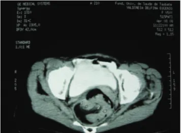
An abdominal and pelvic CT scan showed a target image in the rectosigmoid region, which su-ggested a loop inside a loop (Figure 1). The
colo-noscopy conirmed the presence of a vegetating le -sion of the sigmoid, hard with friable surface 20 cm from the anal border. The lesion was blocking 90% of the light and preventing the entrance of the de-vice. A new biopsy was conducted and showed the presence of a tubular pattern adenocarcinoma with strong atypia.
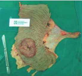
The patient was admitted for surgery and un-derwent a radical anterior upper rectosigmoidec-tomy, with primary manual termino-terminal anasto-mosis. At intraoperative, a vegetating sigmoid tumor of about 7.0x5.0 cm was observed, which was inva-ginated within the sigmoid and the rectum, thus allo-wing its exteriorization through the anus (Figure 2).
The presence of a main ganglion was identiied, with
2 cm in diameter, hard and located at the emergency of the inferior mesenteric artery. The anatomopatho-logical examination of the resected piece showed
that the lesion was microscopically iniltrated to the
serous and, out of the 17 dissected lymph nodes in the pericolic adipose tissue, only one was compro-mised. It was close to the inferior mesenteric artery (main ganglion) (Figures 3 and 4). The patient evol-ved without intercurrences, and was discharged from the hospital on the third postoperative day.
Figure 1. Pelvic computed tomography showing lesion in the rectal region (target image).
DIsCUssION
Intussusception can usually be classiied accor -ding to the compromised intestinal segment; it can be called enteric (small intestine), ileocolic (penetration of the ileum in the ileocecal valve), ileocecal (when the ileocecal valve is the intussusception point), colo-colic (colon) and colorectal4. In the studied case, the
sigmoid was exteriorized through the anal oriice.
The general clinical Picture is variable, but ab-dominal pain is the most common symptom, present in 100% of the studied cases2,5,7-9. Other symptoms are
nausea, vomit, hematochezia, changes in intestinal ha-bit, distension and palpable abdominal mass10,11.
Ho-wever, the abdominal mass is not a common inding
related to intussusceptions among adults, occurring in 7 to 42% of the cases2,7. In the studied case, the patient
presented with moderate abdominal pain before eva-cuating and at the moment of digital reduction of the mass that was exteriorized through the anus, hemato-chezia and changes in the intestinal habit.
The certain diagnosis is based on surgical indin -gs. However, imaging tests and minimally invasive procedures can be useful in cases like this, in which the diagnosis can be established before surgery.
Simple contrast abdomen x-rays, ultrasonogra-phy, abdominal CT scan and colonoscopy can reveal the segment that is affected by the disease1.
Barium studies like intestinal transit and enema may help the diagnosis; however, in cases of
com-plications, such as ischemia or intestinal perforation, they are contraindicated.
Ultrasonography is the choice due to the accu-racy to diagnose intussusceptions, both for adults and for children, showing the “target” image or the “onion skin” in the cross-sectional view, and the “pseudokidney sign” or “double kidney” in the lon-gitudinal view, which may not be pathognomonic, but very suggestive5.
Abdominal and pelvic CT have also been impor-tant for the preoperative diagnosis of this condition1,5.
The density of the mass generated by the compromi-sed segment, which is associated to the edema of the intestinal wall and the mesenteric, creates a characte-ristic signal in the CT, which is also called the “target sign”1. However, the tomography is not reliable
con-cerning the differentiation between neoplasm and the
nonspeciic thickening of the intestinal wall. Besides,
this examination is still limited since it is not availa-ble in all the emergency services and due to the need of contrast administration5. Colonoscopy may help in
cases of colonic obstruction.
The comparison between the different
examina-tions in order to deine the diagnosis, such as x-ray, ul -trasound, barium studies, colonoscopy and CT, shows that CT is the test with the most diagnostic sensitivity,
proving to be eficient and 88.6% more recommended
to diagnose intussusceptions among adults12,13. Our
patient was investigated with colonoscopy and
tomo-graphy, which conirmed the intussusception and its
Figure 3. Surgical piece with tumor lesion and the main affected ganglion.
REsUmO: A intussuscepção intestinal é uma doença rara em adultos, sendo na maior parte dos casos causada por neoplasia maligna. Os sintomas são geralmente inespecíicos e crônicos, na maioria das vezes sugerindo obstrução intestinal. O tratamento consiste na re -moção oncológica do tumor. Este artigo relata o caso de uma paciente com quadro de hematoquezia e exteriorização de massa através do ânus que foi submetido à retossigmoidectomia anterior alta em bloco e conirmado o diagnóstico de adenocarcinoma de sigmoide.
Palavras-chave: intussuscepção intestinal; adenocarcinoma de cólon; protocolectomia.
REFERENCEs
1. Wang N, Cui XY, Liu Y, Long J, Xu YH, Guo RX, et al. Adult intussusception: a retrospective review of 41 cases. World J Gastroenterol 2009;15(26):3303-8.
2. Yakan S, Calıskan C, Makay O, Denecli AG, Korkut MA.
Intussusception in adults: Clinical characteristics, diagnosis and operative strategies. World J Gastroenterol 2009;15(16):1985-9. 3. Butte BJM, Iniguez CA, Torres MJ. Intususcepción de colon
por lipoma. Rev Chi Cir 2006;58(2):151-4. etiology. It was possible to perform the preoperative
abdominal staging.
The treatment for the intussusceptions in adults demands an individual and systematic approach. La-parotomy is mandatory, once it can identify an organic lesion that could be neoplastic. The theoretical possi-bility to implant malignant cells indicates the resec-tion of the lesion. The need and the extension of this resection are controversial, since there is the risk of an unnecessary intestinal resection2,7.
In cases of colocolonic intussusceptions, it is ne-cessary to resect the segment with an oncologic pur-pose due to the high risk of malignity2,7,14, which could
be observed in this study; we had already diagnosed the sigmoid adenocarcinoma, and the patient presen-ted a sigmoido-anal insussusception. She was submit-ted to a radical rectosigmoidectomy, which was essen-cial, since the main lymphatic ganglion had metastatic compromise, in the root of the inferior mesenteric ar-tery.
As to the surgical approach, laparoscopy perfor-med by a trained team can be used with several ad-vantages; however, the conventional path is still more common15. In this case, the conventional approach
was used, and the patient did not present with any postoperative complication, being discharged early.
Nowadays, the patient has inished the chemotherapy
cycles, and is asymptomatic
The incidence of colorectal malignant neoplasm, which is the main organic cause of intussusception, has
been increasing in Brazil and represents the ifth most
common cause of death by cancer16. It is more
fre-quent among white males, especially those aged more than 40 years, with mean age of 60 and 70 years17,18.
In this case, the patient was female, black, at the ifth
decade of life, and her age was within the prevalent age group.
The malignant lesions of the colon are adenocar-cinomas in 95% of the cases, more commonly located in the rectosigmoid segment, which can be observed in the present case, in which the patient had a tumor affecting the sigmoid, which was the “head” of the in-vagination, that presented as a mass that was exterio-rized by the anal canal17,18.
At the postoperative staging of the disease pro-posed by Dukes, which considers the tumor depth in the intestinal wall and the compromise of regional
lymphatic ganglia, the case was classiied as Dukes
C for presenting a compromised regional lymphatic ganglion2,13. Imperfections in this classiication sys
-tem led to the creation of new classiications; TNM
is the most appropriate and the most used one, even though its accuracy is around 65%, which leads to a
law when estimating the evolution of patients11,13. The
stage of our patient was T3 N1 M0, stage IIIa. The involvement of lymphatic nodules is considered to be the most important discriminating factor when related to the short survival of patients13,19,20.
FINAL CONsIDERAtIONs
Intussusception is a rare condition, and, in this case, the “head” of the invagination was formed by a malignant sigmoid neoplasm, which was exteriori-zed through the anus. It was diagnosed at the
4. Marinis A, Yiallourou A, Samanides L, Dafnios N, Anastasopoulos G, Vassiliou I, et al. Intussusception of the bowel in adults: a review. World J Gastroenterol 2009;15(4):407-11
5. Korkmaz O, Yilmaz HG, Taçyildiz HH, Akgün Y.
Intussusception in adults. Ulus Travma Acil Cerrahi Derg 2009;15(2):154-8.
6. Gordon PH, Nivatvongs S. Principles and practice of surgery for the colon, rectum, and anus. 2nd ed. Missouri: Quality Medical Publishing; 1999. p. 900-1097.
7. Dell’abate P, Del Rio P, Sommaruga L, Arcuri MF, Sianesi M. Laparoscopic treatment of sigmoid colon intussusception by large malignant tumor. Case report. G Chir 2009;30(8-9):374-6.
8. Zissin R, Gayer G, Konen O, Shapiro-Feinberg M. Transient
colocolic intussusception. J Clin Imaging 2000;24(1):8-9.
9. Chen CF, Chuang CH, Lu CY, Hu C, Kuo TL, Hsieh JS. Adult
intussusception secondary to lymphangioma of the cecum: a
case report. Kaohsiung J Med Sci 2009;25(6):347-52.
10. Martin-Lorenzo JG, Torralba-Martinez A, Liron-Ruiz R. Intestinal invagination in adults. Int J Colorectal Dis 2004;19(1):68-72.
11. Warshauer DM, Lee JKT. Adult intussusception detected at
CT or MR imaging: clinical-imaging correlation. Radiology 1999;212(3):853–60.
12. Pisano G, Manca A, Farris S, Tatti A, Atzeni J, Calò PG. Adult idiopathic intussusception: a case report and review of the literature. Chir Ital 2009;61(2):223-9.
13. Chang CC, Chen YY, Chen YF, Lin CN, Yen HH, Lou HY. Adult intussusception in Asians: clinical presentations, diagnosis and treatment. J Gastroenterol Hepatol 2006;22(11):1767-71.
14. Hanan B, Diniz TR, da Luz MM, da Conceição SA, da Silva RG, Lacerda-Filho A. Intussusception in adults. Colorectal Dis 2010;12(6):574-8.
15. Chuang CH, Hsieh CB, Lin CH, Yu JC. Laparoscopic management of sigmoid colon intussusception caused by a malignant tumor: case report. Rev Esp Enferm Dig 2007;99(10):615-6.
16. Priolli DG, Cardinalli IA, Piovesan H, Margarido NF, Martinez CAR. Proposta para estadiamento do câncer colorretal baseada em critérios morfofuncionais. Correlação com níveis séricos do antígeno carcinoembrionário. Rev Bras Coloproct 2007;27(4):374-83.
17. Cruz GMG, Santana JL, Santana SKAA, Constantino JRM,
Chamone BC, Ferreira RMRS, et al. Câncer colônico - epidemiologia, diagnóstico, estadiamento e gradação tumoral de 490 pacientes. Rev Bras Coloproct 2007;27(2):139-53. 18. Roediger WEW. Estadiamento TNM. Trad. Marcio
Constantino Mimessi. 6a ed. São Paulo: Fundação Oncocentro de São Paulo; 2006. p. 347-59.
19. Mahmoud N, Rombeau J, Ross HM, Fry RD. Colon e reto. In: Sabiston DC. Tratado de Cirurgia: a base biológica da moderna prática cirúrgica. Rio de Janeiro: Elsevier; 2005. p. 1443-66
20. Araújo PHJ, Rangel MF, Batista TP. Intussuscepção íleo-cólica em adulto. Rev Bras Coloproct 2008;28(4):470-3.
Correspondence to: