Mammary Invasive Micropapillary Carcinoma in Cats:
Clinicopathologic Features and Nuclear DNA Content
F. S
EIXAS, C. P
ALMEIRA, M. A. P
IRES,
ANDC. L
OPESVeterinary Sciences Department, Centre of Animal and Veterinary Sciences, University of Tra´s-os-Montes e Alto Douro, Vila Real, Portugal (FS, MAP); Immunology Department, Portuguese Institute
of Oncology, and Pathology Department, Fernando Pessoa University, Porto, Portugal (CP); and Molecular Pathology and Immunology Department, Instituto de Cieˆncias Biome´dicas de Abel Salazar, University of Porto, and Pathology Department, Portuguese Institute of Oncology, Porto, Portugal (CL)
Abstract. Invasive micropapillary carcinoma (IMC) is a variant of infiltrating ductal carcinoma of
the breast associated with poor outcome. In this study, we report 16 carcinomas of the feline mammary gland displaying histologic features that correspond to IMC of the breast in women. The clinicopathologic findings, overall survival time, disease-free survival time, and nuclear DNA content of these cats were compared with 65 more common invasive mammary carcinomas (other feline mammary carcinoma [FMC]) of nonspecified type. IMC was associated with larger tumor size, higher histologic grade (P , .0001), deeper muscle invasion (P 5 .004), and more frequent lymphovascular invasion and nodal metastases (P 5 .009 and P 5 .001, respectively) than other FMCs. The aneuploid pattern was more frequent in IMC lesions. IMCs were also associated with lower survival rates. In summary, all cases of feline IMC were associated with clinicopathologic features of high biologic aggressiveness and should be classified as independent histologic types of FMC.
Key words: Cats; DNA image content; histologic types of neoplasms; invasive micropapillary
carcinomas; mammary neoplasms.
Introduction
Carcinomas are the most common neoplasias of
the feline mammary gland, representing 80–90% of
cases. They have high biologic aggressiveness and
poor prognosis
10,13,18and are an important cause of
mortality, especially in middle-age and elderly
females.
11Mammary carcinomas of female cats
have several types of histologic patterns, a mixture
of which is found in the same tumor. The World
Health Organization (WHO)
13classification of
feline mammary carcinomas (FMCs) is a descriptive
histologic classification that is difficult to match
due to the high number of patterns present in one
lesion.
10Most studies have not found associations
between histologic type and prognosis.
2,4,10In most
cases, looking for the histologic patterns may be
futile. However, there are some cases for which
unique histology does indicate a more or less
aggressive behavior. The micropapillary invasive
pattern is a distinct histologic pattern that we
observed in 16 female cat tumors. In addition to
histology, they also presented with DNA content,
behavior, and survival rates that justify the
in-dependent classification of invasive micropapillary
carcinoma (IMC).
Materials and Methods
Eighty-one feline IMCs surgically resected from 73 queens and observed in the Histopathology Laboratory of Tra´s-os-Montes e Alto Douro University were selected for this study. Tissues were fixed in 10% buffered formalin and embedded in paraffin. Three-micrometer–thick sections were stained with hematox-ylin and eosin (HE). All cases were reviewed and reclassified independently by two pathologists to detect micropapillary growth patterns without knowledge of the clinical outcome. We defined as IMCs all carcino-mas with more than 50% infiltrating micropapillary pattern. All cases were graded according to the Elston
and Ellis6 scoring system and staged according to the
WHO clinical staging system TNM for feline mammary
tumors.18To confirm the presence of myoepithelial cells
and to evaluate the tumor growth fraction, additional tumor sections were immunostained with a panel of
antibodies using the streptavidin-biotin-peroxidase
method. The markers used were cytokeratin (CK) 14 (1:20; NCL-LL 002; Novocastra Laboratories Ltd., Newcastle Upon Tyne, UK), p63 (1:150; 4A4; Neomar-kers, Fremont, CA), calponin (1:400; CALP, Dako Corp, Carpinteria, CA), and antigen Ki-67 (1:100; Mib-1; Dako Corp). Immunohistochemical data were
eval-uated as positive (+) or negative (2) for p63 (nuclear)
Mi-totic and Ki-67 indices were assessed in 8 to 10 representative areas at the periphery of the tumors, in the most mitotically active areas, or areas that presented the highest Mib-1 positivity at high-power magnification
(403). The numbers of immunopositive cells per 1,000
cells examined were expressed as percentages.
Automated image analysis was performed on 6-mm sections cut from the paraffin block used for histologic analysis. Slides were stained with the Feulgen DNA staining kit (Cell Analysis Systems, Elmhurst, IL), and analysis was performed using a CAS 200 image analyzer (Becton-Dickinson, San Jose, CA). In each lesion, a minimum of 100 nonoverlapping and well-preserved tumor nuclei and 20 to 30 normal lymphocytes (used as internal reference diploid cells) were measured. The resultant DNA histograms were analyzed and classified as diploid or aneuploid according to previously
de-scribed methods.14 The 5c exceeding rate (5cER),
defined as the percentage of tumor nuclei with DNA
content above 5n, was also evaluated.20
Statistical analysis was performed using SPSS soft-ware (version 11.5; SPSS Inc., Chicago, IL). The numeric parameters were tested by analysis of variance, Kruskal-Wallis, or Mann-Whitney tests. Other clinico-pathologic factors, including nuclear or histologic grade,
were evaluated by a x2 or Fisher test (2-sided). The
follow-up was performed by the referring surgeons for a 2-year period. Overall survival time was defined as the period between surgery and death due to cancer. Queens that died of other causes were censored at the time of death. Disease-free survival (DFS) was defined as the period of time between surgery and recurrent or metastatic disease. Survival rate was calculated by the Kaplan-Meier method, and statistical significance was examined using log-rank test. P , .05 was considered statistically significant.
Results
The micropapillary pattern, a common
architec-tural pattern in feline mammary carcinomas, was
observed in 70 of 81 (86.4%) analyzed invasive
carcinomas. With respect to the established
pro-portion of the micropapillary component, 16
(19.7%) carcinomas were classified as IMCs
be-cause more than 50% of these tumors had an
infiltrating micropapillary pattern.
Microscopically, IMC lesions were mainly
com-posed of atypical cells arranged in small tufts and
avascular papillae devoid of fibrovascular cores
(Figs. 1, 2) or in cell clusters floating in clear empty
spaces, mimicking extensive lymphatic invasion
(Fig. 3). Tumor cells, cuboidal or polygonal in
shape, had eosinophilic or amphophilic abundant
cytoplasm and vesicular pleomorphic nuclei with
prominent nucleoli. The cell clusters in the lacunar
spaces had solid or tubular configurations and were
arranged so that the polarities of the cells were
reversed, with abundant cytoplasm facing the clear
spaces on high-power magnification (Fig. 4). These
spaces were not lined by endothelial cells. In most
cases, stroma was fine reticular fibrocollagenous
and lacked desmoplasia.
Tumors with pure micropapillary patterns were
fairly unusual (1/16), and more frequently (15/16)
the infiltrating micropapillary areas were seen in
association with solid areas and small papillary
areas. Micropapillary carcinoma cells massively
invaded the thoracic or abdominal muscle (12/16)
and lymphatic vessels (16/16). The micropapillary
pattern was preserved in the emboli. Myoepithelial
cells were not identifiable in this tumor type using
HE-stained slides or basal/myoepithelial markers.
Fig. 1. Mammary gland; cat. Micropapillary
carci-noma composed of small papillary clusters lined by
polygonal cells showing high-grade nuclei. HE. Bar 5
60mm.
Fig. 2. Mammary gland; cat. Most papillae are
abortive and lack true fibrovascular cores. HE. Bar 5
The clinicopathologic features of IMCs and
other FMCs are summarized in Tables 1 and 2.
The DNA ploidy pattern was determined in 7
IMCs and 65 other FMCs (Table 1). There were no
significant differences between IMCs and other
FMCs with respect to the queens’ ages. The ages
for IMCs and other FMCs ranged from 7 to 15
years (mean age, 11.6
6 2.7 years) and from 6 to 19
years (mean age, 11.6
6 3.1 years), respectively.
IMC cases were associated with larger tumor sizes,
although this difference did not achieve statistical
significance. IMC lesions varied from 1 to 7 cm,
whereas other carcinomas varied from 0.55 to
5.5 cm. IMCs were associated with higher nuclear
grade (P 5 .001) and histologic grade (P , .0001)
than other more common invasive carcinomas of
nonspecified type. All IMC cases were high-grade
carcinomas (grade III), whereas 3 (4.6%) other
FMCs were classified as grade I, 36 (55.4%) as
grade II, and 26 (40.0%) as grade III.
Micropapil-lary carcinomas were mostly high-stage lesions,
although this difference did not achieve statistical
significance.
Deep-muscle invasion (P 5 .004),
lymphovascu-lar tumor emboli (P 5 .009), and lymph node
metastases (P 5 .001) were more common in IMCs.
Metastases showed the same arrangement found in
the primary tumors (Fig. 5): a pure micropapillary
or mixed solid micropapillary pattern. No
signifi-cant differences were observed in mitotic and Ki-67
labeling indices between the 2 tumor groups
featured in the study. Carcinomas in situ with pure
micropapillary or micropapillary and solid
cribri-form patterns were observed in 10 IMCs (62.5%)
and in 20 (30.8%) other FMCs.
With regard to DNA image analysis, no
signif-icant differences between IMCs and other FMCs
were observed. However, the aneuploid pattern
frequency and the percentage of true aneuploid
cells with DNA content higher than 5c were higher
in the IMC group.
Follow-up data is summarized in Table 3.
Over-all survival data was available in 8 queens
di-agnosed with IMC and 44 queens didi-agnosed with
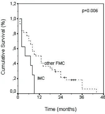
other FMCs of nonspecified type. Kaplan-Meier
survival curves revealed that queens with
nonmi-cropapillary tumors had better survival rates than
those with IMCs. All queens presenting IMC
lesions died or were euthanized due to oncologic
disease in a 9-month period after surgery. The
median overall survival was 4 months for IMCs,
whereas it was 10 months for queens presenting
with other FMCs (P 5 .006; Fig. 6).
Data concerning DFS were available in 7 queens
presenting with IMCs: 4 (57.1%) queens were
censored from the study because they were not
free of oncologic disease after mastectomy. All of
the other 3 queens developed disease within a
5-month period. On the other hand, just 2 of 30
queens presenting with other FMCs showed signs
of metastatic disease shortly after surgery and were
censored from analysis. The median DFS in this
group was 10 months (P 5 .04; Fig. 7).
Discussion
Mammary neoplasias are the third most frequent
neoplasias in cats, accounting for 12% of all feline
tumors and for 17% of all neoplasias of queens.
12,18Although the WHO classification of FMCs is
based on morphologic criteria,
13it does not
recognize the micropapillary pattern, which is
Fig. 3. Mammary gland; cat. Micropapillary
carci-noma showing neoplastic cell clusters floating in
a spongy loose stroma. HE. Bar5 120 mm.
Fig. 4. Mammary gland; cat. The cell clusters are
arranged so that the polarity of the cells is reversed. HE.
included in the spectrum of papillary tumors.
However, the presence of papillae devoid of
stromal fibrovascular cores was first described in
FMCs by Carpenter et al.,
2and there is a report of
2 cases in canine mammary glands.
3IMCs are defined as carcinomas composed of
small papillary structures without stroma or small
clusters of tumor cells lying within an artifactually
created clear space simulating vascular
chan-nels.
3,7,19,22,23,24IMC was first reported in women’s
breasts as a distinctive variant of infiltrating ductal
carcinoma. Subsequent studies have demonstrated
that this tumor, with its unique histologic features,
has a high incidence of lymphatic invasion and
lymph node metastases on clinical presentation,
resulting in poor outcome.
19Recently in humans,
carcinomas demonstrating histologic features
sim-ilar to IMC of the breast have been reported to
occur in various organs, including urinary bladder,
lung,
7,16,19ureter, salivary gland, colon,
19and
ovary.
7,16All cases were associated with poor
prognosis.
7,16,19All the cases reported in this series have similar
morphologic features to cases observed in women’s
breast tumors.
7,8,15,16,23,24They were characterized
by small micropapillary structures or cell
aggre-Table 1. Clinicopathologic features and nuclear DNA content of feline mammary IMCs compared with other
FMCs of nonspecified type.
IMCs (n 5 16) Other FMCs (n 5 65) P
Mean age (years) (n 5 76) 11.66 2.7 11.66 3.1 .99
Tumor size median (cm; minimum–
maximum) 2.8 (1.0–7.0) 2.5 (0.55–5.5) .17 Breed (n [%]) Domestic Shorthair 8 (50.0) 39 (60.0) Siamese 8 (50.0) 22 (33.8) Persian 0 (0.0) 1 (1.5) Crossbreed 0 (0.0) 3 (4.6) .55 Cutaneous ulceration (n [%]) Absent 8 (50.0) 48 (73.8) .08 Present 8 (50.0) 17 (26.2) Nuclear grade (n [%]) II 0 (0.0) 28 (43.1) .001 III 16 (100.0) 37 (56.9) Histologic grade (n) I 0 (0.0) 3 (4.6) II 0 (0.0) 36 (55.4) ,.0001 III 16 (100.0) 26 (40.0) Stage (TNM; n [%]) 1 1 (6.3) 19 (29.2) 2 3 (18.8) 18 (27.7) .06 3 12 (75.0) 28 (43.1) Invasion (n [%]) Stroma 4 (25.0) 43 (66.2) .004 Muscle 12 (75.0) 22 (33.8)
Lymphovascular tumour emboli (n [%])
Absent 0 (0.0) 20 (30.8) .009
Present 16 (100.0) 45 (69.2)
Lymph node metastases (n [%])
Absent 5 (31.3) 51 (78.5) .001 Present 11 (68.8) 14 (21.5) DNA ploidy (n [%]) (n 5 72) Diploid 1 (14.3) 21 (32.3) .43 Aneuploid 5 (85.7) 44 (67.7) 5cER (n [%]) (n 5 72) ,1 1 (14.3) 7 (10.8) .49 0 0 (0.0) 11 (16.9) .1 6 (85.7) 47 (72.3)
gates floating in clear spaces, mimicking vascular
invasion. These feline IMCs were associated with
larger tumor sizes and with higher histologic grade
and stage than nonmicropapillary carcinomas; our
data also confirmed the lymphotropic nature of
IMCs. Nodal involvement is a significant feature of
this tumor, and in this series, 10/17 (58.8%) IMC
tumors were associated with lymph node
metasta-ses, whereas just 14/65 (21.5%) other FMCs
presented lymph node metastases. All cases
re-ported here had aggressive clinical behavior and
shortened overall survival times than
nonmicropa-pillary invasive tumors. DFS was also low.
The association among aneuploidy, 5cER, and
IMC also agrees with reports that associate
abnormal DNA content
5,17,21and 5cER with
in-creased biologic aggressiveness.
1,9,20,21As reported above, the micropapillary pattern is
frequently observed in FMCs; foci of the
micro-Table 2. Comparison of mitotic and Ki-67 rates between IMCs and other FMCs.
IMCs (n 5 16) Other carcinomas (n 5 65) P
Mitotic index (%)
Median (minimum–maximum) 0.77 (0.28–3.69) 1.09 (0.18–3.45) .11
Ki-67 index (%)
Median (minimum–maximum) 34.75 (19.40–70.35) 41.10 (20.35–77.70) .07
IMC5 invasive micropapillary carcinoma; FMC 5 feline mammary carcinoma.
Table 3. Corrected survival rate in cats with available follow-up data.*
Overall survival (n 5 52) Disease-free survival (n 5 31)
n
Mean survival (months)
Average 1-year survival
rate (n [%]) n
Mean survival (months)
Average 1-year survival rate (n [%])
IMC 8 4 0 (0.0) 3 5 0 (0.0)
Other FMC 44 10 22 (50.0) 28 10 9 (32.1)
P .006 .04
IMC5 invasive micropapillary carcinoma; FMC 5 feline mammary carcinoma.
* Survival values are mean percentages for cumulative survival obtained by Kaplan-Meier test.
Fig. 5. Lymph node; cat. The lymph node
metas-tases show micropapillary patterns. HE. Bar5 120 mm.
Fig. 6. Comparison of overall survival time
be-tween invasive micropapillary carcinoma and other more common feline mammary carcinomas.
papillary pattern, less than 50% of the tumor area,
were detected in 53/65 (81.5%) other FMCs.
Because this tumor type is an aggressive variant
of carcinoma irrespective of the extent of IMC
7,16and its site of origin,
23the poor prognosis
classically associated with feline carcinomas may
be linked to this growth pattern. Although
speculative, this fact deserves more investigation.
In summary, this report described some clinical
and pathologic features of 16 cats with histologic
features that correspond to IMCs of the mammary
gland in women. To the best of our knowledge, this is
the first report describing the clinicopathologic
features of this tumor type in feline mammary glands.
Conclusion
All cases of feline IMC of the mammary gland
herein reported were associated with
clinicopatho-logic features of high bioclinicopatho-logic aggressiveness and
low survival rate and should be classified as an
independent histologic type of FMC. These results
also suggest that FMC, on the basis of histologic
and clinical outcome, is a good animal model to
study women’s breast cancer, especially IMC.
Acknowledgements
We thank Mrs. Lı´gia Bento for excellent technical assistance. This work was supported by a PhD grant
from the EU/Fundo Social Europeu, reference 2/5.3/ PRODEP/2001.
References
1 Auer GU, Caspersson TO, Wallgren AS: DNA content and survival in mammary carcinoma. Anal Quant Cytol 2:161–165, 1980
2 Carpenter JL, Andrews LK, Holzworth J: Tumours and tumour-like lesions. In: Diseases of the Cat: Medicine and Surgery, ed. Holzworth J, vol. 1, pp. 527–537. WB Saunders Co., Philadelphia, PA, 1987
3 Cassali GD, Serakides R, Ga¨rtner F, Schmitt FC: Invasive micropapillary carcinoma of the dog mammary gland. A case report. Arq Bras Med Vet Zootec 24:366–369, 2002
4 Castagnaro M, Casalone C, Ru G, Nervi GC, Bozzetta E, Caramelli M: Argyrophilic nucleolar organiser regions (AgNORs) count as indicator of post-surgical prognosis in feline mammary carcino-mas. Res Vet Science 64:97–100, 1998
5 Destexhe E, Bicker E, Coignoul F: Image analysis evaluation of ploidy, S-phase fraction and nuclear area in canine mammary tumours. J Comp Pathol 113:205–216, 1995
6 Elston CW, Ellis IO: Assessment of histological grade. In: Systemic Pathology—The Breast, ed. Elston CW and Ellis IO, 3rd ed., vol. 13, pp. 365– 384. Churchill and Livingstone, London, UK, 1998 7 Kim MJ, Gong G, Joo HJ, Ahn S-H, Ro JY: Immunohistochemical and clinicopathologic charac-teristics of invasive ductal carcinoma of breast with micropapillary carcinoma component. Arch Pathol Lab Med 129:1277–1282, 2005
8 Kuroda H, Sakamoto G, Ohnisi K, Itoyama S: Clinical and pathologic features of invasive micro-papillary carcinoma. Breast Cancer 11:169–174, 2004
9 Lorenzato M, Abboud P, Masure M, Bouttens D, Visseaux-Coletto B, Quereux C, Adnet JJ: Image
cytometry detection of breast cancer cells with.5C
DNA content and minor DNA stemlines. Anal Quant Cytol Histol 22:199–205, 2000
10 Madewell BR, Theilen GH: Tumors of the mamma-ry gland. In: Veterinamamma-ry Cancer Medicine, ed. Theilen GH and Madewell BR, 2nd ed., pp. 327– 340. Lea & Febiger, Philadelphia, PA, 1987
11 Millanta F, Lazzeri G, Mazzei M, Vannozzi I, Poli A: MIB-1 labelling index in feline dysplastic and neoplastic mammary lesions and its relationship with postsurgical prognosis. Vet Pathol 39:120–126, 2002 12 Misdorp W: Tumors of the mammary gland. In: Tumors in Domestic Animals, ed. Meuten DJ, 4th ed., pp. 575–606. Iowa State Press, Ames, IA, 2002 13 Misdorp W, Else RW, Hellme´n E, Lipscomb TP:
Histological classification of mammary tumors of the dog and cat. In: WHO International Histological Classification of Tumors of Domestic Animals, 2nd ed., vol. 7, pp. 11–29. World Health Organization
Fig. 7. Comparison of disease-free survival between
invasive micropapillary carcinoma and other mammary carcinomas.
and Armed Forces Institute of Pathology, Washing-ton, DC, 1999
14 Oliveira PA, Palmeira C, Lourenc¸o L, Lopes C: Evaluation of DNA content in preneoplastic changes of mouse urinary bladder induced by N-butyl-N-(4-hydroxybutyl)nitrosamine. J Exp Clin Cancer Res 24:609–616, 2005
15 Paterakos M, Watkin WG, Edgerton SM, Moore DH, Thor AD: Invasive micropapillary carcinoma of the breast: a prognostic study. Hum Pathol 30:1459–1463, 1999
16 Ramalingam P, Middleton LP, Tamboli P, Troncoso P, Silva EG, Ayala AG: Invasive micropapillary carcinoma of the breast metastatic to the urinary bladder and endometrium: diagnostic pitfalls and review of the literature of tumours with micropapil-lary features. Ann Diagn Pathol 7:112–119, 2003 17 Rutteman GR, Cornelisse CJ, Dijkshoorn NJK,
Poortman J, Misdorp W: Flow cytometric analysis of DNA ploidy in canine mammary tumors. Cancer Res 48:3411–3417, 1988
18 Rutteman GR, Withrow SJ, MacEwen EG: Tumors of the mammary gland. In: Small Animal Clinical Oncology, ed. Withrow SW and MacEwen EG, 3rd ed., pp. 467–473. WB Saunders Co., Philadelphia, PA, 2001
19 Sakamoto K, Watanabe M, De la Cruz C, Honda H, Ise H, Mitsui K, Namiki K, Mikami Y, Moriya
T, Sasano H: Primary invasive micropapillary carcinoma of the colon. Histopathology 47:479– 484, 2005
20 Santos L, Lameiras C, Afonso J, Palmeira C, Pereira S, Costa C, Amaro T, Bento MJ, Morais A, Criado B, Lopes C: Is DNA content alteration a consequence of proliferative and differentiation changes in urothelial bladder tumours? Acta Urol 20:9–17, 2003
21 Sidoni A, Cavaliere A, D’Amico GA, Brachelente G, Bucciarelli E: Biopathological significance of single cell DNA aneuploidy measured by static cytometry in breast cancer. Breast 10:325–329, 2001
22 Thor AD, Eng C, Devries S, Paterakos M, Watkin WG, Edgerton S, Moore DH, Etzell J, Waldman FM: Invasive micropapillary carcinoma of the breast is associated with chromosome 8 abnormalities detected by comparative genomic hybridization. Hum Pathol 33:628–631, 2002
23 Walsh MM, Bleiweiss IJ: Invasive micropapillary carcinoma of the breast: eight cases of an under-recognized entity. Hum Pathol 32:583–589, 2001
24 Zekioglu O, Erhan Y, Bayramoglu H, O¨ zdemir N:
Invasive micropapillary carcinoma of the breast: high incidence of lymph node metastasis with extranodal extension and its immunohistochemical profile compared with invasive ductal carcinoma. Histopathology 44:18–23, 2004
Request reprints from Dr. Fernanda Seixas Travassos, Laborato´rio de Histologia e Anatomia Patolo´gica, Clı´nicas Veterina´rias, UTAD, 5001-801 Vila Real (Portugal). E-mail: fseixas@utad.pt.