Immunohistochemical profile of high-grade ductal
carcinoma
in situ
of the breast
Amanda Arantes Perez,IRafael Malagoli Rocha,IIDe´bora Balabram,IA´tila da Silva Souza,IHelenice GobbiI IFaculdade de Medicina da Universidade Federal de Minas Gerais, Breast Pathology Laboratory, Belo Horizonte/MG, Brazil.IIA.C. Camargo Cancer
Hospital, Sa˜o Paulo/SP, Brazil.
OBJECTIVE:To determine the frequency of the immunohistochemical profiles of a series of high-grade ductal carcinomain situof the breast.
METHODS:One hundred and twenty-one cases of high-grade ductal carcinomain situ, pure or associated with invasive mammary carcinoma, were identified from 2003 to 2008 and examined with immunohistochemistry for estrogen receptor, human epidermal growth factor receptor 2, cytokeratin 5, and epidermal growth factor receptor. The tumors were placed into five subgroups: luminal A, luminal B, HER2, basal-like, and ‘‘not classified’’.
RESULTS: The frequencies of the immunophenotypes of pure ductal carcinoma in situ were the following: luminal A (24/42 cases; 57.1%), luminal B (05/42 cases; 11.9%), HER2 (07/42 cases; 16.7%), basal-like phenotype (00/42 cases; 0%), and ‘‘not classified’’ (06/42 cases; 14.3%). The immunophenotypes of ductal carcinomain situ
associated with invasive carcinoma were the following: luminal A (46/79 cases; 58.2%), luminal B (10/79 cases; 12.7%), HER2 (06/79 cases; 7.6%), basal-like (06/79 cases; 7.6%), and ‘‘not classified’’ (11/79 cases; 13.9%). There was no significant difference in the immunophenotype frequencies between pure ductal carcinomain situand ductal carcinoma in situ associated with invasive carcinoma (p.0.05). High agreement was observed in immunophenotypes between both components (kappa = 0.867).
CONCLUSION:The most common immunophenotype of pure ductal carcinomain situwas luminal A, followed by HER2. The basal-like phenotype was observed only in ductal carcinoma in situ associated with invasive carcinoma, which had a similar phenotype.
KEYWORDS: Ductal Carcinomain situ; Molecular Profile; Immunoprofile; Molecular Phenotype.
Perez AA, Rocha RM, Balabram D, Souza A´ S, Gobbi H. Immunohistochemical profile of high-grade ductal carcinomain situof the breast. Clinics. 2013;68(5):674-678.
Received for publication onDecember 21, 2012;First review completed onJanuary 4, 2013;Accepted for publication onFebruary 4, 2013
E-mail: [email protected]
Tel.: 55 31 34099118
& INTRODUCTION
Breast cancer has a heterogeneous natural history and varying morphological and immunohistochemical profiles and prognoses. Until recently, the classification and therapeutic decisions regarding breast carcinoma were based on histological features and classical prognostic and predictive factors.
Recent cDNA microarray studies have identified distinct groups of tumors with disparate prognoses, resulting in a new classification of invasive breast carcinomas. Breast tumors are categorized into five subgroups based on their molecular profile: luminal A, luminal B, HER2, basal-like,
and normal breast-like (1–3). Gene expression signatures and protein expression profiles through immunohistochem-istry correlate well in invasive breast cancers (4–8).
The basal-like subtype has attracted the attention of researchers and physicians because it has been associated with poor clinical outcomes. These outcomes likely reflect this subtype’s high proliferative capacity and the lack of directed therapies, as basal-like tumors do not typically express estrogen receptor or overexpress HER2 (2–3). The basal-like phenotype is more frequent among invasive tumors that have a high histological grade (9–11).
Ductal carcinoma in situ (DCIS) represents a precursor lesion to invasive breast cancer, in which most molecular alterations are already present (12). Based on this model, basal-like invasive ductal carcinomas, which are primarily high grade, arise from high-grade DCIS.
Many studies have evaluated invasive mammary carci-nomas (IMCs), but few studies have examined the molecular profile of DCIS of the breast through immuno-histochemistry. Assuming that DCIS is a precursor of invasive carcinoma, we expect that the molecular phenotypes
Copyrightß2013CLINICS– This is an Open Access article distributed under the terms of the Creative Commons Attribution Non-Commercial License (http:// creativecommons.org/licenses/by-nc/3.0/) which permits unrestricted non-commercial use, distribution, and reproduction in any medium, provided the original work is properly cited.
No potential conflict of interest was reported.
previously described for IMC will also be identified among cases of DCIS.
The purpose of this study was to determine the frequency of the basal-like phenotype and other immunophenotypes in a series of cases of high-grade DCIS of the breast, either pure or associated with invasive carcinoma, to compare the frequency of immunohistochemical profiles in pure or IMC-associated DCIS cases and to assess the agreement of immunophenotypes between in situ and invasive compo-nents in DCIS cases that are associated with invasive components. We chose a specific subset of high-grade DCIS because the basal-like phenotype is more frequent among invasive tumors that have a high histological grade (9–11). By identifying basal-like DCIS, these tumors can be treated more aggressively than other DCIS subtypes to improve the prognosis.
& MATERIALS AND METHODS
Specimen selection
In total, 202 cases of high-grade DCIS, pure or associated with invasive carcinoma, were consecutively identified from the histopathology files of the Breast Pathology Laboratory, School of Medicine, Federal University of Minas Gerais, Brazil, from 2003 to 2008. Seventy-one cases (35%) were excluded: 14 cases showed autolysis (7%), 32 cases received neoadjuvant chemotherapy (16%), 11 cases were a local recurrence following breast-conserving surgery (5%), and 14 cases had insufficient tumor tissue for sectioning (7%). The original hematoxylin and eosin-stained sections from 131
cases were reviewed to confirm the diagnosis of high-grade DCIS and select a representative block for immunostaining. Formalin-fixed, paraffin-embedded blocks were not found in 10 cases (5% of the total). Thus, immunohistochemistry was performed in 121 high-grade DCIS cases (60% of the total).
The criteria defined by the World Health Organization (2012) were used for the histopathological diagnosis of DCIS (13). The DCIS histological grade was determined using the criteria of Scott et al., 1997 (14).
Clinical, tumor, and treatment features
The age at diagnosis, menopausal status, tumor size, primary surgical treatment, and adjuvant therapy were retrospectively evaluated.
Menopausal status was defined based upon in-person interview data.
Immunohistochemistry
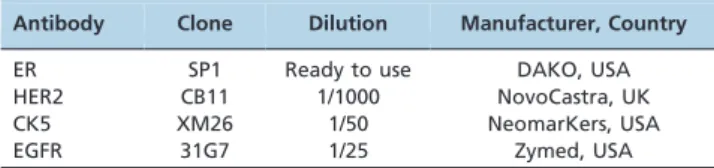
Estrogen receptor (ER), HER2 overexpression, cytokeratin 5 (CK5), and epidermal growth factor receptor (EGFR) were assessed. The reactions were performed with automated equipment (BenchMark XT/LTTM – Ventana, USA) using the UltraView Universal REF 760–500 DAB kit (Ventana, USA) according to the manufacturer’s instructions. The sources and dilutions of the primary antibodies are listed in Table 1.
Allred’s scoring system was used to evaluate estrogen receptor status; cases were considered positive when at least
Figure 1 - A)High-grade ductal carcinomain situassociated with invasive mammary carcinoma positive for estrogen receptor (100x). Arrows indicate thein situcomponent.B)High-grade ductal carcinomain situpositive for HER2 (400x).C)High-grade ductal carcinoma
1% of neoplastic cells showed moderate or strong nuclear staining (15). HER2 overexpression was analyzed according to the American Society of Clinical Oncology and College of American Pathologists (16). Any degree of cytoplasmic staining for CK5 and any degree of distinct membranous staining for EGFR were considered positive expression (17).
Immunohistochemical profile
The tumors were divided into five subgroups according to their immunohistochemical profile: luminal A (ER+/
HER2-), luminal B (ER+/HER2+), HER2 (ER-/HER2+),
basal-like (ER-/HER2-/EGFR+ and/or CK5+), and ‘‘not
classified’’ (all markers negative) (17,18). The basal-like phenotype was defined according to Nielsen’s criteria (6).
Statistical analysis
Pearson’s asymptotic and exact chi-square tests were used to compare proportions. The Mann-Whitney test was used to compare medians. A p-value,0.05 was considered statistically significant. The kappa test was used to assess the concordance between phenotypes. Kappa values greater than 0.80 demonstrated excellent agreement (19). This study was approved by the Research Ethics Committee of the Federal University of Minas Gerais (protocol 655/08).
& RESULTS
Pure DCIS was detected in 46/131 cases (35% of the total), and 85/131 cases (65% of the total) were DCIS associated with invasive carcinoma. Immunohistochemistry was per-formed for 121 cases, including 42 cases of pure DCIS (35% of the total) and 79 cases of DCIS associated with invasive carcinoma (65% of the total).
The median age at diagnosis was 51 years (standard deviation ¡14 years) among cases of pure DCIS and 53 years (standard deviation¡19 years) among cases of IMC-associated DCIS (p= 0.913). The median DCIS size was 13 mm. There was no significant difference in menopausal status (p= 0.779) or median tumor size (p= 0.836) between pure and IMC-associated DCIS cases. There was a sig-nificant difference in primary surgical treatment and adjuvant therapy between pure and IMC-associated DCIS cases (p,0.05). Cases with DCIS associated with IMC were treated with more extensive surgery and more often received adjuvant therapy.
The frequencies of the molecular immunophenotypes of DCIS are shown in Table 2 and Figure 1. Among samples of pure DCIS, the luminal A phenotype was the most common (24/42 cases; 57.1%), followed by the HER2 phenotype (07/ 42 cases; 16.7%), the ‘‘not classified’’ phenotype (06/42 cases; 14.3%), and the luminal B phenotype (5/42 cases; 11.9%). The basal-like phenotype was not identified among the pure DCIS cases. The immunophenotypes of DCIS associated with invasive carcinoma were the following: luminal A (46/79 cases; 58.2%), luminal B (10/79 cases;
12.7%), HER2 (06/79 cases; 7.6%), basal-like (06/79 cases; 7.6%), and ‘‘not classified’’ (11/79 cases; 13.9%).
There was no significant difference in frequency between immunophenotypes in pure and IMC-associated DCIS samples (p.0.05). Excellent agreement was observed between in situ and invasive components with regard to immunophenotypes (kappa = 0.867).
& DISCUSSION
Breast cancer comprises a heterogeneous group of diseases with regard to presentation, morphology, biologi-cal characteristics, clinibiologi-cal behavior, and response to therapy (6,9,20). In the past 20 years, concomitant with the wide use of screening mammography, the DCIS incidence has risen dramatically (21–22). The understanding of the biology and clinical behavior of DCIS is currently limited. Molecular profiling through gene array studies is likely to have a major impact on breast cancer classification and management, and it is important that similar approaches are taken to advance the understanding of DCIS.
The immunohistochemical staining of paraffin sections using antibody panels has been shown to be a reliable surrogate for the molecular classification of invasive breast cancers through gene expression profiling studies. Antibodies against estrogen receptor, progesterone receptor, HER2, cytokeratin 5/6, and EGFR have been particularly useful for this purpose (4–8). In fact, this approach to molecular classification (that is, using immunostaining as a surrogate for expression profiling) is arguably the most practical approach to phenotyping a large number of archived specimens for which fresh tissue is not available for expression array analysis.
Recent advances have led to an emerging molecular classification for invasive breast cancer based on the biological characteristics of the tumor rather than being limited to morphological analysis. Much less attention has been focused on dissecting the biological subtypes of DCIS, the immediate precursor to invasive breast cancer. There have been discrepancies in the results of those studies. There have also been discrepancies in the relative frequency of subtypes betweenin situand invasive disease (17,20,23– 26).
In our series, we showed that DCIS can be classified into the five immunophenotypes that have been described for
Table 2 - Immunohistochemical profile of high-grade DCIS (pure or associated with invasive mammary carcinoma).
Pure DCIS DCIS+IMC
Phenotype N (%) N (%) p-value*
Luminal A 24 (57.1%) 46 (58.2%) 0.264 Luminal B 05 (11.9%) 10 (12.7%)
HER2 07 (16.7%) 06 (7.6%)
Basal-like 00 (0%) 06 (7.6%) ‘‘Not classified’’ 06 (14.3%) 11 (13.9%)
TOTAL 42 (100%) 79 (100%)
DCIS = ductal carcinomain situ; IMC = invasive mammary carcinoma Luminal A: ER+/HER2-; Luminal B: ER+/HER2+; HER2: ER-/HER2+; Basal: ER-/
HER2-/EGFR+and/or CK5+; ‘‘Not classified’’: ER-/HER2-/EGFR-/CK5-.
p= significance level.
*= Exact Pearson’s chi-square test; refers toin situcomponent (pure or associated with invasive carcinoma).
Table 1 -Sources and dilutions of the primary antibodies.
Antibody Clone Dilution Manufacturer, Country
ER SP1 Ready to use DAKO, USA
HER2 CB11 1/1000 NovoCastra, UK
CK5 XM26 1/50 NeomarKers, USA
invasive breast carcinomas using a panel of four markers. Molecular classification improves the current morphological classification and provides insight into the biology under-lying DCIS heterogeneity.
Previous studies have evaluated the immunoprofiles of DCIS independently of histological grade (17,20,23). Bryan et al. restricted their study to high-nuclear-grade DCIS lesions because basal-like invasive carcinomas are poorly differentiated tumors in histopathological studies (24). In our study, we chose a specific subset of high-grade DCIS of the breast to determine the frequency of the basal-like phenotype because this subtype is more frequent among invasive tumors that have a high histological grade (9– 11,24).
Basal-like tumors have attracted the attention of pathol-ogists, surgeons, and oncologists and constitute a prognostic group of breast cancers with aggressive behavior. These tumors affect younger patients, are more prevalent in African-American women, and exist more often as interval cancers (18). Basal-like tumors are candidates for specific targeted therapy. By identifying basal-like DCIS, surgeons and oncologists can likely treat these tumors more aggres-sively to improve the prognosis.
There is no consensus regarding markers that define basal-like tumors by immunohistochemistry (11). Some groups have suggested that basal-like tumors are triple negative, i.e., negative for estrogen and progesterone receptors and HER2. However, the triple-negative pheno-type is not synonymous with the basal-like phenopheno-type (9,27). Other authors consider the basal-like phenotype as showing positivity for basal cytokeratins, regardless of the expression of other markers (5). EGFR positivity, which is associated with positivity for basal cytokeratins and negativity for estrogen receptor and HER2, defines basal-like breast cancers for other authors (6,17–18). EGFR gene amplification and/or high EGFR expression are biological predictors of poor prognosis in breast carcinomas. EGFR has also been used as a marker of the basal phenotype and has been investigated as a potential target therapy for human breast cancer (17–18). In our study, we classified the basal-like phenotype according to Nielsen’s criteria (6), which includes EGFR evaluation.
Our data demonstrated good agreement between the molecular profile of DCIS and synchronous IMC with regard to immunohistochemical phenotypes. Contemporary models suggest that high- and low-grade invasive ductal cancers arise through disparate pathways: high-grade IMC develops directly from poorly differentiated DCIS rather than low-grade IMC or low-low-grade DCIS. DCIS represents a stage in the development of breast cancer in which most molecular alterations are already present [12,28). Based on this model, basal-like invasive ductal carcinomas, which are primarily high grade, arise from high-grade DCIS. Until recently, however, a basal-likein situcomponent was not known to exist.
Livasy et al., Paredes et al., Bryan et al., and Clark et al. observed the following frequencies of the basal phenotype in pure DCIS: 8%, 10.1%, 6%, and 4.2%, respectively (17,23– 24,26). In our study, the basal-like phenotype was not identified among pure DCIS. We identified the basal phenotype in 7.6% of IMC-associated DCIS cases. Tamimi et al. observed a similar frequency (7.7%) of the basal phenotype for thein situcomponent (20). This difference in frequencies might be related to the criteria used to classify
tumors as well as variables from the preanalytical and analytical phases of immunohistochemical reactions, such as the choice of primary antibodies. According to the tumor type-specific evolution from DCIS to invasive carcinoma (28), another possible interpretation of these differences in frequencies is that the DCIS lesions in the cited papers were diagnosed at different stages of progression.
Our data showed an increased frequency of the HER2 phenotype in pure high-grade DCIS, which is consistent with previous studies demonstrating a higher prevalence of HER2 protein overexpression and gene amplification among DCIS in comparison to invasive breast cancers and suggesting that HER2/neu gene amplification is inversely related to invasive progression in DCIS patients (28–29).
We did not observe a significant difference in the frequencies of molecular phenotypes in pure or IMC-associated DCIS. Tamimi et al. showed differences in the frequencies of luminal A, luminal B, and HER2 phenotypes in pure DCIS versus invasive breast cancers, but there was no difference in the basal-like phenotype and ‘‘not classified’’ cases (20).
We did not observe the basal-like phenotype in pure DCIS, despite the presence of this profile in IMC-associated DCIS cases. Based on the tumor type-dependent model of breast cancer progression from DCIS to invasive cancer, triple-negative cancers may progress much faster than the other three tumor types, suggesting that some unrecognized mechanisms or features might help these tumors progress. At the time of breast tumor diagnosis, more aggressive types will have fewer DCIS lesions in comparison to the less aggressive types, with more tumors still in the DCIS phase. With regard to the speed of becoming invasive breast cancers, the fastest are the triple-negative lesions, while pure HER2-positive tumors are almost three times slower. Luminal A and luminal B DCIS are two tumor types that show intermediate probabilities of progression to invasive carcinoma (28). Therefore, our results are in agreement with the tumor type-dependent model of breast cancer progres-sion from DCIS to invasive cancer.
In conclusion, immunophenotypes that were previously identified among invasive mammary carcinomas were also observed among cases of DCIS. The most common immunophenotype of pure DCIS was luminal A, followed by the HER2 phenotype. The basal-like phenotype was observed only in DCIS associated with invasive carcinoma, which had a similar phenotype. No significant difference was identified between pure DCIS and IMC-associated DCIS phenotypes. There is a critical need for prospective analyses of new and known breast cancer molecular markers in large cohorts of patients with DCIS to differ-entiate indolent from aggressive DCIS and better tailor the need and extent of current therapies.
& ACKNOWLEDGMENTS
We are grateful to Sandra J. Olson, MBs, for editing the manuscript for language. This work was supported in part by grants from Fundac¸a˜o de Amparo a Pesquisa de Minas Gerais (FAPEMIG), Fundac¸a˜o de Amparo a` Pesquisa de Sa˜o Paulo (FAPESP), Conselho Nacional de Desenvolvimento Cientı´fico e Tecnolo´gico (CNPq), and Coordenac¸a˜o de Aperfeic¸oamento de Pessoal de Nı´vel Superior (CAPES).
& AUTHOR CONTRIBUTIONS
Balabram D performed the statistical analysis. Souza AS separated the original slides and blocks for immunohistochemistry. Gobbi H participated in the design and coordination of the study, review of original slides, and analysis of the immunohistochemical reactions and helped drafting the manuscript. All of the authors have read and approved the final version of the manuscript.
& REFERENCES
1. Perou CM, Sorlie T, Eisen MB, van de Rijn M, Jeffrey SS, Rees CA, et al. Molecular portraits of human breast tumors. Nature. 2000;406(6797):747-52, http://dx.doi.org/10.1038/35021093.
2. Sorlie T, Perou CM, Tibshirani R, Aas T, Geisler S, Johnsen H, et al. Gene expression patterns of breast carcinomas distinguish tumor subclasses with clinical implications. Proc Natl Acad Sci USA. 2001;98(19):10869-74, http://dx.doi.org/10.1073/pnas.191367098.
3. Sorlie T, Tibshirani R, Parker J, Hastie T, Marron JS, Nobel A, et al. Repeated observation of breast tumor subtypes in independent gene expression data sets. Proc Natl Acad Sci USA. 2003;100(14):8418-23, http://dx.doi.org/10.1073/pnas.0932692100.
4. Rakha EA, Putti TC, Abd El-Rehim DM, Paish C, Green AR, Powe DG, et al. Morphological and immunophenotypic analysis of breast carcino-mas with basal and myoepithelial differentiation. J Pathol. 2006; 208(4):495-506, http://dx.doi.org/10.1002/path.1916.
5. Rakha EA, El-Sayed ME, Green AR, Paish EC, Lee AH, Ellis IO. Breast carcinoma with basal differentiation: a proposal for pathology definition based on basal cytokeratin expression. Histopathology. 2007;50(4):434-38, http://dx.doi.org/10.1111/j.1365-2559.2007.02638.x.
6. Nielsen TO, Hsu FD, Jensen K, Cheang M, Karaca G, Hu Z, et al. Immunohistochemical and clinical characterization of the basal-like subtype of invasive breast carcinoma. Clin Cancer Res. 2004;10(16):5367-74, http://dx.doi.org/10.1158/1078-0432.CCR-04-0220.
7. Livasy CA, Karaca G, Nanda R, Tretiakova MS, Olopade OI, Moore DT, et al. Phenotypic evaluation of the basal-like subtype of invasive breast carcinoma. Mod Pathol. 2006;19(2):264-71, http://dx.doi.org/10.1038/ modpathol.3800528.
8. Matos I, Dufloth R, Alvarenga M, Zeferino LC, Schmitt F. p63, cytokeratin 5, and P-cadherin: three molecular markers to distinguish basal phenotype in breast carcinomas. Virchows Arch. 2005;447(4):688-94, http://dx.doi.org/10.1007/s00428-005-0010-7.
9. Reis-Filho JS, Tutt AN. Triple negative tumors: a critical review. Histopathology. 2008;52(1):108-18.
10. Silva F, Carvalho S, Milanezi F, Schmitt FC. Basal-like carcinoma of the breast. Acta Med Port. 2008;21(4):373-78.
11. Fadare O, Tavassoli FA. The phenotypic spectrum of basal-like breast cancers: a critical appraisal. Adv Anat Pathol. 2007;14(5):358-73, http:// dx.doi.org/10.1097/PAP.0b013e31814b26fe.
12. Buerger H, Otterbach F, Simon R, Poremba C, Diallo R, Decker T, et al. Comparative genomic hybridization of ductal carcinoma in situ of the breast – evidence of multiple genetic pathways. J Pathol. 1999;187(4):396-402, http://dx.doi.org/10.1002/(SICI)1096-9896(199903)187:4,396::AID-PATH286.3.0.CO;2-L.
13. Schnitt SJ, Allred C, Britton P, Ellis IO, Lakhani SR, Morrow M, et al. Ductal carcinoma in situ. In: Lakhani SR, Ellis IO, Schnitt SJ, Tan PH, van de Vijver MJ, editors. WHO Classification of Tumours of the Breast. IARC: Lyon; 2012.
14. Scott MA, Lagios MD, Axelsson K, Rogers LW, Anderson TJ, Page DL. Ductal carcinoma in situ of the breast: reproducibility of histological
subtype analysis. Hum Pathol. 1997;28(8):967-73, http://dx.doi.org/10. 1016/S0046-8177(97)90013-7.
15. Hammond ME, Hayes DF, Dowsett M, Allred DC, Hagerty KL, Badve S, et al. American Society of Clinical Oncology/College of American Pathologists guideline recommendations for immunohistochemical test-ing of estrogen and progesterone receptors in breast cancer. Arch Pathol Lab Med. 2010;134(7):907-22.
16. Wolff AC, Hammond ME, Schwartz JN, Hagerty KL, Allred DC, Cote RJ, et al. American Society of Clinical Oncology/College of American Pathologists guideline recommendations for human epidermal growth factor receptor 2 testing in breast cancer. J Clin Oncol. 2007;25(1):118-45. 17. Livasy CA, Perou CM, Karaca G, Cowan DW, Maia D, Jackson S, et al. Identification of a basal-like subtype of breast ductal carcinoma in situ. Hum Pathol. 2007;38(2):197-204, http://dx.doi.org/10.1016/j.humpath. 2006.08.017.
18. Carey LA, Perou CM, Livasy CA, Dressler LG, Cowan D, Conway K, et al. Race, Breast Cancer Subtypes, and Survival in the Carolina Breast Cancer Study. JAMA. 2006;295(21):2492-502, http://dx.doi.org/10.1001/ jama.295.21.2492.
19. Landis JR, Koch GG. The measurement of observer agreement for categorical data. Biometrics. 1977;33(1):159-74, http://dx.doi.org/10. 2307/2529310.
20. Tamimi RM, Baer HJ, Marotti J, Galan M, Galaburda L, Fu Y, et al. Comparison of molecular phenotypes of ductal carcinoma in situ and invasive breast cancer. Breast Cancer Res. 2008;10(4):R67, http://dx.doi. org/10.1186/bcr2128.
21. Burstein HJ, Polyak K, Wong JS, Lester SC, Kaelin CM. Ductal carcinoma in situ of the breast. N Engl J Med. 2004;350(14):1430-41.
22. O’Sullivan MJ, Morrow M. Ductal carcinoma in situ – current manage-ment. Surg Clin N Am. 2007;87(2):333-51.
23. Paredes J, Lopes N, Milanezi F, Schmitt FC. P-cadherin and cytokeratin 5: useful adjunct markers to distinguish basal-like ductal carcinomas in situ. Virchows Arch. 2007;450(1):73-80, http://dx.doi.org/10.1007/ s00428-006-0334-y.
24. Bryan BB, Schnitt SJ, Collins LC. Ductal carcinoma in situ with basal-like phenotype: a possible precursor to invasive basal-like breast cancer. Mod Pathol. 2006;19(5):617-21, http://dx.doi.org/10.1038/modpathol.38005 70.
25. Dabbs DJ, Chivukula M, Carter G, Bhargava R. Basal phenotype of ductal carcinoma in situ: recognition and immunohistologic profile. Mod Pathol. 2006;19(11):1506-11.
26. Clark SE, Warwick J, Carpenter R, Bowen RL, Duffy SW, Jones JL. Molecular subtyping of DCIS: heterogeneity of breast cancer reflected in pre-invasive disease. Br J Cancer. 2011;104(1):120-27, http://dx.doi.org/ 10.1038/sj.bjc.6606021.
27. Carvalho FM, Bacchi LM, Santos PPC, Bacchi CE. Triple-negative breast carcinomas are a heterogeneous entity that differs between young and old patients. Clinics. 2010;65(10):1033-36, http://dx.doi.org/10.1590/ S1807-59322010001000019.
28. Kurbel S. In search of triple-negative DCIS: tumor-type dependent model of breast cancer progression from DCIS to the invasive cancer. Tumor Biol. 2012. Dec 4. [Epub ahead of print].