Panoramic radiographs for visualization of
upper airway narrowing
Florence Mitsue Sekito,1 Mariana Ribeiro de Moraes Rego,1 Mayra Cardoso,1 Plínio Senna,1 Hilda Maria Montes Ribeiro de Souza,1 Daniel de Moraes Telles1 1Department of Prosthodontics, School of Dentistry, State University of Rio de Janeiro (UERJ), Rio de Janeiro, RJ, Brazil
• Conflicts of interest: none declared.
AbstrAct
Objective: the study evaluated the utilization of closed-mouth panoramic radiographies (PR) to the visualization of soft tissues of upper airways, from nasopharynx to oropharynx. The symmetry of sides, and narrowing sites were also evaluated. We established parameters for evaluation of the potentially obstructive soft tissues in the upper airways. Material and Methods: this study analyzed PR of 65 subjects (54 women and 11 men). We used closed-mouth panoramic radiographies to visualize the structures from the nasal cavity to the hypopharynx region, including upper and lower jaws, UA and soft palate. Results: the UA panoramic view, taken by the right and left sides proved to be advantageous, since it facilitated the detection of possible irregularities and asymmetry in these airways. The narrowest sites were seen in velopharynx (near to the oropharynx). The maximum and minimum distance measured at nasopharynx level were 44.8 mm and 17.3 mm; and at velopharynx level were 22.2 mm and 1.4 mm, respectively. The length of the soft palate ranged from 78.3 mm to 31.4 mm. Conclusion: it has been shown that PR can be a useful exam to preliminary investigation of narrowing sites of UA, and to suggest if the patient requests additional exams.
Keywords: Panoramic radiography; Intrinsic sleep disorder; Obstructive sleep apnea.
Introduction
T
he participation of dentists has been progressively increasing in the study of the upper airways (UA), as well as their possible sites of collapse, in the ther-apies associated with Obstructive Sleep Apnea Hypopnea Syndrome (OSAHS), which is characterized by recurrent el-evation of airflow obstruction due to total or partial colapse in the UA.1 The OSAHS form a part of a spectrum of sleepdisordered breathing affecting a significant proportion of the general population2 and can lead to sleep fragmentation,
sleep deprivation and the known sequelae of disturbed sleep architecture, including associated daytime tiredness and al-terations in normal behavior patterns.3 The regulation of the
pharyngeal airway would occur due to four muscle groups, which could be classified as muscles regulating the soft pal-ate position, tongue, hyoid bone apparatus, and posterolpal-ater- posterolater-al pharyngeposterolater-al wposterolater-all.4 The size and position of the tongue and
soft palate are particularly important for the maintenance of the pharyngeal airway. Both are highly mobile structures that could occlude this passage.4 The type of respiration also
seemed to influence air travel, such as oral breathing, which may further narrow the oro / velopharynx, since a 1.5 cm oral opening can produce a posterior displacement of the tongue with a decrease of 1.0 cm of the diameter of the oro-pharynx, mainly in dorsal decubitus.4,5 A prevalence of 81%
of collapsing in the velopharynx was observed, while 50% of the patients presented areas of narrowing or secondary collapse in the oropharynx regions. Soft palate was the most affected region in patients with OSAHS.6 Among the high
technology techniques and tests available for evaluation of UA, panoramic radiographs (PR) have been neglected,
de-spite the general visualization of the lower two thirds of the face, the information provided, and its widespread use in dentistry. They are valuable as a diagnostic aid in general practice and in various dental specialties, and can be further used to visualize UA.7 This study aims to propose an
analy-sis of the upper airways using panoramic radiographs, with teeth in habitual occlusion (maximum habitual intercuspa-tion - MHI). The emphasis of this study is on the visibility and symmetry of air passages of naso, velo and oropharynx and detection of potentially obstructive regions. This study tested the hypothesis that panoramic radiographs allow the visualization and initial evaluation of narrowing of the upper airway that can influence the airflow of this region, through relative linear measurements.
Material and Methods
The sample consisted of 65 subjects, 54 females and 11 males, aged between 19 and 75 years. The individuals were randomly selected among patients and students in spon-taneous demand at the dental care clinics of the School of Dentistry of the University of the State of Rio de Janeiro (FO-UERJ). All patients received and signed Free and In-formed Consent forms. The protocol of this research was ap-proved by the Research Ethics Committee of the Pedro Er-nesto University Hospital, under number 887-CEP/HUPE. Patients with a history of previous lung diseases, smokers, a history of systematic use of topical nasal medication and those with cardiorespiratory syndromes were excluded. The anamnesis collected documentary data such as name, ad-dress, telephone, age and sex. A clinical examination of the oral cavity was performed.
Panoramic Radiography (PR) Analysis
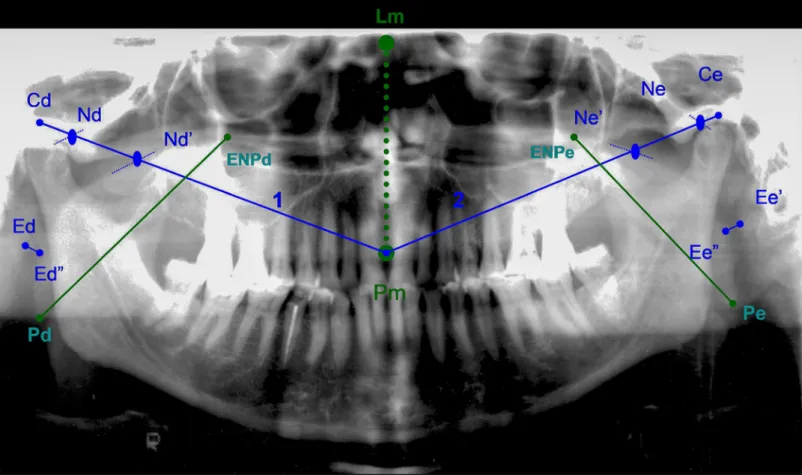
Panoramic Radiographs were taken with the same device (Orthophos Plus, Sirona Dental System, Bernsheim, Germa-ny) with program P1. They were performed with the head of the patient in an orthostatic position, at maximum habitual intercuspation (MHI), without the use of the interocclusal support device. The evaluation of UA spaces was performed by mapping the anatomical structures, using linear mea-sures established by morphometric points proposed by the authors (Figure 1). All points and lines are described in
Ta-ble 1. Panels of transparent acetate paper were made on a negatoscope (Konex Radiological Accessories, São Paulo, Brazil). The linear distances were measured with a digital caliper (model 727-6 / 150, Starrett, São Paulo, Brazil). All the analysis of the tracing were performed by the same ex-aminer from modifications of some of the parameters orig-inally idealized by the analysis of Levandoski8, proposed to
diagnose asymmetries of hard tissues in panoramic radio-graphs.
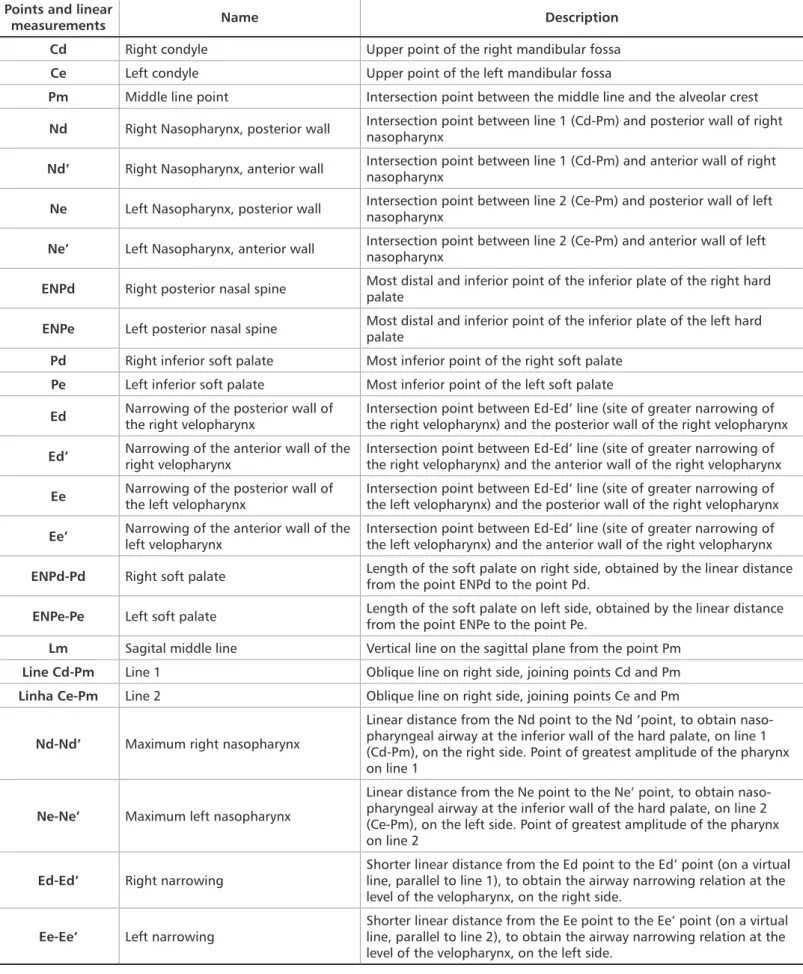
Table 1. Reference points and linear measurements for the PR analysis
Points and linear
measurements Name Description
Cd Right condyle Upper point of the right mandibular fossa
Ce Left condyle Upper point of the left mandibular fossa
Pm Middle line point Intersection point between the middle line and the alveolar crest
Nd Right Nasopharynx, posterior wall Intersection point between line 1 (Cd-Pm) and posterior wall of right nasopharynx
Nd’ Right Nasopharynx, anterior wall Intersection point between line 1 (Cd-Pm) and anterior wall of right nasopharynx
Ne Left Nasopharynx, posterior wall Intersection point between line 2 (Ce-Pm) and posterior wall of left nasopharynx
Ne’ Left Nasopharynx, anterior wall Intersection point between line 2 (Ce-Pm) and anterior wall of left nasopharynx
ENPd Right posterior nasal spine Most distal and inferior point of the inferior plate of the right hard palate
ENPe Left posterior nasal spine Most distal and inferior point of the inferior plate of the left hard palate
Pd Right inferior soft palate Most inferior point of the right soft palate
Pe Left inferior soft palate Most inferior point of the left soft palate
Ed Narrowing of the posterior wall of
the right velopharynx Intersection point between Ed-Ed’ line (site of greater narrowing of the right velopharynx) and the posterior wall of the right velopharynx
Ed’ Narrowing of the anterior wall of the right velopharynx Intersection point between Ed-Ed’ line (site of greater narrowing of the right velopharynx) and the anterior wall of the right velopharynx
Ee Narrowing of the posterior wall of the left velopharynx Intersection point between Ed-Ed’ line (site of greater narrowing of the left velopharynx) and the posterior wall of the right velopharynx
Ee’ Narrowing of the anterior wall of the left velopharynx Intersection point between Ed-Ed’ line (site of greater narrowing of the left velopharynx) and the anterior wall of the right velopharynx
ENPd-Pd Right soft palate Length of the soft palate on right side, obtained by the linear distance from the point ENPd to the point Pd.
ENPe-Pe Left soft palate Length of the soft palate on left side, obtained by the linear distance from the point ENPe to the point Pe.
Lm Sagital middle line Vertical line on the sagittal plane from the point Pm
Line Cd-Pm Line 1 Oblique line on right side, joining points Cd and Pm
Linha Ce-Pm Line 2 Oblique line on right side, joining points Ce and Pm
Nd-Nd’ Maximum right nasopharynx
Linear distance from the Nd point to the Nd ‘point, to obtain naso-pharyngeal airway at the inferior wall of the hard palate, on line 1 (Cd-Pm), on the right side. Point of greatest amplitude of the pharynx on line 1
Ne-Ne’ Maximum left nasopharynx
Linear distance from the Ne point to the Ne’ point, to obtain naso-pharyngeal airway at the inferior wall of the hard palate, on line 2 (Ce-Pm), on the left side. Point of greatest amplitude of the pharynx on line 2
Ed-Ed’ Right narrowing
Shorter linear distance from the Ed point to the Ed’ point (on a virtual line, parallel to line 1), to obtain the airway narrowing relation at the level of the velopharynx, on the right side.
Ee-Ee’ Left narrowing Shorter linear distance from the Ee point to the Ee’ point (on a virtual line, parallel to line 2), to obtain the airway narrowing relation at the level of the velopharynx, on the left side.
In order to calculate the degree of narrowing of the UA, the percentage distance between the shorter distance and the greater distance of the pharynx, for the right and left sides, were as follows:
1. Relative narrowing of the right pharynx: Ratio between the narrower region (Ed-Ed’) and the wider region (Nd-Nd’) of the inner walls of the velo and nasopharynx, respectively, on the right side, as a percentage: Ed-Ed’/ Nd-Nd’.
2. Relative narrowing of the left pharynx: Ratio between the narrower region (Ee-Ee’) and the wider region (Ne-Ne’) of the inner walls of the velo and nasopharynx, respectively, on the left side, in percentage: Ee-Ee’/Ne-Ne’.
Mean measurements of each variable were calculated, corresponding to the arithmetic means between the left and right sides. Example: maxillary nasopharynx = (maximum right nasopharynx + maximum left nasopharynx) / 2. The distances of the soft palate were also measured.
The measures were analyzed using descriptive statistical analysis and Student-t test, using the SPSS 20.0 program (SPSS, Chicago, USA).
The agreement between two measurements performed by the same observer (intraobserver reliability) was evaluated by the Intraclasses Correlation Coefficient (ICC)9 with a
sig-nificance level of 0.05.
Results
An intraobserver agreement between the first and second evaluation was above 90% (p <0.0001) for all measurements.
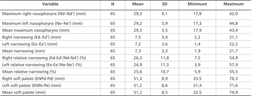
Comparing the right and left sides, the radiographic measurements were similar in the larger air spaces (Nd-Nd’, Ne-Ne’) (p=0.78) and greater narrowing (Ed-Ed’, Ee-Ee’) (p=0.21) of the pharynx in the images of right and left sides. Also, no differences were found between measurements of soft palate length on the left and right sides (p=0.51). These results are presented in Table 2.
The mean relative narrowing was 25.6%. The relative nar-rowing was 26.3% for the right side and 24.9% for the left side.
Discussion
This study demonstrated the possibility of using pan-oramic radiographs to visualize the upper airways. In all the radiographs of this study, the nasopharynx, velopharynx, oropharynx and hypopharynx, as well as the regions of nar-rowing of these passages, were visualized without difficul-ties. The same occurred with the nasal cavity, air passages between the inferior nasal conchae, and the nasal septum.
The medical-dental specialties have numerous restric-tions in exploring the potential of panoramic radiographs as an auxiliary diagnostic examination. This is due to a pre-es-tablished concept that the PR present many distortions, which is not confirmed in the literature, through compar-ative studies between measurements of dry skulls and PR,
and between these and computed tomography.10 Akcam et
al.11 analyzed the correlation and level of prediction between
PR and lateral cephalometric radiographs, using regression equations. They showed that the correlations and the level of prediction obtained were significantly corresponding be-tween them, when using the horizontal plane of Frankfurt.
Nowadays professionals are giving more importance to PR, for the ease of simultaneous visualization of the struc-tures on both sides, verification of the symmetry of the jaws and jaw, correlating the angular and linear measurements of the PR with other types of radiographs.12 The literature has
shown that PR can be used in many forms of research, pro-vided that technical criteria for the acquisition of their im-age are carefully obeyed. It is the first radiography requested by most dentists, and therefore the areas of constriction of the UA could be observed quickly and simplified in the first
Variable N Mean SD Minimum Maximum
Maximum right nasopharynx (Nd–Nd’) (mm) 65 29,3 6,1 17,8 42,0
Maximum left nasopharynx (Ne–Ne’) (mm) 65 29,2 5,9 17,3 44,8
Mean maximum nasopharynx (mm) 65 29,3 5,5 17,9 43,4
Right narrowing (Ed–Ed’) (mm) 65 7,5 3,4 2,2 21,1
Left narrowing (Ee–Ee’) (mm) 65 7,2 3,6 1,4 22,2
Mean narrowing (mm) 65 7,3 3,3 1,9 21,7
Right relative narrowing (Ed-Ed’/Nd-Nd’) (%) 65 26,3 11,8 7,5 54,9
Left relative narrowing (Ee-Ee’/Ne-Ne’) (%) 65 24,9 11,3 3,9 57,9
Mean relative narrowing (%) 65 25,6 10,7 5,9 55,5
Right soft palate (ENPd-Pd) (mm) 65 51,3 8,9 33,5 78,3
Left soft palate (ENPe-Pe) (mm) 65 51,2 8,6 31,4 71,6
clinical visits. This would enable the early detection of po-tential candidates for OSAHS.
The evaluation of images through two different aqui-sitions of the same anatomical structure, during the same exam, favors the analysis of the symmetry. The lack of sym-metry between both sides would already be indicative of a morphological alteration of the pharyngeal light.
In this research we opted for the acquisition of the im-ages in maximum intercuspation (MHI), since the man-dibular protrusion could affect the posterior air space.13-15
In MHI we can visualize the maxillomandibular structures and the UA in a functional state closer to the habitual po-sition, which mean that the temporomandibular joints and the masticatory musculature are at lower levels of muscular activity, minimizing its influence on the airflow of the UA.
The analysis of the sample (Table 2) showed similar val-ues between the right and left side linear measurements of the same UA region (Nd-Nd’ and Ne-Ne’, Ed-Ed’ and Ee-Ee’), suggesting uniform dimensions, although their image was not symmetrical. However, the comparison of the rela-tive narrowings between naso and velopharynx in percent-age values showed that there was a 26.3% relation for the right side (Ed-Ed’/Nd-Nd’), 24.9% for the left side (Ee-Ee’/ Ne-Ne’), and 25.6% for mean values. This means that the right and left nasopharynx measurements (Nd-Nd’ and Ne-Ne’, respectively) reduced their linear distance by approxi-mately 75% in the regions of greater narrowing in the right and left (Ed-Ed’ and Ee-Ee’). The maximum diameter of the pharynx reduced from a mean value of 29.3 mm to a mean
value of 7.3 mm, demonstrating the severity of the collapses observed.
According to Hudgel6 the sites of collapse may be
pri-mary or secondary, depending on the extent of pharyngeal light reduction, commonly occurring in the velo and oro-pharynx. He considered primary when the reduction was greater than 75% of the normal value and secondary when the reduction occurred between 25% and 75% of the normal value.6 In this study, the maximum value of mean relative
narrowing was 55.5%. We have found substantial reductions reaching 75.35%, mainly in the velopharynx (bordering the oropharynx), which is suggestive of primary obstruc-tions, in agreement with Hudgel.6 The authors suggest that
patients with a history of daytime drowsiness, Mallampati 3 index, and reduction of UA above 50% in the PR would deserve attention. These findings could indicate the need of further examinations for the investigation of respiratory obstructions such as OSAHS. The magnification effect and the distance to the cut plane of the PR did not influence the evaluation of the sites of narrowing and the recognition of the anatomical structures involved in this study.
Conclusion
According to our results, we can affirm that the analysis of PR allowed the evaluation of upper airway symmetry and its possible narrowing sites, once the regions of interest were fully visualized and evaluated, without difficulties, through-out the entire sample.
9. Bartko JJ, Carpenter WT Jr. In: On the Methods and Theory of Rehabili-tation. The J Nerv Mental Dis. 1976;163(5):307-16.
10. Catić A, Celebić A, Valentić-Peruzović M, Catović A, Jerolimov V, Muretić I. Evaluation of the precision of dimensional measurements of the mandible on panoramic radiographs. Oral Surg Oral Med Oral Pathol Oral Radiol Endod. 1998;86(2):242-8.
11. Akcam MO, Altiok T, Ozdiler E. Panoramic radiographs: a tool for investigating skeletal pattern. Am J Orthod Dentofacial Orthop. 2003; 123(2):175-81.
12. Langland OE, Sippy FH. Anatomic structures as visualized on the Or-thopantomogram Oral Surg. 1968;26(4):475-84.
13. Smith AM, Battagel JM. Non-apneic snoring and the orthodontist: ra-diographic pharyngeal dimension changes with supine posture and man-dibular protrusion. J Orthod. 2004;31(2):124-31.
14. Battagel JM, Johal A, Kotecha B. A cephalometric comparison of subjects with snoring and obstructive sleep apnoea. Eur J Orthod. 2000;22(4):353-65.
15. Johal A, Battagel JM. An investigation into the changes in airway di-mension and the efficacy of mandibular advancement appliances in subjects with OSA. Br J Orthod. 1999;26(3):205-10.
References
1. Farre R, Rigau J, Montserrat JM, Ballester E, Navajas D. Evaluation of a simplified oscillation technique for assessing airway obstruction in sleep apnoea. Eur Respir J. 2001;17(3):456-61.
2. Cistulli PA, Gotsopoulos H, Marklund M, Lowe AA. Treatment of snor-ing and obstructive sleep apnea with mandibular repositionsnor-ing appliances. Sleep Med Rev. 2004;8(6):443-57.
3. Olsen KD, Kern EB. Nasal influences on snoring and obstructive sleep apnea. Mayo Clin Proc.1990;65(8):1095-105.
4. Kuna ST, Remmers JE. Anatomy and physiology of upper airway obstruc-tion. In: Principles and practice of sleep medicine. 3rd ed. Philadelphia: WB Saunders; 2000.p. 840-58.
5. Lopatiéne K, Babarskas A. Malocclusion and upper airway obstruction. Medicine. 2002;38(39):277-83.
6. Hudgel DW. Variable site of airway narrowing among obstructive sleep apnea patients. J Appl Physiol. 1986;61(4):1403-9.
7. Luz JG, Miyazaki LT, Rodrigues L. Verification of the symmetry of the mandibular ramus in patients with temporomandibular disorders and asymptomatic individuals: a comparative study. Bull Group Int Rech Sci Stomatol Odontol. 2002;44(3):83-7.
8. Piedra I. The Levandoski Panoramic Analysis in the diagnosis of facial and dental asymmetries. J Clin Ped Dent. 1995;20(1):15-21.
Submitted: 10/20/2018 / Accepted for publication: 11/10/2018 Corresponding Author
Florence Mitsue Sekito E-mail: fmsekito@gmail.com
Mini Curriculum and Author’s Contribution
1. Florence Mitsue Sekito - DDS and MSc. Contribution: intellectual idea of the study, technical procedures, data acquisition, data interpretation, preparation of the manuscript. ORCID: 0000-0003-3625-8735
2. Mariana Ribeiro de Moraes Rego - DDS, MSc and PhD. Contribution: data interpretation, preparation of the manuscript, critical review and final approval. OR-CID: 0000-0003-2992-6002
3. Mayra Cardoso - DDS, MSc and PhD. Contribution: data interpretation, preparation of the manuscript, critical review and final approval. ORCID: 0000-0002-1232-8551
4. Plínio Senna - DDS, MSc and PhD. Contribution: preparation of the manuscript, critical review and final approval. ORCID: 0000-0003-0743-5376
5. Hilda Maria Montes Ribeiro de Souza - DDS, MSc and PhD. Contribution: preparation of the manuscript, critical review and final approval. ORCID: 0000-0001-5283-1146
6. Daniel de Moraes Telles - DDS, MSc and PhD. Contribution: data interpretation, preparation of the manuscript, critical review and final approval. ORCID: 0000-0001-9576-4342