Faculdade de Medicina da Universidade de Lisboa Trabalho Final de Mestrado Integrado em Medicina
Use of Dexamethasone Intravitreal Implant for the
treatment of Diabetic Macular Edema: Analysis of
Randomized Clinical Trials
Maria Inês Sanches Silva nº12917 Orientador: Dr. Mário Canastro
Clínica Universitária de Oftalmologia do Hospital de Santa Maria 2015/2016
2 Abstract
The aim of this study is to review the best clinical evidence concerning the use of dexamethasone intravitreal implant in diabetic macular edema (DME). Main outcomes assessed included change in best corrected visual acuity (BCVA) and change in central macular thickness (CMT). Main adverse events, such as, cataract and increased intraocular pressure (IOP) were evaluated. This article reviews six randomised clinical trials (RCTs) searched through several databases. Dexamethasone intravitreal implant are effective compared to laser, anti-VEGF and placebo, and seems to be more effective in combination to anti-VEGF and laser in lowering CMT. It was associated with increased incidence of cataracts (after the first year of treatment) and IOP rise but it required fewer injections and, thus, a final lower cost. This implant may be a promising treatment option for eyes with persistent DME and can be a treatment option in eyes that do not respond to anti-VEGF therapy, pseudophakic eyes or in those eyes about to undergo cataract surgery, in vitrectomized eyes or in patients who have barriers to monthly treatment regimen.
Resumo
O objetivo deste estudo é providenciar a melhor evidência cientifica relativa ao uso do implante intraocular de dexametasona, no edema macular diabético. Os principais parâmetros avaliados foram a acuidade visual e a espessura macular central. Os principais efeitos adversos, como a catarata e o aumento da pressão intraocular, foram igualmente avaliados. Este artigo analisa seis estudos randomizados e controlados, os quais foram retirados de várias bases de dados cientificas. O implante intraocular de dexametasona demonstrou ser efetivo comparativamente à terapia com laser, agentes anti-VEGF e placebo, e demonstrou, inclusivamente, ser mais efetiva em combinação com agentes anti-VEGF e laser na diminuição da espessura macular central. No entanto, esta terapia está associada a um aumento da incidência de cataratas (após o primeiro ano de tratamento) e a um aumento da pressão intraocular, porém, requer um número menor de injeções, o que determina, um custo final inferior. Este implante pode ser considerado uma alternativa inovadora e uma opção possível para olhos que não respondem a terapia com agentes anti-VEGF, olhos pseudofáquicos ou com cirurgia de catarata programada, olhos vitrectomizados ou em pacientes que não suportam o regime de tratamento mensal.
3 Key words: adverse effect, anti-VEGF, cataract, central macular thickness,
corticosteroids, dexamethasone, diabetic macular edema, increased intraocular pressure, laser photocoagulation, Ozurdex®, visual acuity.
Abbreviations: BCVA (best corrected visual acuity); BRVO (branch retinal vein
occlusion); CRVO (central retinal vein occlusion); CMT (Central Macular Thickness); CRT (Central Retinal Thickness); DDS (drug delivery system); Dex (dexamethasone); DME (diabetic macular edema); DR (diabetic retinopathy); ETDRS (Early Treatment Diabetic Retinopathy Study); EU (European Union); FA (fluocinolone acetonide); IOP (intraocular pressure); ME (macular edema); PLGA (poly D,L-lactide-co-glycolide); RCT (Randomized Clinical Trial); RVO (retinal vein occlusion); TA (triamcinolone acetonide), VA (Visual Acuity); VEGF (vascular endothelial growth factor).
Introduction
The major cause of impaired vision in patients between 25 and 74 years of age is Diabetic ocular disease.[1] Visual impairment is mainly originated by complications related to diabetic maculopathy and neovascularization, as retinal detachment or neovascular glaucoma. Causes of visual impairment and blindness in developing countries (such as cataracts and trachoma) are declining, but the prevalence of Diabetic retinopathy (DR) is growing.[2] Diabetic macular edema (DME) affects around 21 million people worldwide. [3]
The 10-year incidence of DME was different among different types of diabetes or insulin patients use, with 20.1% in patients with type 1 diabetes, 25.4% in patients with type 2 diabetes who used insulin and 13.9% in patients with type 2 diabetes who did not use insulin. [4]
Risks factors comprise the type of diabetes, level of glycemic control and associated conditions such as hypertension, dyslipidemia, smoking, nephropathy and pregnancy. Thus, the several preventive and therapeutic interventions, such as intravitreal pharmacological therapies, are crucial to minimize the morbidity associated with DR.
4
What is DME?
Macular Edema (ME) can occur at any stage of DR and involve both the macular region and peripheral retina. Clinically significant DME is characterized by capillary leakage, fluid accumulation, and macular thickening within 500 microns of the fovea, circinate hard exudates within 500 microns of the fovea if associated with adjacent retinal thickening, or one or more areas of retinal thickening at least 1500 microns in diameter that is within one disc diameter (1500 microns) of the fovea. DME leads to breakdown of the blood retinal barrier [5], and consequent visual acuity (VA) loss. [6]
The pathophysiology of ME is complex and involves an increased hydrostatic venous pressure, inflammation, endothelial dysfunction, and increased vascular permeability factors, such as vascular endothelial growth factor (VEGF) with a range of factors implicated. [7]
Through several years, the investigation of the pathophysiology of ME led to a better understanding of the role of inflammatory mediators that are responsible for the accumulation of fluid within the retina and cellular damage. [8] Several factors are involved in the inflammation derived from DME and ME secondary to RVO, including the expression of multiple inflammatory factors like vascular endothelial growth factor (VEGF), intercellular adhesion molecule-1, interleukin-6, and monocyte chemotactic protein-1, and also leukostasis and alterations in endothelial tight junction proteins.[5] The inflammation magnitude in the ME progressively increases as long as time went by. For the past decades, laser photocoagulation has been the treatment of choice for DME, however in recent years, the treatment of clinically significant ME has been a rapidly evolving area and several alternative options have emerged. Today, intravitreal anti-VEGF pharmacotherapy is the considered the first-line treatment but other options are available as, vitreoretinal surgery or intravitreal corticosteroids, including dexamethasone (Dex).
Roll of Inflammation and Corticosteroids
Corticosteroids can help to reduce many of the inflammatory processes in the development of ME, since they are potent and selective agonists of the glucocorticoid receptor, which are expressed in ocular tissues and are sensitive to corticosteroids in
5
ophthalmic treatments, as Dex, triamcinolone acetonide (TA), and fluocinolone acetonide (FA). [9] [3]
Several mechanisms have been proposed. They have potent anti-inflammatory effects by expression of several anti-inflammatory proteins, including IL-10, adenosine, and IκBα, the natural inhibitor of NFκB. [3] They also reduce vascular permeability and promote retinal fluid clearance through their effects on transcellular aquaporin-4 (AQP4) and potassium channels, the two main channels controlling retinal fluid movement on retinal Müller cells. [3]
At the outer blood retinal barrier, corticosteroids keep tight junction integrity and they stabilize the existing retinal vasculature, have antiangiogenic effects by suppressing the production of paracrine angiogenic factors, and also, reducing the growth of new blood vessels. [3] They can also inhibit fibrin deposition and leukocyte trafficking, repress homing and migration of inflammatory cells, and inhibit the synthesis of prostaglandins and other cytokines.[9]
In the past, intraocular or periocular corticosteroids administration produced just a transient treatment effect. As a result, repeated injections were necessary. However, Ozurdex® deliver Dex over an extended time frame, with less injections and favorable outcomes (mean VA and CRT). Nevertheless, it is also associated with high rates of cataract formation and glaucoma. [8]
Use of Ozurdex®
Ozurdex® is approved by the United States Food and Drug Administration and by the European Medicines Agency, and is already available throughout the European Union as a treatment licensed for the treatment of ME following branch or central retinal vein occlusion (BRVO or CRVO) and treatment of non-infectious uveitis affecting the posterior segment of the eye since September 2010 and for DME since July 2014.[10][11] [12]
Ozurdex® is an intravitreal implant containing 0.7 mg (700 mcg) of Dex in the Novadur® solid polymer biodegradable, slow-release drug delivery system (DDS) that is injected through the pars plana by a customized, single-used 22-gauge applicator. [7][12] The Novadur® system contains poly D,L-lactide-co-glycolide polymer matrix
6
without preservative witch slowly degrades to lactic acid and glycolic acid and is designed to deliver drug to the retina over a period of up to 6 months after intravitreal injection.[12] The intravitreal administration bypasses the blood–retinal barrier with minimal systemic absorption, which results in continuous high concentrations of Dex in the retina and vitreous during the first 2 months after the injection, and lower concentrations are sustained up to 6 months. By avoiding the peak vitreous drug concentrations produced by intermittent bolus corticosteroid injections and the need for frequent injections, the implant reduce the risk of adverse steroid-associated ocular effects (cataract, increased intraocular pressure, and glaucoma) and injection-related complications (lens injury, retinal detachment, and infectious endophthalmitis). [3] The intensity of pain associated with the insertion procedure is not substantially different from that produced by intravitreal anti-VEGF treatment, which use a 28 to 30 gauge needle. The implant is biodegradable and subsequent implant(s) can be inserted without the need for surgical removal of the previous implant. The impact velocity of the Dex intravitreal implant, using the applicator, is insufficient to cause retinal damage proved by high-speed, real-time photography of the injection procedure.[3] The injection procedure has some complications, such as accidental lens touch, and splitting or fracture of the implant, and some in situ complications associated with migration into the anterior chamber, potentially leading to development of corneal edema.[3] Intravitreal injections have been associated with endophthalmitis, eye inflammation, increased intraocular pressure (IOP), and retinal detachment. Patients should be monitored following the injection. [10]
Prophylactic treatment of ME that is not clinically important is not recommended, but once DR progresses to macular edema, treatment is indicated to slow the rate of vision loss and improve the long-term prognosis.[3] Treatment of DR is directed both at prevention (strict glycemic control early in the course of diabetes) and at treatment of established disease.[13]
For two decades, laser photocoagulation has been the mainstay of treatment for DME but even if it was useful in preserving vision, it was of limited effect in restoring lost vision, once lost.[14] In most patients with clinically significant ME, anti-VEGF agents are actually first-line therapy in order to their established efficacy, less incidence of complications, and good ability of administration. However, corticosteroids comparable
7
to laser, seems to be less aggressive to retina, and comparable to anti-VEGF agents, it had a long-term effect, and provide less interventions. [14][15]
Purpose
The aim of this paper is to review the best evidence supporting the use Ozurdex® for the treatment of DME. This study focuses on the current evidence for the use of Dex versus anti-VEGF drugs and/or laser, as assessed by change in best corrected visual acuity (BCVA), central retinal thickness (CRT), and their adverse events (cataract and increased IOP).
8 Methods
This review was limited to RCTs looking at the use of dexamethasone intravitreal implant and eligibility criteria are described in flow-diagram 1.
The literature search was performed from August 2015 to February 2016 using the databases Medline, EMBASE, Cochrane Library, World Health Organization International Clinical Trials Registry Platform, clinicaltrials.gov and UpToDate.
The search terms to search all trials registers and databases were: anti-VEGF, corticosteroids, dexamethasone, diabetic macular edema, laser photocoagulation, Ozurdex®.
To be included, studies (1) had to be Randomised Clinical Trials, (2) had a minimum follow-up of 6 months and (3) had to comprise at least two treatment arms or sham control, including Dex as active comparator of interest. Studies were excluded if they (1) had no control group, (2) assessed the effect of the mentioned treatments in ME due to other retinal diseases besides DME (3) were combined with a surgical intervention or (5) published studies in languages other than English.
Data were extracted by three authors (MC, DC, MS) and checked by two of them (MC and MS). Search results were screened by two independent authors (MC and MS). Disagreements were resolved by discussion between the two review authors (MC and MS).
Information was extracted from each included trial on: (1) type of outcome measure (mean change in BCVA, assessed in terms of Early Treatment Diabetic Retinopathy Study (ETDRS) letters; mean decrease CRT; safety analysis of adverse events, as cataract and increased IOP); (2) type of intervention ( dexamethasone intravitreal implant, anti-VEGF agents, laser therapy); (3) trial’s inclusion and exclusion criteria.
9
Flow-diagram 1. Information through the different phases of this review
The RCTs evaluated in this review were: (1) Three-Year, Randomized, Sham-Controlled Trial of Dexamethasone Intravitreal Implant in Patients with Diabetic Macular Edema (MEAD study); (2) A Randomized Clinical Trial of Intravitreal Bevacizumab versus Intravitreal Dexamethasone for Diabetic Macular Edema: The BEVORDEX Study; (3) Dexamethasone Intravitreal Implant in Combination with Laser Photocoagulation for the Treatment of Diffuse Diabetic Macular Edema (PLACID STUDY); (4) A prospective randomised controlled clinical trial comparing a combination of repeated intravitreal Ozurdex® and macular laser therapy versus macular laser only in centre-involving diabetic macular edema (OZLASE study); (5) Randomized Controlled Trial of an Intravitreous Dexamethasone Drug delivery system in Patients With Diabetic Macular Edema (Haller et al); (6) A 12-Month,
Single-Masked, Randomized Controlled Study Of Eyes With Persistent Diabetic Macular Edema After Multiple Anti-Vegf Injections To Assess The Efficacy Of The Dexamethasone-Delayed Delivery System As An Adjunct To Bevacizumab Compared With Continued Bevacizumab Monotherapy (Maturi et al).
94 articles identified through database searching
Exclusion of studies which are not RCTs and
were not related to DME (n=44)
44 of records excluded by primary outcomes and after subanalysis removed
10 Results
RCTs were analyzed concerning type of study, sample, intervention and control group. Data extracted from the RCTs concerning primary outcomes - BCVA, CRT- and adverse events - cataract incidence and IOP elevation - are summarized in tables.
Primary outcomes
Visual Acuity
Table 1 summarizes inclusion and exclusion criteria of RCTs (annexed document) and table 2 overviews the results of BCVA.
Dose and Sham Control
Two included trials compared two doses of Dex administered as an intravitreal implant (0,7 mg and 0,35 mg) with sham control: MEAD study and Haller.
The baseline characteristics of the studies are reasonably similar, except in number of enrolled patients (1048 patients were enrolled in MEAD study and 315 patients in Haller et al) and follow-up period (3 years versus 180 days), however, the comparable results was from baseline to the studies end.
In MEAD study [5], the percentage of patients who achieved ≥15 letter improvement in BCVA from baseline at study end (3 years) was superior with Dex 0,7 mg than Dex 0,35 mg and sham (22,2%, 18,4% and 12,0% respectively) with statistical significance (P ≤ 0.018). The mean average change in BCVA from baseline during the study was 3.5 letters, 3.6 letters and 2.0 letters (Dex 0,7 mg, Dex 0,35 mg and sham, respectively). In spite of follow-up was 3 years, the greater effect of Dex implant in percentage of patients with a ≥15 letter improvement in BCVA from baseline were observed as early as day 21 between each Dex group (P ≤ 0.003) and at the majority of visits.
11 (M a tu ri et a l) 2 0 1 5 F o ll o w u p : 1 2 m o n th s De sig n : 2 a rm s (H a ll er et a l) 2 0 1 0 F o ll o w u p : 1 8 0 d ay s De sig n : 3 a rm s P lac eb o -co n tro ll ed MEAD stu d y (Bo y er et a l) 2 0 1 4 F o ll o w u p : 3 y ea rs (o r 3 9 m o n th s fo r p ati en ts trea ted a t m o n th 3 6 ). De sig n : 3 a rm s S tu d y Tab le 2 . – Ba se li n e ch ara cteristics o f RCTs an d V A o u tco m e re su lts. 3 0 p ati en ts M ea n Ba se li n e BCVA : -Gro u p 1 = 6 5 ; -Gro u p 2 = 6 4 ; M ed ian n u m b er o f re -in jec ti o n s: Gro u p 1 : 8 Gro u p 2 : 9 ( P = 0 .3 ) 3 1 5 p ati en ts M ea n Ba se li n e BCVA : -Gro u p 1 = 5 4 .7 ; -Gro u p 2 = 5 4 .4 ; -Gro u p 3 = 5 4 .4 . 1 0 4 8 p ati en ts M ea n Ba se li n e BCVA : Gro u p 1 = 5 6 ,1; Gro u p 2 = 5 5 ,5 ; Gro u p 3 = 5 6 ,9 . Th e m ed ian n u m b er o f stu d y trea tme n ts: Gro u p 1 : 4 Gro u p 2 : 5 Gro u p 3 : 3 Pa rticip a n ts a n d b a se li n e v a lu es Gro u p 1 : (n = 2 1 ) Be v ac izu m ab 1 ,2 5 m g at b ase li n e p lu s De x 0 ,7 m g a t 1 -m o n th v isit ; Gro u p 2 : (n = 1 9 ) Be v ac izu m ab 1 ,2 5 m g a t b ase li n e an d a t 1 -mo n th . Gro u p 1 : (n = 5 7 ) De x imp lan t 0 .7 m g ; Gro u p 2 : (n = 5 7 ) De x imp lan t 0 .3 5 m g ; Gro u p 3 : (n = 5 7 ) s h am . Gro u p 1 : (n = 3 5 1 ) De x imp la n t 0 .7 m g ; Gro u p 2 : (n = 3 4 7 ) De x imp la n t 0 .3 5 m g ; Gro u p 3 : (n = 3 5 0 ) sh am . Inter v en tio n Th e m ea n c h an g es in v isu al ac u ity : Gro u p 1 : 5 .4 + 1 0 .7 letters Gro u p 2 : 4 .9 + 1 2 .3 P = 0 .9 . P erc en tag e o f pa ti en ts wit h a ≥1 0 -letter i m p ro v em en t in BCVA : At d ay 9 0 : -Gro u p 1 : 3 3 .3 % -Gro u p 2 : 2 1 .1 % -Gro u p 3 : 1 2 .3 % P = 0 .0 0 7 v s 7 0 0 -µg g ro u p ). At d ay 1 8 0 : -Gro u p 1 : 3 0 % -Gro u p 2 : 1 9 % -Gro up 3 : 2 3% (P ≥0 .4 fo r trea ted v s g ro up 3 ). Th e m ea n (sta n d ard d ev iat io n ) av era g e ch an g e in BCVA : -Gro u p 1 : 3 .5 (8 .4 ) -Gro u p 2 : 3 .6 (8 .1 ) -Gro u p 3 : 2 .0 (8 .0 ) P erc en tag e o f pa ti en ts wit h a ≥1 5 -letter i m p ro v em en t in BCVA : -Gro u p 1 : 2 2 .2 % ; -Gro u p 2 : 1 8 .4 % ; -Gro u p 3 : 1 2 .0 % ; P ≤ 0 .0 18 . O u tc o m e (c h a n g e fr o m b a se li n e a t stud y e n d )
12 Pl a cid S tu d y (Ca ll a n a n et al ) 2 0 1 3 F o ll o w -up : 1 2 m o n th s De sig n : p ara ll el -g ro u p O ZLASE stud y (H en g et a l) 2 0 1 5 F o ll o w -up :5 6 we ek s De sig n : 2 arm s Th e BEVO RD EX S tu d y (G il li es et a l) 2 0 1 4 F o ll o w -up : 1 2 m o n th s De sig n : 2 arm s 2 5 3 p ati en ts M ea n Ba se li n e BCVA : Gro u p 1 : 5 7 Gro u p 2 : 5 7 .5 M ea n n u m b er o f in jec ti o n s: Gro u p 1 : 1 .6 7 8 0 p ati en ts M ea n Ba se li n e BCVA : -Gro u p 1 : 6 6 .1 -Gro u p 2 : 6 6 .6 M ea n n u m b er o f in jec ti o n s in 5 6 we ek s: -Gro u p 1 : 3 .5 M ea n n u m b er o f M LT : -Gro u p 1 : 0 .7 7 -Gro u p 2 : 3 .0 5 6 1 p ati en ts M ea n Ba se li n e BCVA : -Gro u p 1 = 5 6 ,3 Gro u p 2 = 5 5 ,5 M ea n n u m b er o f in jec ti o n s in 1 2 m o n th s: -Gro u p 1 : 8 .6 -Gro u p 2 : 2 .7 Gro u p 1 : (n = 1 2 6 ) De x 0 ,7 m g a t b ase li n e p lu s las er at m o n th 1; Gro u p 2 : (n = 1 2 7 ) sh am imp lan t at b ase li n e an d las er at m o n th 1 . Gro u p 1 : (n = 4 0 ) co m b in ed re p ea ted De x am th aso n e 0 ,7 m g an d m ac u lar las er th era p y (M LT ); Gro u p 2 : (n = 4 0 ) M LT a lo n e. Gro u p 1 : (n = 4 2 ) b ev ac izu m ab 1 ,2 5 m g ev ery 4 we ek s Gro u p 2 : (n = 4 6 ) d ex im p lan t 0 ,7 m g ev ery 1 6 we ek s. Bo th p ro -re -n ata. P erc en tag e o f p ati en ts ac h iev in g a t lea st a 1 0 -letter i m p ro v em en t: At m o n th 6 : Gro u p 1 :2 2 .2 Gro u p 2 : 1 7 .3 P = 0 .3 2 6 At m o n th 9 : Gro u p 1 : 3 1 .7 Gro u p 2 : 1 7 .3 P = 0 .0 0 7 At m o n th 1 2 : Gro u p 1 : 2 7 .8 Gro u p 2 : 2 3 .6 ( P = 0 .4 5 3 ) Th e m ea n c h an g e in BCVA : -Gro up 1 : − 0.3 (S D 11 .4 ) -Gro u p 2 : + 0 .4 (S D 9 .5 ) Eff ec t e stim ate 0 .8 8 ( 95 % CI −3 .4 6 t o 5 .2 2). P erc en tag e o f pa ti en ts wit h a ≥1 5 -letter i m p ro v em en t in BCVA : -Gro u p 1 : 1 5 .8 % -Gro u p 2 : 5 .1 % P erc en tag e o f p ati en ts lo sin g ≥3 0 ET DRS letters : -Gro u p 1 : 2 .6 % -Gro u p 2 : 0 .0 % M ea n g ain o f BCVA : Gro u p 1 : 8 .9 Gro u p 2 : 5 .6 P erc en tag e o f pa ti en ts wit h a ≥1 5 -letter i m p ro v em en t in BCVA : Gro u p 1 : 3 1 % ; Gro u p 2 : 2 2 % ; M ea n g ain in BCVA : Gro u p 1 : 8 .9 Gro u p 2 : 5 ,6 P erc en tag e o f p ati en ts lo sin g ≥ 1 0 letters : -Gro u p 1 : 0 % -Gro u p 2 : 1 1 % Th ere wa s n o si g n if ica n t effe ct b ase d o n trea tme n t re ce iv ed fo r th e ch an g e in BCVA fo r th e p se u d o p h ak ic ey es (m ea n c h an g e in BCVA : 7 ,7 letters g ro u p 1 ; 1 0 ,4 letters fo r g ro u p 2 ; P = 0 .4 7 )
13
Subgroup analysis revealed that BCVA results varied between pseudophakic and phakic eyes. The mean average BCVA improvement with Dex implant (either 0,7 mg or 0,35 mg) was greater until the time of cataract development. The mean number of phakic eyes and pseudophakic eyes, enrolled in study, was 257,6 and 91,6 respectively. Cataract formation changed the improvement of BCVA in phakic eyes. MEAD study described results in a subanalysis group of pseudophakic eyes (gain of ≥ 15 letters in BCVA from baseline at the study end of pseudophakic eyes): 23.3% in Dex 0,7 mg, 15,9 % in Dex 0,35 mg and 10,9% in sham group.
In the overall population, the mean change in BCVA decreased after 9 months, but in pseudophakic population these results were more stable for 3 years. After cataract surgery, the improvement in vision was restored in phakic eyes and the results from baseline at study end was equivalent in both eyes, pseudophakic and phakic eyes. Haller et al [16] demonstrated the percentage of patients who achieved ≥10 letters of improvement in BCVA results at day 60, 90 and 180, but the percentage of patients who achieve ≥15 letters are shown in a figure, hence, the percentage (p values are not estimated) were estimated throw image analysis. The estimated percentage of patients who achieve ≥15 letters at day 180 were, approximately, 14% (Dex 0,7 mg), 9% (Dex 0,35mg) and 7% (sham; P=0.22 Dex 0,7 mg versus sham). At day 90, were approximately 11% (Dex0,7 mg), 5% (Dex0,35mg) and 2% (sham; P= 0.05 Dex0,7 mg versus sham). The p values, at day 90, demonstrate that the difference between the Dex 0,7 mg and sham group was statistically significant. This difference was not significant at day 180.
In this study, cataract formation did not affect BCVA, due to the short follow-up period. Bevacizumab
The studies included in this paper which compared Dex to bevacizumad are Maturi et al and Bevordex study. The aim of these RCTs was to determine if Dex implant 0,7 mg combined with bevacizumab 1,25 mg (anti-VEGF agent) provides greater benefit than bevacizumab alone and to determine head-to-head the effect of Dex implant opposed to bevacizumab (Maturi et al and Bevordex, respectively). Baseline characteristics of the groups are reasonably similar, even though, Bevordex study had involved almost twice the number of patients compared to Maturi.
14
In Maturi et al [17], the mean VA changes from baseline to 12 months and the average time to achieve ≥15 letter improvement, were similar between two groups. The improvement in VA was fairly stable in the first 9 months, and then slowly improved until month 12.
Before study, the eyes enrolled had received bevacizumab injections. After enrollment in study, it were randomly assigned to receive combination therapy (bevacizumab plus Dex) or bevacizumab monotherapy. The eyes that received combination treatment and, simultaneously, had received the greatest number of injections (previously), acquired a greater improvement in VA comparable to eyes that received monotherapy and the same previously injections.
Bevordex study [18] demonstrated an improvement in BCVA of ≥10 letters from baseline by the 12 month visit of 40% in bevacizumab group compared with 41% of Dex group. An improvement in BCVA of ≥15 letters from baseline by the 12 months was 31% (bevacizumab group) and 10% (Dex group). The mean improvement in BCVA was 8.9 letters (bevacizumab group) and 5.6 (Dex group), but the difference was not statistically significant (P =0.24).
Cataract formation conditioned a loss of ≥10 letters in 11% in Dex implant eyes, but none in the bevacizumab group. The difference between the two groups was not statistically significant and the mean number of injections over the 12 months was 8.6 and 2.7, for bevacizumab and Dex group, respectively. The percentage of patients that preferred dexamathesone implant was 46% comparable with 33% of patients that preferred bevacizumab implant.
Laser therapy
The studies witch evaluated Ozurdex® 0,7 mg combined with Laser therapy compared with laser monotherapy are OZLASE study and PLACID Study.
OZLASE Study had failed to prove an improvement in mean change in BCVA at 56 weeks in the combination group (table 2), despite of frequent injections (mean of 3.5 Ozurdex® injections in the combination arm). [19]
PLACID Study compared 6 monthly Dex implant 0,7 mg combined to laser therapy versus laser therapy alone, with a mean of 1.67 implants used over a period of 12
15
months. From month 1 to 9, the study proved a significantly improvement in BCVA in the combination arm, mainly among patients who received Dex implant before laser treatment compared with patients who received the sham injection (23.2% versus. 11.9%; P = 0.035). However, at study end (month 12), PLACID did not prove significant different in percentage of patients who gained ≥10 letters in BCVA, between the two treatment groups.
Central retinal thickness
The outcome results and baseline characteristics are shown in table 3. Dose and sham control
MEAD study [5], demonstrated a significantly improvement in mean average reduction in CRT, during the study, in eyes treated with Dex implant 0.7 mg and 0.35 mg compared to sham. Eyes treated with Dex implant, despite cataract and vision loss, showed a decrease in CRT. After cataract surgery, CRT increased in sham group but did not increase in Dex implant groups. Notably, an increase in CRT after cataract surgery was observed in the sham group but it wasn’t detected in the Dex implant groups. Haller et al [16] demonstrated, at day 90, a significantly improvement in mean change of macular thickness in eyes which had received Dex implant 0.7 mg comparable with sham. A significant improvement in fluorescein leakage was observed in the same group. In both parameters (macular thickness and fluorescein leakage), there were a greater response in eyes treated with Dex implant 0.7 mg than 0.35 mg and sham group, suggesting a dose-response relationship.
16 ME AD stud y (Bo y er e t a l) 2 0 1 4 S tu d y Tab le 3 . -CRT b ase li n e ch ara cteristics a n d o u tco m e re su lt s M ea n CRT , µm : Gro u p 1 : 4 6 3 .0 Gro u p 2 : 4 6 6 .8 Gro u p 3 : 4 6 0 .9 Ba se li n e CRT Gro u p 1 : (n = 3 5 1 ) De x imp lan t 0 .7 m g ; Gro u p 2 : (n = 3 4 7 ) De x imp lan t 0 .3 5 m g ; Gro u p 3 : (n = 3 5 0 ) sh am . Inter v en tio n M ea n a v era g e re d u cti o n in CR T: Gro u p 1 : -1 1 1 .6 Gro u p 2 : 1 0 7 .9 Gro u p 3 : -4 1 .9 P < 0 .0 0 1 M ea n (S D) av era g e ch an g e in CR T { fro m b ase li n e ac ro ss th e stu d y , µm : Gro u p 1 : -1 3 1 .8 Gro u p 2 : -1 1 7 .1 Gro u p 3 : -5 0 .8 M ea n (S D) av era g e ch an g e in CR T ( fro m b ase li n e ac ro ss th e st u d y b y lens st a tu s, µm : P h ak ic ey es ( b ase li n e to c atara ct): Gro u p 1 : -1 0 8 .9 Gro u p 2 :-9 3 .2 Gro u p 3 :-3 7 .2 P h ak ic ey es (ca tara ct to c atara ct su rg ery ): Gro u p 1 :-1 1 7 .9 Gro u p 2 :-1 0 7 .2 Gro u p 3 :-6 .6 P h ak ic ey es (ca tara ct su rg ery to la st CRT ): Gro u p 1 :-8 8 .8 Gro u p 2 :-8 4 .8 Gro u p 3 :+7 7 .5 P se u d o p h ak ic ey es: Gro u p 1 : 1 3 1 .8 Gro u p 2 : -1 1 7 .1 Gro u p 3 : 5 0 .0 P v alu e (b etwe en g ro u p 1 a n d 3 ) : < 0 .0 0 1 P v al u e b etwe en g ro u p 2 a n d 3 ): < 0 .0 0 1 O u tc o m e (fr o m b a se li n e d u rin g th e stud y ,( µm )
17 (M a tu ri et al ) 2 0 1 5 (H a ll er e t al ) 2 0 1 0 M ed ian ce n tral su b field th ick n ess , µm : Gro u p 1 : 3 4 4 Gro u p 2 : 4 2 3 Ey es we re eli g ib le fo r re trea tme n t if th e ce n tral su b field th ic k n ess m ea su re d ˃ 25 0 µm . M ea n CRT , µm : Gro u p 1 : 4 2 8 .3 Gro u p 2 : 4 4 6 .5 Gro u p 3 : 4 1 7 .5 Gro u p 1 : (n = 2 1 ) Be v ac izu m ab 1 ,2 5 m g at b ase li n e p lu s De x 0 ,7 m g a t 1 -m o n th v isit ; Gro u p 2 : (n = 1 9 ) Be v ac izu m ab 1 ,2 5 m g a t b ase li n e an d a t 1 -m o n th Gro u p 1 : (n = 1 1 ) De x imp lan t 0 .7 m g ; Gro u p 2 : (n = 1 4 ) De x imp lan t 0 .3 5 m g ; Gro u p 3 : (n = 1 9 ) s h am . M ea n CRT , µm : At m o n th 3 : g ro u p 1 : 2 8 4 g ro u p 2 : 4 1 2 At m o n th 9 : g ro u p 1 : 3 6 7 g ro u p 2 : 3 7 4 At m o n th 1 2 : g ro u p 1 : 3 2 7 g ro u p 2 : 3 8 2 M ea n re d u cti o n in c en tral su b field th ick n ess ( fro m b ase li n e to m o n th 1 2 ): g ro u p 1 : -4 5 + 1 0 7 µm g ro u p 2 : -3 0 + 1 0 0 µm P = 0 .0 3 De cre as ed o f flu o re sc ein lea k ag e: 2 o r m o re lev els: Gro u p 1 : 3 6 .4 % Gro u p 2 : 2 2 .2 % Gro u p 3 : 5 .4 % P v alu e (g ro u p 1 v ersu s g ro u p 3 ) = < 0 .0 0 1 P v alu e( g ro u p 2 v ersu s g ro u p 3 ) = 0 .0 1 3 o r m o re lev els: Gro u p 1 : 2 7 .3 % Gro u p 2 : 1 3 .0 % P v alu e (g ro u p 1 v ersu s g ro u p 3 ) = < 0 .0 0 1 P v alu e( g ro u p 2 v ersu s g ro u p 3 ) = 0 .0 3 At d ay 9 0 : M ea n c h an g e in m ac u lar t h ick n es s, µm : Gro u p 1 :-1 3 2 .2 7 G ro u p 2 :-4 2 .5 7 Gro u p 3 :+ 3 0 .2 1 P v alu e b etwe en g ro u p 1 a n d 3 : < 0 .0 0 1 P v alu e b etwe en g ro u p 2 a n d 3 : 0 .0 7
18 Pl a cid S tu d y (Ca ll a n a n et a l) 2 0 1 3 O ZLASE stud y (H en g e t a l) 2 0 1 5 Th e BEVO RD E X S tu d y (G il li es et al ) 2 0 1 4 M ea n CRT , µm : Gro u p 1 : 4 3 8 Gro u p 2 : 4 3 0 P v alu e= 0 .6 2 6 CRT , µm : Gro u p 1 : 4 5 9 Gro u p 2 : 4 52 M ea n n u m b er o f in jec ti o n s in 5 6 we ek s: Gro u p 1 : 3 .5 M ea n n u m b er o f M LT : -Gro u p 1 : 0 .7 7 -Gro u p 2 : 3 .0 5 CM T , µm : Gro u p 1 : 5 0 3 Gro u p 2 : 4 7 4 .3 P v alu e= 0 .3 8 M ea n n u m b er o f in jec ti o n s in 1 2 m o n th s: -Gro u p 1 : 8 .6 -Gro u p 2 : 2 .7 Gro u p 1 : (n = 1 2 6 ) De x 0 ,7 m g a t b ase li n e p lu s las er at m o n th 1 ; Gro u p 2 : (n = 1 2 7 ) sh am imp lan t at b ase li n e an d las er at m o n th 1 . Gro u p 1 : (n = 4 0 ) co m b in ed re p ea ted De x am th aso n e 0 ,7 m g a n d m ac u lar l ase r th era p y (M LT ); Gro u p 2 : (n = 4 0 ) M LT a lo n e Gro u p 1 : (n = 4 2 ) b ev ac izu m ab 1 ,2 5 m g ev ery 4 we ek s Gro u p 2 : (n = 4 6 ) d ex imp la n t 0 ,7 m g e v ery 1 6 we ek s. Bo th P RN M ed ian Ce n tral S u b field Th ick n ess , µm : At 8 we ek s: Gro u p 1 : 3 4 4 .5 Gro u p 2 : 4 4 3 At 2 4 we ek s: Gro u p 1 : 3 2 6 .5 Gro u p 2 : 4 5 7 At 5 6 we ek s: Gro u p 1 : 3 0 1 .5 Gro u p 2 : 3 8 1 M ea n CM T d ec re ase d , µm : Gro u p 1 : 1 2 2 Gro u p 2 : 1 8 7 (P = 0 .0 1 5 ).
19
Bevacizumab
Baseline characteristics in Maturi [17] revealed lower CRT in eyes of the combination group than bevacizumab treatment (344 µm versus 423 µm).
During 12 months, the mean changes of CRT in combination group, had an irregular configuration, with peaks that corresponded to Dex implant injections at month 1, 5 and 9. In the same treatment group, the average CRT significantly decreased after the first Dex injection through month 3, and increased until month 5. The irregular trace of these results, became more regular, with moderate peaks, after second and third injections. In bevacizumab treatment group, the mean changes in CRT were relatively stable, but declined progressively between month 8 and 12. In conclusion, results in table 3 expressed a greater mean reduction in central subfoveal thickness in combination group compared to bevacizumab group, from baseline until month 12 (-45 µm and -30 µm, respectively).
Bevordex study [18], achieved the same conclusion than Maturi et al, with a mean change in CMT of -122 μm for the bevacizumab group and -187 μm for Dex implant group. In Dex treatment group, the CMT peaked at month 4 and 8. The number of injections in the Dex implant group and in the bevacizumab treatment group was 2.7 and 8.6 injections, respectively.
Laser therapy
OZLASE study [19], at 56 weeks, showed a decrease in central subfield thickness of −113 μm in combination group and −17 μm in laser therapy alone (p<0.001), revealing a better result for the combination group.
PLACID Study [20], provided results about CRT and area of diffuse vascular leakage, measured angiographically. The decrease in CRT was significantly greater in the combination group compared to laser therapy alone group at 4 of the 8 follow-up visits, but there was no difference in the mean reduction in retinal thickness at month 6 or 12. However, changes in area of vascular leakage were statistically significant at all time points, during the study (P≤0.007).
20 Adverse Events
The most prevalent adverse events recognized in RCTs were cataract events and rises of IOP, hence, the following results focuses only this adverse events.
Cataract
The development of cataract were noted in almost studies, after the first year, and it changed results of vision improvement.
Dose and sham control
MEAD study [5] verified that rate of cataract formation was higher in eyes that received Dex 0.7 mg and 0.35 mg implants comparable with sham (67.9, 64.1 and 20.4 respectively). This study divided the safety analyze between two groups: pseudophakic and phakic eyes. Cataract formation was associated with mean improvement in BCVA, as discussed above. Pseudophakic eyes treated with Dex implant had a stable VA gain, while phakic eyes had a decrease in VA at month 15, due to cataract formation. Cataract surgery was perfomed between month 18 and 30, and after surgery, improvement in BCVA was restored in Dex implant groups. An increase in CRT was only observed in sham group and there was no increase in CRT in Dex 0.7 mg or 0.35 mg treatment groups.
Haller [16] reported no significant difference in cataract formation between study groups. However, follow-up period of this study was only 6 months and there was prior cataract extraction in some patients (table 4).
Table 4. -Prior cataract extraction- data from Haller et all. Dex 0.35 mg (n=57), n (%) Dex 0.7 mg (n=57) ), n (%) Observation (n=57), n (%) Prior cataract extraction 11 (19) 12 (21) 11 (19)
21 Pl a cid S tu d y (Ca ll a n a n et a l) 2 0 1 3 O ZLASE stud y (H en g ) 2 0 1 5 Th e BEVO RD E X S tu d y (G il li es et al ) 2 0 1 4 (M a tu ri et al ) 2 0 1 5 MEAD stud y (Bo y er e t al ) 2 0 1 4 S tu d y Tab le 5 . -Ca tara ct sa fe ty p ara m eters in RCTs P h ak ic : Gro u p 1 : 9 1 (7 2 .2 ) Gro u p 2 : 9 5 (7 4 .8 ) P se u d o p h ak ic: Gro u p 1 : 3 5 (2 7 .8 ) Gro u p 2 : 3 2 (2 5 .2 ) Be twe en - Gro u p P Va lu e= 0 .6 4 2 P h ak ic : Gro u p 1 : 2 7 (6 7 .5 % ) Gro u p 2 : 2 7 (6 7 .5 % ) P se u d o p h ak ic: Gro u p 1 : 1 3 (3 2 .5 % ) Gro u p 2 : 1 3 (3 2 .5 % ) P h ak ic ( n = 6 2 ) P se u d o p h ak ic (2 6 ): Gro u p 1 (n = 1 0 ): 3 8 .5 % Gro u p 2 (n = 1 6 ): 6 1 .5 % P h ak ic : Gro u p 1 : 1 3 (6 1 .9 % ) Gro u p 2 : 1 2 (6 3 .2 % ) P se u d o p h ak ic: Gro u p 1 : 8 (3 8 .1 % ) Gro u p 2 : 7 (3 6 .8 % ) P h ak ic: Gro u p 1 : 2 6 5 (7 5 .5 ) Gro u p 2 : 2 5 9 (7 4 .6 ) Gro u p 3 : 2 4 9 (7 1 .1 ) P se u d o p h ak ic: Gro u p 1 : 8 6 (2 4 .5 ) Gro u p 2 : 8 8 (2 5 .4 ) Gro u p 3 : 1 0 1 (2 8 .9 ) Co rti ca l, n u clea r, o r p o ste ri o r su b ca p su lar o p ac it ies a t b ase li n e: 8 5 .0 % o f p ati en ts (6 5 1 /7 6 6 ) Le n s sta tu s, n (%) P h ak ic: Gro u p 1 : 2 2 .2 % Gro u p 2 : 9 .5 % ; P = 0 .0 1 7 . P h ak ic ey es ( n = 2 7 ): Gro u p 1 : 7 7 .7 % Gro u p 2 : 1 4 .8 % In cre ase in ca tara ct b y > 2 g ra d es, n (% ): Gro u p 1 (n = 4 2 ) : 2 (4 .8 ) Gro u p 2 (n = 4 6 ): 6 (1 3 ) In p h ak ic ey es: Gro u p 1 : 6 7 .9 , Gro u p 2 : 6 4 .1 ; Gro u p 3 : 2 0 .4 Ca ta ra ct (%) P h ak ic ey es: G ro u p 1 : 3 .2 % Gro u p 2 : 3 .9 % P h ak ic ey es ( n = 2 7 ): Gro u p 1 : 3 3 % Gro u p 2 : 0 % P h ak ic ey es: Gro u p 1 : 2 .4 Gro u p 2 : 6 .5 Gro u p 1 : 5 9 .2 Gro u p 2 : 5 2 .3 Gro u p 3 : 7 .2 Ca ta ra ct surg er y (%)
22
Bevacizumab
In Maturi study [17] the development of cataract was detected in nine eyes. Decreases in vision gain, in the combination group, was associated with progression of cataract, as described in MEAD study.
Bevordex study [18], such as studies described above, proved that cataract formation was higher with Dex implant comparable to bevacizumab implant (13% versus 4.8%), and it was associated to a higher rate of cataract surgery in the same group (6.5% versus 2.4%). Cataract formation rates increased in the second year of treatment, with intravitreal steroid therapy, as seen in other studies. In Bevordex study, loss of more than 10 letters in BCVA was associated with cataract formation in Dex implant group (11% in Dex implant group versus 0% in bevacizumab implant group). Although rate of cataract formation was higher in Dex implant group, the mean gain in BCVA was not statistically significant different between two groups.
Laser therapy
In OZLASE study [19] phakic eyes treated with combination treatment experienced a higher rate of cataract formation than laser therapy alone (21 patients and 4 patients, respectively) and ,also, a higher rate of cataract surgery (33%). After cataract surgery, eyes in the combination group, showed an improvement in VA at week 56. OZLASE study proved similar cataract-related confounding effects than MEAD study. Despite OZLASE demonstrated no difference in BCVA between the combination group and laser therapy alone arm at week 56, it proved a significant reduction in macular thickness in combination arm.
PLACID Study [20] proved that, phakic eyes treated with Dex implant plus laser therapy, obtained a higher rate of cataract formation comparable to eyes treated with laser therapy alone (22.2% and 9.5%, respectively). Nevertheless, rates of cataract surgery were not different between treatment groups (3.2% and 3.9%).
Increased intraocular pressure Dose and sham control:
In MEAD study [5], there was a transient IOP increase, which peaked 2 months after implant injection and returned to baseline levels 6 months after each intervention.
23
Increases in IOP were controlled with medications, and no patient underwent removal of the implant to control IOP. Increases in IOP of 35 mmHg or more, were confirmed in 6.6 % of eyes treated with Dex0.7 mg, 5.2% of eyes treated with Dex0.35 mg. One eye, in each group of Dex treatment, needed glaucoma filtering surgery. There was no cumulative effect of Dex implants, since IOP did not increased after subsequent treatments or in year 2 or 3, and the rate use of IOP lowering medications remained stable during the study.
Haller [16] described an increased IOP in eyes treated with Dex implant. An IOP of ≥25 mmHg up to day 90 and throw day 180 was seen in Dex treatment groups, and none in the observation group, which mostly occurred during the first week of the study. Unlike MEAD study, no eye required laser or filtering surgery to control IOP, and increases in IOP were controlled with IOP lowering drugs.
Bevacizumab
Both bevacizumab comparative studies demonstrated that eyes treated with Dex developed higher rates of increased IOP. In Maturi study [17], one of the patients developed increased IOP in both eyes, but only one eye received Dex implant.
In Bevordex study [18], throughout the 12 month study, 12 eyes increased IOP of ˃ 25 mmHg at least once, and all were treated with Dex implant. In the same study, one eye from each group, needed laser trabeculoplasty to control IOP. Any case of filtering surgery to control IOP was described in Maturi [17].
Laser therapy
Comparative RCTs between Dex implant plus laser therapy and laser therapy alone, demonstrated that eyes in the first group has higher rates of increased IOP and in all cases, the elevations in IOP were managed with IOP-lowering medication or observation. In both studies, no surgery for elevated IOP were required.
OZLASE study [19] described that the increased frequency of Dex implant injections, did not result in higher intraocular concerns.
24 (H a ll er e t al ) 2 0 1 0 MEAD stud y (Bo y er e t a l) 2 0 1 4 S tu d y Tab le 6 . -IOP ba se li ne c ha ra cteristics a nd RCT’ re su lts No e y e h ad a n IOP g re ater t h an 2 2 m m Hg a t b ase li n e. Gro u p 1 : 1 5 .3 Gro u p 2 : 1 5 .6 Gro u p 3 : 1 5 .3 Ba se li n e IO P (m m H g ) In cre ase d IOP, n (% ): Gro u p 1 (n = 5 3 ): 5 (9 .4 ) Gro u p 2 (n = 5 5 ): 8 (1 4 .5 ) Gro u p 3 (n = 5 7 ): 0 (0 .0 ) Am o n g -Gro u p P Va lu e = 0 .0 0 6 In cre ase in IOP of ≥1 0 m m H g fro m b ase li ne a t an y ti m e th ro u g h d ay 1 8 0 : Gro u p 1 : 1 5 % Gro u p 2 : 1 5 % Gro u p 3 : 2 % IOP of ≥2 5 m m Hg a t a ny st ud y v isit up to d ay 9 0: Gro u p 1 : 7 .5 % Gro u p 2 : 1 2 .7 % Gro u p 3 : 0 % IOP of ≥2 5 m m Hg at an y sc h ed u led fo llo w -u p v isit d u rin g th e stu d y (th ro u g h d ay 1 8 0 ): Gro u p 1 : 1 3 .2 % Gro u p 2 : 1 6 .4 % Gro u p 3 : 0 % In cre ase o f ≥1 0 m m Hg fr om b ase li ne , n (% ): Gro u p 1 : 9 6 (2 7 .7 ) Gro u p 2 : 8 5 (2 4 .8 ) Gro u p 3 : 1 3 (3 .7 ) IOP ≥2 5 m m Hg a t a ny v isit d u rin g th e st u d y , n (% ): Gro u p 1 : 1 1 1 (3 2 .0 ) Gro u p 2 : 9 4 (2 7 .4 ) Gro u p 3 : 1 5 (4 .3 ) IOP ≥3 5 m m Hg a t a ny v isit d ur in g th e st ud y, n ( % ): Gro u p 1 : 2 3 (6 .6 ) Gro u p 2 : 1 8 (5 .2 ) Gro u p 3 : 3 (0 .9 ) Cha n g e o f IO P No e y e re q u ired las er o r su rg ica l in terv en ti o n to co n tro l in tra - o cu lar p re ss u re . Gro u p 1 : 1 4 4 (4 1 .5 ) Gro u p 2 : 1 2 9 (3 7 .6 ) Gro u p 3 : 3 2 (9 .1 ) Us e o f IO P -lo we rin g m ed ica ti o n , n (%)
25 1 No m o re in fo rm ati o n in RCT. Pl a cid S tu d y (Ca ll a n a n et a l) 2 0 1 3 O ZLASE stud y (H en g e t a l) 2 0 1 5 Th e BEVO RD E X S tu d y (G il li es e t al ) 2 0 1 4 (M a tu ri et al ) 2 0 1 5 1 In cre ase in IOP fr o m b ase li n e o f a t lea st 1 0 m m Hg : Gro u p 1 : 1 5 .2 % IOP o f 2 5 m m Hg o r h ig h er a t so m e p o in t d u rin g th e stu d y : Gro u p 1 : 1 6 .8 % IOP o f 3 5 m m Hg o r h ig h er at so m e p o in t d u rin g th e stu d y : Gro u p 1 : 4 .0 % IOP o f 2 5 m m Hg o r h ig h er at 1 2 m o n th : Gro u p 1 : 0 % 1 Ra ise d IOP re q u iri n g a d d it io n al to p ica l an ti g la u co m a trea tme n t: Gro u p 1 : 2 0 % IOP elev ati o n b y a t lea st 5 m m Hg fro m b ase li n e at an y fo ll o w -u p v isit d u rin g y ea r-1 , n (% ): Gro u p 1 (n = 4 2 ): 8 (1 9 ) Gro u p 2 : (n = 4 6 ): 2 1 (4 5 .7 ) IOP elev ati on ≥1 0 m m Hg , n (% ): Gro u p 1 : 0 (0 ) Gro u p 2 : 9 (1 9 .6 ) IOP of ˃ 2 5 m m Hg a t lea st o nc e d uri ng fo llo w -u p v isit s th ro u g h o u t th e 1 2 -m o n th stu d y : Gro u p 1 : 0 (0 ) Gro u p 2 : 1 2 (2 6 ) IOP s o f g re ater t h an 2 1 m m Hg , n (% ): Gro u p 1 (n = 2 1 ): 6 (2 8 .5 ) Gro u p 2 (n = 1 9 ): 1 (5 .3 ) Trea tme n t with IOP -lo we rin g m ed ica ti o n s: Gro u p 1 : 1 5 .9 % Gro u p 2 : 1 .6 % No su rg eries we re re q u ire d fo r e le v ated IOP o v er 1 2 m o n th s. To p ica l th era p y : Gro u p 1 : 8 (2 0 % ) Gro u p 2 : 1 (2 .5 % ) No trab ec u lec to m y o r tu b e sh u n t su rg ery wa s re q u ire d in e it h er arm o f th e stu d y . S elec ti v e las er trab ec u lo p las ty , n (% ): Gro u p 1 : 1 (2 .4 ) Gro u p 2 : 1 To p ica l b rimo n id in e tartra te/ti m o lo l m alea te: Gro u p 1 (n = 2 1 ): 6 (2 8 .5 ) Gro u p 2 (n = 1 9 ): 1 (5 .3 )
26 Discussion
In this study, the effects of Ozurdex® were evaluated for the treatment of DME. The results of this six RCTs proved the benefit of Ozurdex® by improving VA and reducing CMT in patients with DME.
MEAD and Haller provided improvement in vision (at year 3 and day 90, respectively), despite VA was confounded by cataract in phakic patients after the first year only in MEAD study, since follow-up period of Haller was only 180 days and cataract formation was determined in studies witch follow-up was upper than 12 months. Although comparative studies between Bevacizumab and Dex (Maturi and Bevordex) showed no statistically significant difference in VA’s results between the two groups, the CRT had greater decreases with Dex implant and provided less injections than Bevacizumab implant (8.6 versus 2.7 injections - Bevordex Study; combined group received three fewer bevacizumab injections than those in the monotherapy group – Maturi study). There was a dose-response relationship, since the response of 0.7 mg was greater than the 0.35 mg Dex implant, in MEAD and Haller studies.
Mathew showed that Ozurdex® not only is an effective therapy in primary outcomes (visual function and macular thickness) but also reduces leakage in the macula. [21] The retreatment time is indicated in 20 weeks (and shorter than 6 months), since represent a reasonable time in terms of effectiveness (VA and CRT), appearing of side-effects, and clinical efficacy of Ozurdex® peaks at 12- 16 weeks. [21] [20]
Although earlier studies, such as the MEAD study, included patients with persistent EMD after laser treatment, more recent studies include patients who have been refractory to laser therapy and anti-VEGF agents and thus, VA is unlikely to improve despite an amelioration of macular edema. [22]
Laser studies (OZLASE and PLACID) did not prove an improvement in mean change in BCVA in Dex implant plus laser group, despite showing an improvement in anatomical outcome in the combination group. Nevertheless laser therapy can be used selectively for focal lesions and it is an important comparator. [14]
27
Dex, almost uniformly, increased the incidence of cataracts and IOP elevation. Even though, other adverse events were measurable in some RCTs (such as vitreous hemorrhage, retinal tear, retinal detachment, vitreous loss, endophthalmitis, hypotony, and complication of device insertion), it were uncommon. [5] Ozurdex® can be used in patients with recent stroke or heart attack, however, it is contraindicated if the anterior segment is not compartmentalized from the posterior segment, because of implant migration to the anterior chamber in case of disruption of the posterior lens capsule or zonular dehiscence. [23]
Cataract formation happened more in Dex treatment groups, after the first year of treatment, (comparable to sham, bevacizumab alone and laser alone). Cataract surgery was performed between 18 and 30 months in MEAD study, and after surgery marked mean visual gain was seen, in almost studies. OZDRY study (fixed-dose versus PRN) showed that percentage of cataract surgery was higher in PRN group (10.2% versus 2.9%). [22]
Increases in IOP was consistent in every RCT and it peaked 2 months after each injection of Dex. [5][22] One case of glaucoma filtering surgery was described in MEAD study and laser trabeculoplasty was needed in one eye from each group (Dex and Bevacizumab group) in Bevordex Study. In other studies, the elevations in IOP were managed with IOP-lowering medication or observation. Additionally, MEAD study proved that there was no cumulative effect of Dex implant.
Comparative studies between TA plus laser and laser alone demonstrated that combined group had greater improvements in BCVA than laser alone, and proved that TA plus laser may be a more cost-effective therapy than ranibizumab and laser in pseudophakic patients with DME. [20] [14]
Different corticosteroids (Dex, FA and TA) have different pharmacologic and pharmacokinetic profiles, since they activate different patterns of gene expression in human trabecular meshwork cell lines. The reason why Dex may have reduced risk of IOP increases and cataract progression than FA and TA is related with its less lipophilic profile and does not accumulate to the same extend in trabecular meshwork and lens. [5] Anti-VEGF agents (such as ranibizumab or bevacizumab) have greater results in BCVA than corticosteroids agents, and less cataract formation or increased IOP results.
28
However, Dex implant provide less injections for the treatment of DME and some studies, which included patients with both eyes enrolled (one treated with Dex implant and the other with bevacizumab implant), proved that patients preferred Dex implant, despite cataract surgery (33% preferred bevacizumab, 46% preferred the Dex implant, and 21% had no preference (P>0.1) ). [24][14] A higher cost of Dex implants compared to anti-VEGF implants is balanced by the reduced need of Dex injections, thus, leading to a lower final cost. Despite the collateral cost of cataract surgery, Dex treatment is probably more cost-effective than anti-VEGF.
This review has some limitations, such as a small number of studies involved and the validity of studies wasn’t improved by assessing the quality of trials using the Cochrane risk of bias tables. This review is not a meta-analysis or a systematic review, then, is less effective to compare different studies. It included six studies that compared Dex implant with sham, bevacizumab and laser therapy, but did not included comparative studies between different corticosteroids (Dex, FA and TA) or between other steroid and anti-VEGF agent or laser therapy. Furthermore, the studies are fairly heterogeneous, with inclusion and exclusion different criteria, different baseline characteristics, follow-up periods, methods for recording IOP rise, cataract formation and also threshold values of cataract surgery. Therefore, strict data treatment are needed to compare different studies stringently and produce reliable and comparable results.
Nowadays, Ozurdex® may be a promising new treatment option for eyes with persistent DME and can be useful in eyes with chronic edema or eyes that do not respond to anti- VEGF therapy. It may also be preferred in pseudophakic eyes or in those eyes about to undergo cataract surgery, in vitrectomized eyes or in patients who have barriers to monthly treatment regimen.
29 Annexed documents Pl a cid S tu d y (Ca ll a n a n e t a l) 2 0 1 3 O ZLASE stud y (H en g e t a l) 2 0 1 5 Th e BEVO RD EX S tu d y (G il li es et a l) 2 0 1 4 (M a tu ri et a l) 2 0 1 5 (H a ll er e t a l) 2 0 1 0 MEAD stu d y (Bo y er e t a l) 2 0 1 4 S tu d y T ab le 1 . I n clu sio n an d E x cl u sio n C riter ia o f R C T s In clu sio n c rit eria : ≥3 4 a nd ≤7 0 letters (2 0/2 00 2 0 /4 0 ). Ex clu si o n c rit eria: Us e o f sy ste m ic c o rti co ste ro id with in 1 2 we ek s p rio r to b ase li n e o r a n ti ci p ated use d uri ng th e stu dy ; Gla uc om a; Histo ry o f an IOP in cre ase ≥1 0 m m Hg o r to ≥2 5 m m Hg in re sp on se to c orti co ste ro id tre atme n t th at re q u ired m u lt ip le IOP -lo we rin g m ed ica ti o n s o r las er o r su rg ica l trea tme n t; Ap h ak ia o r p se u d o p h ak ia ((u n les s th ere is an g io g ra p h ic ev id en ce o f DR a n d c atara ct su rg ery wa s p erfo rm ed m o re th an 3 m o n th s p ri o r to b ase li n e). In clu sio n c rit eria : 54 an d 7 8 letter s (2 0 /8 0– 2 0 /3 0 ); IOP < 3 0 m m Hg . Ex clu si o n c rit eria: Hist o ry o f tre atme n t fo r DMO at a n y ti m e in th e p ast 3 m o n th s (su ch a s fo ca l/ g ri d m ac u lar p h o to co ag u lati o n , in trav it re al o r p eri b u lb ar co rt ico st ero id s, an ti -VEGF d ru g s, o r a n y o th er trea tme n t) in th e st u d y e y e; S u b sta n ti al c atara ct; Ha em o g lo b in A1 c > 1 1 .0 % ; Un co n tro ll ed g lau co m a. In clu sio n c rit eria : 1 7 to 7 2 letters (S n ell en e q u iv alen t, 2 0 /4 0 0 -2 0 /4 0 ); DME a ffe cti n g th e ce n tral fo v ea a t lea st 3 m o n th s after at lea st 1 se ss io n o f las er trea tme n t, o r fo r wh o m las er trea tme n t wo u ld b e u n h elp fu l. Ex clu si o n c rit eria: u n co n tr o ll ed g lau co m a o r g lau co m a co n tro ll ed with m o re th an 1 m ed ica ti o n , l o ss o f v isio n b ec au se o f o th er ca u se s, in terc u rre n t se v ere sy ste m ic d ise ase , o r an y c o n d it io n a ffe cti n g fo ll o w -u p o r d o cu m en tatio n . In clu sio n c rit eria: 2 4 a n d 7 8 letter s (2 0 /3 2– 2 0 /3 2 0 ). Ex clu si o n c rit eria: in trav itrea l an ti -VEGF in jec ti o n with in th e p re v io u s 4 we ek s, in trav it re al co rti co ste ro id in jec ti o n (tri am cin o lo n e) with in th e p re v io u s 8 we ek s, an d las er p h o to co ag u latio n o f th e re ti n a with in th e p re v io u s 1 6 we ek s; p o o rly c o n tr o ll ed d iab etes m ell it u s (Hb A1 c > 1 0 m g /d L); h isto ry o f IOP in cre ase b ec au se o f co rti co ste ro id s t h at wo u ld n o t b e ad eq u ately c o n tr o ll ed with 2 to p ica l g lau co m a m ed ica ti o n s. In clu sio n c rit eria: 3 5 to 6 7 letters ( 2 0 /2 0 0 - 2 0 /4 0 ); M E p ersistin g fo r 9 0 d ay s o r m o re a fter l ase r trea tme n t o r m ed ica l th era p y . Ex clu si o n c rit eria: u se o f sy ste m ic, p erio cu lar, o r in tra o cu lar co rti co ste ro id s wi th in 3 0 d ay s o f e n ro ll m en t; p o o rly c o n tr o ll ed d iab etes (d efin ed a s a h em og lo bin A 1c lev el ˃ 13 % ); m od era te o r se ve re g lau co m a in th e stu dy ey e. In clu sio n c rit eria: BCVA b etwe e n 3 4 a n d 6 8 letters (2 0 /2 0 0 -2 0 /5 0 ); fo v ea -in v o lv ed ME a n d h ad b ee n p re v io u sly trea ted with m ed ica l o r las er th era p y we re e n ro ll ed in th e stu d y . Ex clu si o n c rit eria: trea tme n t wit h in trav it re al an ti-VEGF wit h in 3 m o n th s o f stu d y e n tr y , trea tme n t with in tra v it re al tri am cin o lo n e with in 6 m o n th s o f stu d y e n try , c u rre n t u se o r an ti ci p ated u se o f sy ste m ic ste ro id s d u ri n g th e stu d y ; u n co n tro ll ed d ia b etes (g ly co sy lat ed h em o g lo b in [Hb A 1 c] > 1 0 % ) o r o th er sy ste m ic d ise ase ; g lau co m a. Inclu sio n a nd E x clus io n c rit er ia
30
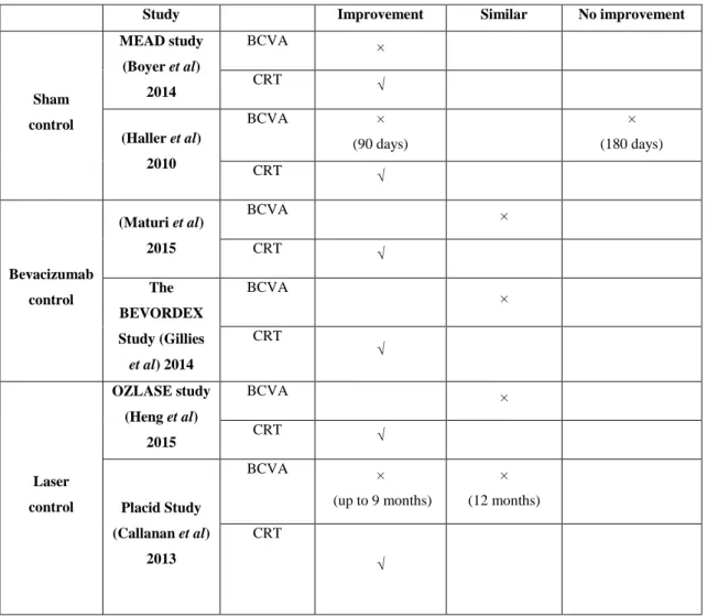
Study Improvement Similar No improvement
Sham control MEAD study (Boyer et al) 2014 BCVA × CRT √ (Haller et al) 2010 BCVA × (90 days) × (180 days) CRT √ Bevacizumab control (Maturi et al) 2015 BCVA × CRT √ The BEVORDEX Study (Gillies et al) 2014 BCVA × CRT √ Laser control OZLASE study (Heng et al) 2015 BCVA × CRT √ Placid Study (Callanan et al) 2013 BCVA × (up to 9 months) × (12 months) CRT √ Table 7. Overview of results
31
Figure 1. Treatment of DME flow diagram.
1
IOP check after 4–8 weeks of initial treatment, especially in eyes with established glaucoma or ocular hypertension or previous history of steroid-induced ocular hypertension. [22]
Diagnostic of DME Focal lesion? Yes Laser therapy No Anti-Vegf Steroids1 Pseudophakic lens? Yes Steroids No Anti-Vegf (> injections) refractory or incomplete response Steroids (< injections)
Fenofibrate Control of blood pressure, blood sugars
32 Acknowledgement message
I would like to express my sincere gratitude to my advisor Dr. Mário Canastro, for the continuous support and his personal availability, for encouraging my research and helped me writing this thesis.
Besides my advisor, I would like to thank to Dr. Daniel Caldeira for the helpful research, which ensured a better result.
A special thanks to Prof. Doctor António Castanheira Dinis, for the remarkable classes which inspired me to follow this area.
33 Bibliography
[1] F. Care, “Introduction,” Diabetes Care, vol. 38, no. Supplement_1, pp. S1–S2, 2015.
[2] R. Lee, T. Y. Wong, and C. Sabanayagam, “Epidemiology of diabetic
retinopathy, diabetic macular edema and related vision loss.,” Eye Vis. (London,
England), vol. 2, p. 17, 2015.
[3] P. U. Dugel and F. Bandello, “Dexamethasone intravitreal implant in the treatment of diabetic macular edema,” pp. 1321–1335, 2015.
[4] R. Klein, M. D. Knudtson, K. E. Lee, R. Gangnon, and B. E. K. Klein, “The Wisconsin Epidemiologic Study of Diabetic Retinopathy XXII. The Twenty-Five-Year Progression of Retinopathy in Persons with Type 1 Diabetes,”
Ophthalmology, vol. 115, no. 11, pp. 1859–1868, 2008.
[5] D. S. Boyer, Y. H. Yoon, R. Belfort, F. Bandello, R. K. Maturi, A. J. Augustin, X.-Y. Li, H. Cui, Y. Hashad, and S. M. Whitcup, “Three-year, randomized, sham-controlled trial of dexamethasone intravitreal implant in patients with diabetic macular edema.,” Ophthalmology, vol. 121, no. 10, pp. 1904–14, 2014. [6] Aiello LM, “Perspectives on diabetic retinopathy. ;,” Am J Ophthalmol 2003, vol.
136:122., 2003.
[7] E. Moisseiev, M. Goldstein, M. Waisbourd, a Barak, and a Loewenstein, “Long-term evaluation of patients treated with dexamethasone intravitreal implant for macular edema due to retinal vein occlusion.,” Eye (Lond)., vol. 27, no. 1, pp. 65–71, 2013.
[8] W.-C. Lam, D. A. Albiani, P. Yoganathan, J. C. Chen, A. Kherani, D. Al Maberley, A. Oliver, T. Rabinovitch, T. G. Sheidow, E. Tourville, L. A. Wittenberg, C. Sigouin, and D. C. Baptiste, “Real-world assessment of
intravitreal dexamethasone implant (0.7 mg) in patients with macular edema: the CHROME study.,” Clin. Ophthalmol., vol. 9, pp. 1255–68, 2015.
[9] J. A. Haller, F. Bandello, R. Belfort, M. S. Blumenkranz, M. Gillies, J. Heier, A. Loewenstein, Y.-H. Yoon, M.-L. Jacques, J. Jiao, X.-Y. Li, and S. M. Whitcup, “Randomized, Sham-Controlled Trial of Dexamethasone Intravitreal Implant in Patients with Macular Edema Due to Retinal Vein Occlusion,” Ophthalmology, vol. 117, no. 6, pp. 1134–1146.e3, 2010.
[10] Allergan, “OZURDEX® (dexamethasone intravitreal implant) For Intravitreal Injection,” Packag. Inser., 2014.
[11] T. Das, F. Da, and R. Diabética, “Tratamento das formas da retinopatia diabética associadas à perda de visão,” 2014.
[12] F. Drugs, “Ozurdex FDA labels,” pp. 4–16, 2014.
[13] P. Claire E Fraser, MD and M. Donald J D’Amico, “Diabetic retinopathy: Prevention and treatment,” UpToDate.
[14] J. A. Ford, N. Lois, P. Royle, C. Clar, D. Shyangdan, and N. Waugh, “Current treatments in diabetic macular oedema: systematic review and meta-analysis.,”
BMJ Open, vol. 3, no. 3, 2013.
[15] P. U. Dugel, A. J. Capone, M. A. Singer, R. F. Dreyer, D. G. Dodwell, D. B. Roth, R. Shi, J. G. Walt, L. C. Scott, D. A. Hollander, and S. S. Group, “Two or more dexamethasone intravitreal implants in treatment-naive patients with
macular edema due to retinal vein occlusion: subgroup analysis of a retrospective chart review study.,” BMC Ophthalmol., vol. 15, p. 118, 2015.
[16] J. A. Haller, B. D. Kuppermann, M. S. Blumenkranz, G. A. Williams, D. V Weinberg, C. Chou, S. M. Whitcup, and D. D. D. S. P. I. I. S. Group,
34
“Randomized controlled trial of an intravitreous dexamethasone drug delivery system in patients with diabetic macular edema.,” Arch. Ophthalmol. (Chicago,
Ill. 1960), vol. 128, no. 3, pp. 289–296, 2010.
[17] R. K. Maturi, L. Bleau, J. Saunders, M. Mubasher, and M. W. Stewart, “A 12-MONTH, SINGLE-MASKED, RANDOMIZED CONTROLLED STUDY OF EYES WITH PERSISTENT DIABETIC MACULAR EDEMA AFTER MULTIPLE ANTI-VEGF INJECTIONS TO ASSESS THE EFFICACY OF THE DEXAMETHASONE-DELAYED DELIVERY SYSTEM AS AN
ADJUNCT TO BEVACIZUMAB COMPARED WITH CONTIN,” Retina, vol. 35, no. 8, pp. 1604–1614, 2015.
[18] M. C. Gillies, L. L. Lim, A. Campain, G. J. Quin, W. Salem, J. Li, S. Goodwin, C. Aroney, I. L. McAllister, and S. Fraser-Bell, “A Randomized Clinical Trial of Intravitreal Bevacizumab versus Intravitreal Dexamethasone for Diabetic
Macular Edema,” Ophthalmology, vol. 121, no. 12, pp. 2473–2481, 2014. [19] L. Z. Heng, S. Sivaprasad, R. Crosby-Nwaobi, Z. Saihan, M. Karampelas, C.
Bunce, T. Peto, and P. G. Hykin, “A prospective randomised controlled clinical trial comparing a combination of repeated intravitreal Ozurdex and macular laser therapy versus macular laser only in centre-involving diabetic macular oedema (OZLASE study),” Br. J. Ophthalmol., pp. bjophthalmol–2015–307136, 2015. [20] D. G. Callanan, S. Gupta, D. S. Boyer, T. a Ciulla, M. a Singer, B. D.
Kuppermann, C.-C. Liu, X.-Y. Li, D. a Hollander, R. M. Schiffman, and S. M. Whitcup, “Dexamethasone intravitreal implant in combination with laser photocoagulation for the treatment of diffuse diabetic macular edema.,”
Ophthalmology, vol. 120, no. 9, pp. 1843–51, 2013.
[21] R. Mathew, E. Pearce, R. Muniraju, A. Abdel-Hay, A. Abdul-Hey, and S.
Sivaprasad, “Monthly OCT monitoring of Ozurdex for macular oedema related to retinal vascular diseases: re-treatment strategy (OCTOME Report 1).,” Eye
(Lond)., vol. 28, no. 3, pp. 318–326, 2014.
[22] J. Ramu, Y. Yang, G. Menon, C. Bailey, N. Narendran, C. Bunce, A. Quartilho, A. T. Prevost, P. Hykin, and S. Sivaprasad, “A randomized clinical trial
comparing fixed vs pro-re-nata dosing of Ozurdex in refractory diabetic macular oedema (OZDRY study).,” Eye (Lond)., vol. 29, no. 12, pp. 1603–1612, 2015. [23] H. Mehta, M. Gillies, and S. Fraser-Bell, “Perspective on the role of Ozurdex
(dexamethasone intravitreal implant) in the management of diabetic macular oedema.,” Ther. Adv. Chronic Dis., vol. 6, no. 5, pp. 234–45, 2015.
[24] J. Ramu, Y. Yang, G. Menon, C. Bailey, N. Narendran, C. Bunce, A. Quartilho, A. T. Prevost, P. Hykin, and S. Sivaprasad, “A randomized clinical trial
comparing fixed vs pro-re-nata dosing of Ozurdex in refractory diabetic macular oedema (OZDRY study).,” Eye (Lond)., vol. 29, no. 12, pp. 1603–1612, 2015.