Patterns of Lymph Node Pathology; Fine
Needle Aspiration Biopsy as an Evaluation
Tool for Lymphadenopathy: A Retrospective
Descriptive Study Conducted at the Largest
Hospital in Africa
Denasha Lavanya Reddy1*, Willem Daniel Francois Venter2, Sugeshnee Pather3
1Department of Internal Medicine, Chris Hani Baragwanath Academic Hospital, University of the Witwatersrand, Johannesburg, South Africa,2Wits Reproductive Health and HIV Institute (RHI) Associate Professor, Department of Medicine, Faculty of Health Sciences, University of the Witwatersrand,
Johannesburg, South Africa,3Division of Anatomical Pathology, School of Pathology, National Health Laboratory Service, Chris Hani Baragwanath Academic Hospital, Faculty of Health Sciences, University of the Witwatersrand, Johannesburg, South Africa
Abstract
Background
Lymphadenopathy is a common clinical presentation of disease in South Africa (SA), partic-ularly in the era of Human Immunodeficiency Virus (HIV) and tuberculosis (TB) co-infection.
Methods
Data from 560 lymph node biopsy reports of specimens from patients older than 12 years at Chris Hani Baragwanath Academic Hospital (CHBAH) between 1 January 2010 and 31 De-cember 2012 was extracted from the National Health Laboratory Service (NHLS), division of Anatomical Pathology. Cytology reports of lymph node fine needle aspirates (FNAs) per-formed prior to lymph node biopsy in 203 patients were also extracted from the NHLS. Con-sent was not obtained from participants for their records to be used as patient information was anonymized and de-identified prior to analysis.
Results
The majority of patients were female (55%) and of the African/black racial group (90%). The median age of patients was 40 years (range12–94). The most common indication for biopsy was an uncertain diagnosis (more than two differential diagnoses entertained), followed by a suspicion for lymphoma, carcinoma and TB. Overall, malignancy constituted the largest biopsy pathology group (39%), with 36% of this group being carcinoma and 27% non-Hodg-kin lymphoma. 22% of the total sampled nodes displayed necrotizing granulomatous inflam-mation (including histopathology and cytology demonstrating definite, and suspicious for
OPEN ACCESS
Citation:Reddy DL, Venter WDF, Pather S (2015) Patterns of Lymph Node Pathology; Fine Needle Aspiration Biopsy as an Evaluation Tool for Lymphadenopathy: A Retrospective Descriptive Study Conducted at the Largest Hospital in Africa. PLoS ONE 10(6): e0130148. doi:10.1371/journal. pone.0130148
Academic Editor:Clive M. Gray, University of Cape Town, SOUTH AFRICA
Received:December 19, 2014
Accepted:May 17, 2015
Published:June 19, 2015
Copyright:© 2015 Reddy et al. This is an open access article distributed under the terms of the
Creative Commons Attribution License, which permits unrestricted use, distribution, and reproduction in any medium, provided the original author and source are credited.
Data Availability Statement:All relevant data are within the paper and its supporting information files.
mycobacterial infection), 8% comprised HIV reactive nodes; in the remainder no specific pa-thology was identified (nonspecific reactive lymphoid hyperplasia). Kaposi sarcoma (KS) accounted for 2.5% of lymph node pathology in this sample. Concomitant lymph node thology was diagnosed in four cases of nodal KS (29% of the subset). The co-existing pa-thologies were TB and Castleman disease. HIV positive patients constituted 49% of this study sample and the majority (64%) of this subset had CD4 counts less than 350 cells/ul. 27% were HIV negative and in the remaining nodes, the HIV status of patients was un-known. The most common lymph node pathologies in HIV positive patients were Mycobac-terial infection (31%), HIV reactive nodes (15%), non-Hodgkin lymphoma (15%) and nonspecific reactive lymphoid hyperplasia (15%). Only 8.7% were of Hodgkin lymphoma. In contrast, the most common lymph node pathologies in HIV negative patients were nonspe-cific reactive lymphoid hyperplasia (45%), carcinoma (25%) and Mycobacterial infection (11%). In this group, non-Hodgkin lymphoma and Hodgkin lymphoma constituted 9% and 8%, respectively. There were more cases of high-grade non-Hodgkin lymphoma in the HIV positive group compared to the HIV negative group. FNA and lymph node biopsy had statis-tically significant good agreement with regard to Hodgkin lymphoma (K 0.774, SE 0.07, 95% CI 0.606-0.882, p=0.001), non-Hodgkin lymphoma (K 0.640, SE 0.07, 95% CI 0.472-0.807, p=0.001), carcinoma (K 0.723, SE 0.069, 95% CI 0.528-0.918, p=0.001), and myco-bacterial infection (K 0.726, SE 0.07, 95% CI 0.618-0.833, p=0.001).
Conclusions
The most common lymph node pathologies in CHBAH are malignancies, nonspecific reac-tive lymphoid hyperplasia, necrotizing granulomatous inflammation and HIV reacreac-tive nodes. The distribution of disease differs in HIV positive patients. Overall, adequate FNA samples of lymph nodes have been found to have good correlation with lymph node biopsy findings in our setting.
Introduction
Lymphadenopathy is a common presentation of disease in South Africa (SA), particularly in the era of Human Immunodeficiency Virus (HIV) and tuberculosis (TB) co-infection. There are only a limited number of studies that have described lymph node pathology in Southern Africa. Chris Hani Baragwanath Academic Hospital (CHBAH) which is a provincial tertiary hospital in Soweto, Johannesburg, contains close to 3300 beds and is the largest hospital in Af-rica [1].
In a post-mortem study performed in Johannesburg, TB was found to be the cause of death in the majority of patients (69%) with advanced Acquired Immunodeficiency Syndrome (AIDS), both before and after starting antiretroviral therapy [2]. Lymph nodes were the fourth most common site of positive mycobacterial cultures in this study (16%), following liver, spleen and lung [2]. The study also suggested that TB often goes unrecognized in these patients, and can be accompanied by other infections or neoplasms [2].
Excisional biopsy of a clinically appropriate lymph node is traditionally favored as a diag-nostic procedure for a multitude of infectious and neoplastic disorders. However, fine-needle Competing Interests:Professor Venter is supported
aspiration (FNA) is replacing it as a first line diagnostic procedure, reserving excisional biopsy for non-diagnostic FNA results that require further investigation [3–4].
We conducted a retrospective review of the lymph node pathology identified at CHBAH to contribute to medical knowledge of common causes of lymphadenopathy requiring biopsy in South Africa, and the role of FNA as an adjunct to excisional lymph node biopsy.
Methods
The study was a retrospective descriptive audit conducted at Chris Hani Baragwanath Academ-ic Hospital, Johannesburg, South AfrAcadem-ica. The study population consisted of all patients over the age of 12 years who underwent lymph node biopsies at CHBAH between 1 January 2010 and 31 December 2012. Lymph node biopsy reports were extracted from the database of the Na-tional Health Laboratory Service (NHLS) Division of Anatomical Pathology.
Patient demographic details were obtained from the laboratory requisition forms submitted by attending clinicians and from pathology reports. There was no review of patient charts or files. Further information regarding HIV status (determined with antibody testing using 4th generation HIV enzyme-linked immunosorbent assay), CD4 count (cells/ul), if relevant, and FNAs performed (smears in our setting) were accessed through the NHLS DisaLab system, using the patients’name and hospital number. Consent was not obtained from participants for their records to be used as patient information was anonymized and de-identified prior to analysis.
All lymph node biopsy and FNA biopsy specimens were analyzed by pathologists employed by the NHLS at CHBAH, including one of the authors. Unfortunately, the authors reviewed not all the slides during this retrospective study. Histologic diagnoses were based on morpho-logic findings and, in the appropriate context, ancillary tests including Mycobacterial staining and culture, immunohistochemistry, FISH (fluorescent in-situ hybridization) techniques, and flow cytometry. The World Health Organization diagnostic criteria were applied to the histo-pathology demonstrated on lymph node biopsies. The diagnosis of tuberculous lymphadenitis was confirmed by Ziehl-Neelsen stain for acid fast bacilli and positive Mycobacterial culture. Mycobacterial culture and Mycobacterial PCR testing using Xpert MTB/RIF are performed in cases of clinically and histopathologically (granulomatous) suspected Mycobacterial lymphade-nitis. Ziehl-Neelsen stains for acid fast bacilli were routinely performed on all lymph node bi-opsies and FNA bibi-opsies that displayed granulomatous inflammation (necrotizing and non-necrotizing). HIV lymphadenitis was graded as follows: grade 1 if associated with hyperplastic features and enlargement of germinal centres with increased apoptosis and phagocytosis by macrophages; grade 2 if there was a reduction in lymphoid follicles and mature lymphocytes, but an increase in plasma cells and perifollicular blood vessels; and grade 3 if the germinal cen-tres were sclerotic. HIV lymphadenitis was not used as a surrogate marker for HIV infection, as it describes a histological pattern not entirely specific to HIV.
Statistical analysis was performed using STATA version 12 for Windows (StataCorp LP, Texas;www.stata.com) and GraphPadQuickCalcs (GraphPad Software Inc, California;www. graphpad.com). A statistician was consulted. The Human Research Ethics committee at the University of the Witwatersrand granted ethical approval of this study (Clearance Certificate number M130626).
Results
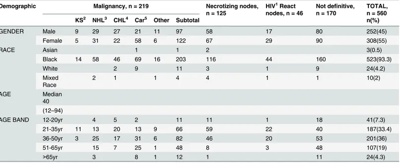
The majority of patients (55%) who had lymph node biopsies were female (Table 1). Over 90% of patients were of the African/black racial group, in keeping with the racial demographic dis-tribution in SA. The median age of patients was 40 years, with a minimum of 12 and a maxi-mum of 94. The most common indication for biopsy was an uncertain diagnosis (more than two differential diagnoses entertained), followed by a suspicion for lymphoma, carcinoma and TB (Table 2).
The most frequent site of biopsy was cervical, followed by“other”which included laparo-scopic biopsies (of intra-abdominal nodes), axillary and inguinal nodal biopsies. Laparolaparo-scopic biopsies were performed as part of a staging or curative process for patients known to have car-cinoma. In other instances, lymph nodes were found incidentally at laparotomy or laparoscopy and sampled.
Interestingly, of the suspected lymphoma cases, 28% were diagnosed with non-Hodgkin lymphoma, and 23% with Hodgkin lymphoma. Of the suspected TB cases, 62% displayed nec-rotizing granulomatous inflammation on biopsy, while 53% of staging carcinoma had con-firmed carcinoma on biopsy.
Overall, malignancy was the largest biopsy pathology group (39%), with 36% of this group having carcinoma and 27% non-Hodgkin lymphoma. 22% of the total sample comprised nodes that displayed necrotizing granulomatous inflammation (including histopathology dem-onstrating definite mycobacterial infection and suspicious for mycobacterial infection). 8% of the sample comprised HIV reactive nodes and in the remainder no specific pathology was identified (including nonspecific reactive lymphoid hyperplasia).
KS accounted for 2.5% of lymph node pathology in the sample. Concomitant lymph node pathology was diagnosed within four cases of nodal KS (29% of the subset). The co-existing pa-thologies were mycobacterial infection in one patient, Castleman disease in two patients, and a combination of mycobacterial infection and Castleman disease in the fourth patient.
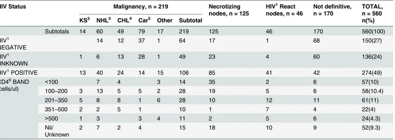
HIV positive patients constituted 49% of the sample, with the majority of patients in this subset (64%) having a CD4 count below 350 cells/ul. 27% were HIV negative and in the re-maining nodes, the HIV status of patients was unknown. The most common lymph node pa-thologies in HIV positive patients were mycobacterial infection (31%), HIV reactive nodes (15%), non-Hodgkin lymphoma (15%) and non-specific reactive lymphoid hyperplasia (15%) (Table 3). Only 8.7% were of Hodgkin lymphoma. In contrast, the most common lymph node pathologies in HIV negative patients were nonspecific reactive lymphoid hyperplasia (45%), carcinoma (25%) and Mycobacterial infection (11%). In this group, non-Hodgkin lymphoma and Hodgkin lymphoma constituted 9% and 8%, respectively.
Table 1. Incisional, Excisional and Core Biopsy Diagnosis in relation to Demographics.
Demographic Malignancy, n = 219 Necrotizing nodes,
n = 125
HIV1React nodes, n = 46
Not definitive, n = 170
TOTAL, n = 560
KS2 NHL3 CHL4 Car5 Other Subtotal n(%)
GENDER Male 9 29 27 21 11 97 58 17 80 252(45)
Female 5 31 22 58 6 122 67 29 90 308(55)
RACE Asian 1 1 2 3(0.5)
Black 14 58 46 69 16 203 116 44 160 523(93.3)
White 2 9 11 3 1 9 24(4.2)
Mixed Race
2 1 1 4 4 1 1 10(2)
AGE Median
40 (12–94)
AGE BAND 12-20yr 4 5 2 11 11 1 18 41(7.3)
21-35yr 11 13 20 13 9 66 59 22 40 187(33.4)
36-50yr 3 25 17 31 6 82 46 20 53 201(36)
51-65yr 15 7 25 1 48 8 3 48 107(19)
>65yr 3 8 1 12 1 11 24(4.3)
1HIV: Human Immunode
ficiency Virus 2KS: Kaposi sarcoma
3
NHL: non-Hodgkin Lymphoma 4CHL: Hodgkin Lymphoma 5Car: carcinoma.
doi:10.1371/journal.pone.0130148.t001
Table 2. Incisional, Excisional and Core Biopsy Diagnosis in relation to Indications and site of biopsy.
Malignancy, n = 219 Necrotizing
nodes, n = 125
HIV1React nodes, n = 46
Not definitive, n = 170
TOTAL, n = 560
KS2 NHL3 CHL4 Car5 Other Subtotal n(%)
INDICATION FOR BIOPSY
Suspect TB6 1 5 2 2 1 11 46 9 8 74(13)
Suspect lymphoma
2 33 28 4 4 71 17 13 19 120(22)
Staging Car5 60 60 54 114(20)
Uncertain Diagnosis
9 16 14 12 9 60 45 20 77 202(36)
Unknown 2 6 5 1 3 17 17 4 12 50(9)
SITE OF LYMPH NODE BIOPSY
Cervical 8 26 37 18 9 98 73 16 31 218(40)
Inguinal 2 10 2 10 1 25 5 9 19 58(10)
Axilla 2 11 7 35 6 61 18 12 38 129(23)
Other 2 13 3 16 1 35 29 9 82 155(27)
1HIV: Human Immunode
ficiency Virus 2KS: Kaposi sarcoma
3NHL: non-Hodgkin Lymphoma 4CHL: Hodgkin Lymphoma 5Car: carcinoma
6TB: Tuberculosis.
HIV lymphadenitis is a histological pattern not entirely specific to HIV and therefore, cannot be used as a surrogate marker for HIV infection.
There were 203 (36%) patients in our study sample who also underwent FNAs prior to biop-sy. Of the FNAs, 23% were inadequate for assessment, 76% were adequate and in 1% adequacy was not commented on. When compared with the histopathology diagnosis as per lymph node biopsies, FNA was found to have statistically significant good agreement/reliability with re-gards to Hodgkin lymphoma (K 0.774, SE 0.07, 95% CI 0.606–0.882, p = 0.001), non-Hodgkin lymphoma (K 0.640, SE 0.07, 95% CI 0.472–0.807, p = 0.001), carcinoma (K 0.723, SE 0.069, 95% CI 0.528–0.918, p = 0.001), and mycobacterial infection (K 0.726, SE 0.07, 95% CI 0.618–
0.833, p = 0.001) (Table 4).
Table 3. Incisional, Excisional and Core Biopsy diagnosis in relation to HIV status.
HIV Status Malignancy, n = 219 Necrotizing
nodes, n = 125
HIV1React nodes, n = 46
Not definitive, n = 170
TOTAL, n = 560
KS2 NHL3 CHL4 Car5 Other Subtotal n(%)
Subtotals 14 60 49 79 17 219 125 46 170 560(100)
HIV1 NEGATIVE
14 12 37 1 64 17 1 68 150(27)
HIV1 UNKNOWN
1 6 13 28 1 49 23 4 60 136(24)
HIV1POSITIVE 13 40 24 14 15 106 85 41 42 274(49)
CD46BAND (cells/ul)
<100 7 4 3 14 35 2 6 57(10)
100–200 3 13 5 5 2 28 19 5 6 58(10.4)
201–350 5 8 8 1 6 28 10 12 11 61(11)
351–500 2 2 5 1 10 1 7 4 22(4)
>500 1 3 3 4 11 2 5 6 24(4.3)
Nil/ Unknown
2 7 2 4 15 18 10 9 52(9.3)
1HIV: Human Immunode
ficiency Virus 2KS: Kaposi sarcoma
3
NHL: non-Hodgkin Lymphoma 4CHL: Hodgkin Lymphoma 5Car: carcinoma
6
CD4: Cluster of Differentiation 4 T-cells.
doi:10.1371/journal.pone.0130148.t003
Table 4. Statistical Agreement between LN FNA and LN Biopsy.
Pathological Diagnosis % observed agreements % agreements expected by chance Kappa SE 95% Confidence Interval P-value
CHL1 94.09 76.92 0.744 0.07 0.606
–0.882 0.001
NHL2 92.61 79.50 0.640 0.07 0.472–0.807 0.001
Car3 96.55 87.55 0.723 0.069 0.528
–0.918 0.001
Granulom inflam NOS4 97.04 95.20 0.385 0.07 -0.005
–0.775 0.001
MI5 89.16 60.49 0.726 0.07 0.618
–0.833 0.001
Non-specific reactive LNH6 80.30 67.28 0.398 0.067 0.249–0.547 0.001
1CHL: Hodgkin Lymphoma 2
NHL: non Hodgkin Lymphoma 3Car: carcinoma
4Granulom in
flam NOS: Granulomatous Inflammation not otherwise specified 5
MI: Mycobacterial Infection 6Non-speci
There were two cases where a single patient had more than one lymph node biopsy during the period of the study. Both patients were HIV positive with CD4 counts below 200 cells/ul. FNAs were performed prior to biopsy in both cases and were not definitive. The sites of the bi-opsies were, respectively, axillary and cervical in the first patient, and cervical both times, in the second patient. The definitive biopsies were both repeated due to a high index of clinical suspi-cion, and subsequently confirmed to be non-Hodgkin lymphoma.
Discussion
Our study affirmed local demographic patterns, in that the majority of patients who underwent lymph node biopsy at CHBAH were middle-aged African/black females. We have demonstrat-ed that nodal pathology diagnoses at CHBAH are inclusive of malignancy, necrotizing granulo-matous inflammation and reactive nodes (due to HIV or other). The most common lymph node pathologies at CHBAH as per biopsy are in keeping with local African literature [5–7]. However, our data is unique in that it also describes differences in pathology between HIV pos-itive and negative patients, the former further stratified by CD4 count.
Our study highlights the known significant correlation between HIV incidence and inci-dence of aggressive B-cell lymphomas and Hodgkin lymphoma [8–9]. Associated risk factors for the development of these malignancies are high HIV viral loads and low CD4 counts, al-though malignancies may occur at any CD4 count. The pathogenesis is al-thought to be immune dysregulation with loss of T-cell immunity against onco-viruses like Epstein-Barr Virus and human herpesvirus 8 [8].
The most common indications for requesting a lymph node biopsy in our setting were an uncertain diagnosis and the suspicion of malignancy or TB, which reflects the relative domi-nance of the latter two diseases in Southern Africa. The global prevalence and death rates from TB are on the decline. However, in contrast, Africa is showing an increase in the tuberculous burden [10].
In a study performed at CHBAH, TB was diagnosed in 1291 patients over a period of two months [1]. The association between HIV infection and TB is well described in South Africa. The high prevalence of TB is also reflected by HIV statistics for SA [11]. South Africa carries the highest global burden of HIV with an estimated 5.6 million people infected, with TB being the most common serious opportunistic infection [11–13].
The previous CHBAH study showed that of the patients diagnosed with TB, 74% had pul-monary TB and of the patients with extrapulpul-monary TB, pleural and miliary were the most common forms [1]. However, more recent publications suggest that TB lymphadenitis is the most common form of extrapulmonary TB [3–4,14]. The high prevalence of TB and TB suspi-cious lymphadenitis at CHBAH has been described, notably in the age band 25–44 years [15]. In our study, 22% of lymph node biopsies and 27% of FNAs comprised nodes that displayed necrotizing granulomatous inflammation (including histopathology and cytology demonstrat-ing definite mycobacterial infection and suspicious for mycobacterial infection), confirmdemonstrat-ing the high prevalence of TB in CHBAH.
The study by Martinson et al stated that the risk of TB increases at lower CD4 counts, and suggested that early antiretroviral therapy reduced the population prevalence of TB in HIV in-fected patients [12]. This suggestion may also be evident in our study as the majority of HIV positive patients in our sample had CD4 counts less than 350 cells/ul and the commonest lymph node pathology in this group was confirmed and suspected TB.
months [16]. The dangers of misdiagnosing TB, and subsequent empiric TB treatment, are progression of underlying disease (malignancy or other infection); toxicity of TB therapy; and development of drug-resistant therapy [11,17].
A Zambian study suggested that primary HIV lymphadenopathy was a significant cause of superficial lymphadenopathy [18]. This finding is also highlighted in our study, as HIV reactive nodes comprised 8% of the total sample, and was amongst the largest nodal pathology group in HIV positive patients (15%).
HIV-associated lymphadenitis (reported in our setting as HIV reactive nodes) is a well-characterized pattern of histological findings in lymph nodes of many HIV-infected individu-als. It is likely due to the lymphotropism of the HI virus. Grade 1 is associated with hyperplastic features, enlargement of germinal centres with increased apoptosis and phagocytosis by macro-phages. In Grade 2 there is a reduction in lymphoid follicles and mature lymphocytes, but an increase in plasma cells and perifollicular blood vessels. In Grade 3, the germinal centres be-come sclerotic [19].
Rare causes of lymphadenopathy in our study included one case of Cryptococcosis
(microbiologically confirmed) in an HIV positive male patient who had profound immunosup-pression (CD4 count of 8 cells/ul). In addition, there was a case of myeloid sarcoma (extrame-dullary counterpart of chronic myeloid leukaemia) in an HIV positive female who had a CD4 count of 1626 cells/ul.
The association between KS and multicentric Castleman disease due to the common causal agent human herpesvirus 8 (HHV8) is well described in the literature [20]. This association has been demonstrated by the co-existence of both these pathologies in lymph nodes in our study. Interestingly, two patients in our study with coexisting KS and Castleman disease had CD4 counts above 350 cells/ul. CD4 counts were not available for the remaining two patients who had concomitant lymph node pathology. A possible explanation for the relatively high CD4 counts may be immune reconstitution inflammatory syndrome.
We then evaluated the outcomes of fine needle aspiration biopsy in our setting. FNA is widely regarded as the diagnostic modality of choice in diagnosing TB lymphadenitis [10,21]. Our study affirms a good correlation between FNA diagnosis and biopsy diagnosis on lymph node specimens. In the event of inadequate FNA samples, the result should be treated with re-serve. If there is a clinical concern for nodal-based mycobacterial disease or malignancy despite a non-contributory FNA, a clinically appropriate lymph node should be submitted for histo-pathologic assessment.
In the event of a persistent clinical concern, despite a negative histopathology result, the nodal biopsy should be repeated with emphasis on selection of a clinically appropriate node. The importance of a repeat biopsy where there is a high clinical suspicion for disease, particu-larly in HIV positive patients, was highlighted in the two patients from our study subsequently diagnosed with non-Hodgkin lymphoma [16]. Our findings suggest that negative biopsy find-ings in HIV positive patients should be treated with reserve if there is a high index of clinical suspicion for lymphoma.
Conclusions
Our study showed that the most common lymph node pathologies occurring in patients who underwent biopsies at CHBAH are malignancies, nonspecific reactive lymphoid hyperplasia, necrotizing granulomatous inflammation due to mycobacterial infection, and HIV reactive nodes. The distribution of disease differs in HIV positive patients. FNA was found to have good overall correlation with histopathology biopsy diagnoses in our setting. The diagnosis specified in an FNA report should be interpreted in conjunction with the comment/s about ad-equacy of the aspirated specimen for assessment. Due to the coexistence of nodal KS with other pathologies, patients who have suspected nodal KS would benefit from proceeding directly to have biopsies for histopathological assessment.
Supporting Information
S1 Dataset. FNA Stratification.(XLS)
Acknowledgments
We would like to acknowledge the staff at the NHLS for their help during data collection, and the masters in epidemiology students who assisted with data analysis. Dr Alison Bentley pro-vided invaluable academic guidance. And finally, we would like to acknowledge our patients at Chris Hani Baragwanath Academic Hospital who inspire us daily.
Author Contributions
Conceived and designed the experiments: DLR WDFV SP. Performed the experiments: DLR. Analyzed the data: DLR SP. Contributed reagents/materials/analysis tools: DLR WDFV SP. Wrote the paper: DLR WDFV SP.
References
1. Edginton ME, Wong ML, Phofa R, Mahlaba D, Hodkinson HJ (2005) Tuberculosis at Chris Hani Barag-wanath Hospital: numbers of patients diagnosed and outcomes of referrals to district clinics.
Interna-tional Journal of Tuberculosis and Lung Disease: 9(4):398–402. PMID:15830744
2. Wong EB, Omar T, Setlhako GJ, Osih R, Feldman C, Murdoch DM et al (2012) Causes of Death on An-tiretroviral Therapy: A Post-Mortem Study from South Africa.Public Library of Science (PLoS) ONE: 7 (10):e47542. doi:10.1371/journal.pone.0047542PMID:23094059
3. Handa U, Mundi I, Mohan S (2012) Nodal tuberculosis revisited: a review.Journal of Infection in
Devel-oping Countries: 6(1):6–12. PMID:22240421
4. Fontanilla JM, Barnes A, Fordham von Reyn C (2011) Current Diagnosis and Management of Peripher-al Tuberculous Lymphadenitis.Clinical Infectious Diseases: 53(6):555–562. doi:10.1093/cid/cir454
PMID:21865192
5. Muthuphei MN (1998) Cervical lymphadenopathy at Ga-Rankuwa Hospital (South Africa): a histological review.Central African Journal of Medicine: 44(12):311–2. PMID:10921203
6. Sibanda EN, Stanczuk G (1993) Lymph node pathology in Zimbabwe: a review of 2194 specimens.
Quarterly Journal of Medicine: 86(12):811–817. PMID:8108537
7. Olu-eddo AN, Omoti CE (2011) Diagnostic evaluation of primary cervical adenopathies in a developing country.Pan African Medical Journal: 10:52. PMID:22384298
8. Kaplan LD (2012) HIV-associated lymphoma.Best Practice and Research Clinical Haematology: 25:101–117. doi:10.1016/j.beha.2012.01.001PMID:22409827
9. Wiggill TM, Mantina H, Willem P, Perner Y, Stevens WS (2011) Changing Pattern of Lymphoma Sub-groups at a Tertiary Academic Complex in a High-Prevalence HIV Setting: A South African Perspective.
Journal of Acquired Immune Deficiency Syndromes: 56(5):460–466. doi:10.1097/QAI.
10. Department of Health. Republic of South Africa. National Tuberculosis Management Guidelines (2009).
11. Schutz C, Ismail Z, Proxenos CJ, Marais S, Burton R, Kenyon C et al (2013) Burden of antituberculosis and antiretroviral drug-induced liver injury at a secondary hospital in South Africa.South African
Medi-cal Journal: 102(6):506–511.
12. Martinson NA, Hoffmann CJ, Chaisson RE (2011) Epidemiology of Tuberculosis and HIV: Recent ad-vances in Understanding and Responses.Proceedings of the American Thoracic Society: 8:288–293. doi:10.1513/pats.201010-064WRPMID:21653530
13. Churchyard GJ, Mametja LD, Mvusi L, Ndjeka N, Hesseling AC, Reid A et al (2014) Tuberculosis con-trol in South Africa: Successes, challenges and recommendations.South African Medical Journal: 104 (3):244–248. PMID:24893501
14. Ligthelm LJ, Nicol MP, Hoek KGP, Jacobson R, van Helden PD, Marais BJ et al (2011) Xpert MTB/RIF for Rapid Diagnosis of Tuberculous Lymphadenitis from Fine-Needle-Aspiration Biopsy Specimens.
Journal of Clinical Microbiology: 49(11):3967. doi:10.1128/JCM.01310-11PMID:21880965
15. Karstaedt AS (2014) Extrapulmonary tuberculosis among adults: Experience at Chris Hani Baragwa-nath Academic Hospital, Johannesburg, South Africa.South African Medical Journal: 104(1):22–24. doi:10.7196/samj.6374PMID:24388080
16. Puvaneswaran B, Shoba B (2013) Misdiagnosis of tuberculosis in patients with lymphoma.South
Afri-can Medical Journal: 103(1):32–33. doi:10.7196/samj.6093PMID:23237121
17. Streicher FM, Muller B, Chihota V, Mlambo C, Tait M, Pillay M et al (2012) Emergence and treatment of multidrug resistant (MDR) and extensively drug-resistant (XDR) tuberculosis in South Africa.Infection,
Genetics and Evolution: 12(4):686–694. doi:10.1016/j.meegid.2011.07.019PMID:21839855
18. Bem C, Patil PS, Bharucha H, Namaambo K, Luo N (1996) Importance of human immunodeficiency virus-associated lymphadenopathy and tuberculous lymphadenitis in patients undergoing lymph node biopsy in Zambia.British Journal of Surgery: 83(1):75–78. PMID:8653372
19. Mahe E, Ross C, Sur M (2011) Lymphoproliferative Lesions in the Setting of HIV Infection: A Five-Year Retrospective Case Series and Review. Pathology Research International. Volume 2011. doi:10. 4061/2011/618760
20. Dupin N, Fisher C, Kellam P, Ariad S, Tulliez M, Franck N et al (1999). Distribution of human herpesvi-rus-8 latently infected cells in Kaposi’s Sarcoma, multicentric Castleman’s disease, and primary effu-sion lymphoma.Proceedings of the National Academy of Sciences of the United States of America: 96 (8):4546–4551. PMID:10200299
21. Razack R, Louw M, Wright CA (2014) Diagnostic yield of fine needle aspiration biopsy in HIV-infected adults with suspected mycobacterial lymphadenitis.South African Medical Journal: 104(1):27–28. doi: