ORIGINAL ARTICLE
J of Evidence Based Med & Hlthcare, pISSN- 2349-2562, eISSN- 2349-2570/ Vol. 2/Issue 10/Mar 09, 2015 Page 1359
CYTOLOGICAL EVALUATION OF MALE BREAST LESIONS IN
GREATER GWALIOR: A FIVE YEAR RETROSPECTIVE STUDY
Jagannath Jatav1, Rajesh Gaur2, Vidyanand Pandit3, Bharat Jain4
HOW TO CITE THIS ARTICLE:
Jagannath Jatav, Rajesh Gaur, Vidyanand Pandit, Bharat Jain. ”Cytological Evaluation of male Breast Lesions in Greater Gwalior: A Five Year Retrospective Study”. Journal of Evidence based Medicine and Healthcare; Volume 2, Issue 10, March 09, 2015; Page: 1359-1364.
ABSTRACT: BACKGROUNDS: Fine needle aspiration cytology is an effective modality for diagnosis of breast lesions. Usually male breast lesions are benign and affect the young male. Most common lesion is gynaecomastia. Male breast cancer accounts for a small proportion of breast cancers. Male breast cancer usually presents at an advanced age. OBJECTIVE: The aim of this study was to examine the nature of male breast lesions and to determine the cytomorphologic patterns of these lesions. METHODS: five year retrospective study was conducted in our institution and in that 112 patients underwent fine needle aspiration cytology of the palpable breast lump after thorough physical examination. The cytological diagnosis was classified as benign, inflammatory, malignant and others. RESULTS: In 112 male patients diagnosed with breast lesions, the most common lesion was gynecomastia (103/112, 91.9%), followed by breast cancer (6/112, 5.4%), inflammatory (2/112, 1.8%) and apocrine metaplasia (01/112, 0.9%). Gynecomastia was commonly found in male patients less than 40 years of age, while breast cancer is seen in male patients over 40 years of age.
KEYWORDS: Male Breast Carcinoma, Gynaecomastia and Fine Needle Aspiration Cytology (FNAC).
INTRODUCTION: FNAC of breast lumps is an important to assess the palpable breast lumps. It is an accurate, rapid, easy to perform, cost-effective and reproducible diagnostic tool that can be carried out at outpatient department.(1,2,3) It is commonly used in the diagnosis and management
of breast lesions both in female and in male.
FNA has various benefits over the open tissue biopsy.(4,5)
1. Rapid, Reliable, Easy to perform and cost effective. 2. A definite treatment plans can be prepared.
3. Molecular ancillary technique i.e. PR & ER, proliferation antigen (Ki67) & DNA pattern analysis can be performed.
AIMS AND OBJECTIVES: The aim of this study was to examine the nature of male breast lesions and to determine the cytomorphologic patterns of these lesions which were diagnosed by FNAC of patients referred to department of cytopathology during January 2010 to December 2014.
ORIGINAL ARTICLE
J of Evidence Based Med & Hlthcare, pISSN- 2349-2562, eISSN- 2349-2570/ Vol. 2/Issue 10/Mar 09, 2015 Page 1360
using 22-gauge needle and 20 ml syringe with the help of pluger by the cytopathologist. Air dried smears were prepared and stained by the Giemsa stain.
RESULTS: Over a five year period, 112 male patients with palpable breast lumps underwent Fine needle aspiration (FNA) at our department. Out of total 112, 104 (92.9%) patients had a unilateral breast lump (55 patient had right breast lump and 49 patient had left breast lump) and 8 (7.1%) patients had a bilateral breast lump. The age ranged from 09 to 84 yrs with a median age of 43.7 yr. Diagnostic aspirates were obtained in all 112 cases. These aspirates were broadly categorized into two groups: benign 106 (94.6%) and malignant 06 (5.4%).
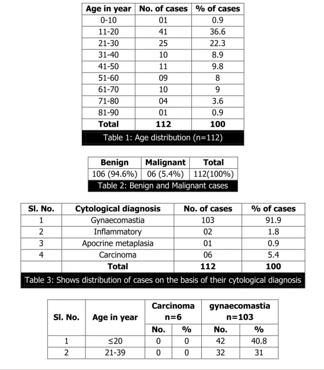
Age in year No. of cases % of cases
0-10 01 0.9 11-20 41 36.6 21-30 25 22.3 31-40 10 8.9 41-50 11 9.8 51-60 09 8 61-70 10 9 71-80 04 3.6 81-90 01 0.9
Total 112 100
Table 1: Age distribution (n=112)
Benign Malignant Total
106 (94.6%) 06 (5.4%) 112(100%)
Table 2: Benign and Malignant cases
Sl. No. Cytological diagnosis No. of cases % of cases
1 Gynaecomastia 103 91.9 2 Inflammatory 02 1.8 3 Apocrine metaplasia 01 0.9 4 Carcinoma 06 5.4
Total 112 100
Table 3: Shows distribution of cases on the basis of their cytological diagnosis
Sl. No. Age in year
Carcinoma n=6
gynaecomastia n=103
No. % No. %
ORIGINAL ARTICLE
J of Evidence Based Med & Hlthcare, pISSN- 2349-2562, eISSN- 2349-2570/ Vol. 2/Issue 10/Mar 09, 2015 Page 1361
3 40-59 2 33.3 18 17.5 4 ≥60 4 66.4 11 10.7
Total 6 100 103 100
Table 4: Age wise comparison of carcinoma and gynaecomasia
The benign lesions constituted the largest number of cases and are further subcategorized into gynaecomastia 103 (91.9%), inflammatory 02 (1.8%) and apocrine metaplasia 01 (0.9%). The prevalence of gynecomastia in different age groups is shown in Table no.1 and 4. Gynecomastia was more common in patients less than 20 years of age (42/103, 40.8%), followed by 20–39 years of age (32/103, 31%), 40–59 years of age (18/103, 17.5%) and over 60 years of age (11/103, 10.7%).
This indicates that gynecomastia occurs more commonly in male patients under 40 years of age and its incidence is decreased as age increases while other male breast lesions occurred in patients over 40 year of age.
The overall proportion of breast cancer was 5.4% (6/112). Breast cancer occurred in about one third (2/6) of 40- 59year-old patients and two third (4/6) over 60 year-old patients. None case of carcinoma seen in patients less than 40 year of age in our study.
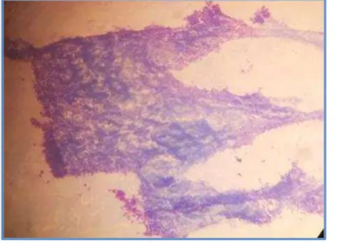
Gynecomastia-smear showing sheets of ductal cells and fragments of loose stroma (×10, giemsa).
ORIGINAL ARTICLE
J of Evidence Based Med & Hlthcare, pISSN- 2349-2562, eISSN- 2349-2570/ Vol. 2/Issue 10/Mar 09, 2015 Page 1362 DISCUSSION: The FNAC of breast lump is worldwide accepted and established method of choice to determine the nature of breast lump. Gynaecomastia was the commonest benign lesion in our study which was concurrent with the findings of Manas Kotepuia et al.(6)
In our study, 103 out of 112cases (91.8%) were Gynecomastia. Gynecomastia was unilateral in 92.2% of the cases (95 out of 103) and more frequent in the right side than left side breast (50 cases were right sided and 45 were in right sided). This was contrast to the studies conducted by Das et al.,(7) and Martin-Bates et al.,(8) who observed it more in the left breast.
The overall prevalence of gynecomastia in male patients with breast lesions during 5 years was 91.9%. This is higher than in Austria (51.2%),(9) Nepal (60%),(10) and Nigeria (61.9%),(11)
Italy (66.07%),(12) USA (67.3%),(13) UK (74.4%),(14) Spain (80.4%),(15) South Africa (82.7%),(16)
and India (84.3%).(17) It was lower than that in Turkey (100%),(18) and Pakistan (100%).(19)
Gynecomastia appeared to be more pronounced in patients less than 20 years of age. This was similar to findings in a study of Manas Kotepuia et al (2014).
In the present study percentage of malignant cases was 5.4% (6/112), this was more or less similar findings to kirana pailoor et al (2014).(20) This percentage was more than Siddiqui MT
(2002),21 and was less than MacIntosh et al (2008),(22) Westend et al (2002)(23) and Wauters et al
(2009).(24)
Ranbeer et al(25) has found (2.5%) cases of inflammation on FNAC of male breast lesions.
MS Gill1 et al.(26) has noted (5.4%) cases of inflammation in their study while Raajul Jain et al.(27)
reported 9% cases of inflammatory lesion. In the present study, 1.8% cases were diagnosed as inflammation.
CONCLUSIONS: Gynecomastia is the most common diagnosis in male patients referred to department of cytology, G R Medical College Gwalior with breast complaints during 2009- 2014. It involved mostly male patients less than 20 years of age.
REFERENCES:
1. Ahmed HG, Ali AS, Almobarak AO (2009). Utility of fine-needle aspiration as a diagnostic technique in breast lumps. Diagn Cytopathol, 37, 881-4.
2. Saleh FM, Ansari NP, Alam O (2012). Comparison between fine needle aspiration cytology with histopathology to validate accurate diagnosis of palpable breast lump. Mymensingh Med J, 21, 450-5.
3. Wang XW, Xiong YH, Zen XQ, Lin HB, Liu QY (2012). Diagnostic accuracy of ultrasonograph guided fine-needle aspiration cytologic in staging of axillary lymph node metastasis in breast cancer patients: a meta-analysis. Asian Pac J Cancer Prev, 13, 5517-23.
4. Silverman JF, Lannin DR, O'Brien K, Norris HT: The triage role of fine needle aspiration biopsy of palpable breast masses. Diagnostic accuracy and cost effectiveness. Acta Cytol; 1987; 31: 731-736.
ORIGINAL ARTICLE
J of Evidence Based Med & Hlthcare, pISSN- 2349-2562, eISSN- 2349-2570/ Vol. 2/Issue 10/Mar 09, 2015 Page 1363
6. Manas Kotepuia, Duangjai Piwkhama, Chaowanee Chupeerachb, Apiram Songsric A retrospective study of gynecomastia in male patients referred to Hatyai Hospital with breast lesions Asian Biomedicine Vol. 8 No. 4 August 2014; 511-515.
7. Martin-Bates E, Krausz T and Phillips I. Evaluation of fine needle aspiration of the male breast for the diagnosis of gynecomastia.Cytopathol.1990; 1: 79-85.
8. Das DK, Junaid TA, Mathews SB, et al. Fine needle aspiration cytology diagnosis of male breast lesions – a study of 185 cases. Acta Cytol.1995; 39: 870-76.
9. Partik B, Mallek R, Rudas M, Pokieser P, Wunderbaldinger P, Helbich TH. [Malignant and benign diseases of the breast in 41 male patients: mammography, sonography and pathohistological correlations]. Rofo. 2001; 173: 1012-8. (In German).
10.Kumar R. A clinicopathologic study of breast lumps in Bhairahwa, Nepal. Asian Pac J Cancer Prev. 2010; 11: 855-8.
11.Olu-Eddo AN, Ugiagbe EE. Benign breast lesions in an African population: a 25-year histopathological review of 1864 cases. Niger Med J. 2011; 52: 211-6.
12.Ambrogetti D, Ciatto S, Catarzi S, Muraca MG. The combined diagnosis of male breast lesions: a review of a series of 748 consecutive cases. Radiol Med.1998.
13.Evans GF, Anthony T, Turnage RH, Schumpert TD, Levy KR, Amirkhan RH, et al. The diagnostic accuracy of mammography in the evaluation of male breast disease. Am J Surg. 2001; 181: 96-100.
14.Al-Allak A, Govindarajulu S, Shere M, Ibrahim N, Sahu AK, Cawthorn SJ. Gynaecomastia: a decade of experience. Surgeon. 2011; 9: 255-8.
15.Munoz Carrasco R, Alvarez Benito M, Munoz Gomariz E, Raya Povedano JL, Martinez Paredes M. Mammography and ultrasound in the evaluation of male breast disease. Eur Radiol. 2010; 20: 2797-805.
16.Michelow P, Dezube BJ, Pantanowitz L. Fine needle aspiration of breast masses in HIV-infected patients: results from a large series. Cancer Cytopathol. 2010; 118: 218-24.
17.Singh R, Anshu, Sharma SM, Gangane N. Spectrum of male breast lesions diagnosed by fine needle aspiration cytology: a 5-year experience at a tertiary care rural hospital in central India. Diagn Cytopathol. 2012; 40:113-7.6; 91: 356-9.
18.Sonmez K, Turkyilmaz Z, Karabulut R, Demirogullari B, Ozen IO, Moralioglu S, et al. Surgical breast lesions in adolescent patients and a review of the literature. Acta Chir Belg. 2006; 106: 400-4.
19.Aslam HM, Saleem S, Shaikh HA, Shahid N, Mughal A, Umah R. Clinico- pathological profile of patients with breast diseases. Diagn Pathol. 2013; 8: 77.
20.Kirana Pailoor, Hilda Fernande, Jayaprakash C, Nisha J Marla, Murali Keshava. Fine needle aspiration cytology of male breast lesions – a retrospective study over a six year period. Journal of clinical and diagnostic research. 2014 oct, vol-8(10): fc13-fc15.
21.Siddiqui MT, Zakowski MF, Ashfaq R, Ali SZ. Breast masses in males: Multiinstitutional experience on fine needle aspiration. Diagn Cytopathol. 2002; 26: 87-91.
ORIGINAL ARTICLE
J of Evidence Based Med & Hlthcare, pISSN- 2349-2562, eISSN- 2349-2570/ Vol. 2/Issue 10/Mar 09, 2015 Page 1364
23.Westend PJ, Jobse C. Evaluation of fine-needle aspiration cytology of breast masses in males. Cancer (Cancer Cytopathol). 2002; 96: 101-04.
24.Wauters CAP, Kooistra BW, Heijden IMK, Strobbe LJA. Is cytology useful in the diagnostic workup of male breast lesions? A retrospective study over a 16-year period and review of the recent literature. Acta Cytol. 2010; 54: 259-64.
25.RanbeerSingh, Anshu, Satish M. Sharma, NitinGangane. Spectrum of Male Breast lesions Diagnosed by Fine Needle aspiration Cytology: A 5- Year Experience at a Tertiary Care Rural Hospital in central India.DiagnCytopathol,Vol 40(2): 113-117.
26.Gill MS, Kayani N, Khan MN, Hasan HS. Breast diseases in males- A morphological review of 150 cases.J Pak Med Assoc 2000; 50; 177-179.
27.Raajul Jain, Smita A. Shah, Tarang B. Kadam, R. N. Gonsai, Karan Vala male breast lesion profile in a tertiary care hospital in western india on fine needle aspiration Int J Cur Res Rev, May 2014/ Vol 06 (10) P 90-95.
AUTHORS:
1. Jagannath Jatav 2. Rajesh Gaur 3. Vidyanand Pandit 4. Bharat Jain
PARTICULARS OF CONTRIBUTORS:
1. 3rd Year Post Graduate, Department of
Pathology, G. R. Medical College, Gwalior, Madhya Pradesh, India. 2. Professor, Department of Pathology, G.
R. Medical College, Gwalior, Madhya Pradesh, India.
3. 2nd Year Post Graduate, Department of
Pathology, G. R. Medical College, Gwalior, Madhya Pradesh, India.
4. Professor & HOD, Department of
Pathology, G. R. Medical College, Gwalior, Madhya Pradesh, India.
NAME ADDRESS EMAIL ID OF THE CORRESPONDING AUTHOR:
Dr. Jagannath Jatav,
Flat No. 101, Arihant Apartment, Naya Bazaar, Lashkar,
Gwalior, Madhya Pradesh. E-mail: [email protected]