Fibroblasts Influence Survival and
Therapeutic Response in a 3D Co-Culture
Model
Meher Majety*, Leon P. Pradel, Manuela Gies, Carola H. Ries
Discovery Oncology, Roche Innovation Center Penzberg, Pharmaceutical Research and Early Development, Penzberg, Germany
*meher.majety@roche.com
Abstract
In recent years, evidence has indicated that the tumor microenvironment (TME) plays a sig-nificant role in tumor progression. Fibroblasts represent an abundant cell population in the TME and produce several growth factors and cytokines. Fibroblasts generate a suitable niche for tumor cell survival and metastasis under the influence of interactions between fi-broblasts and tumor cells. Investigating these interactions requires suitable experimental systems to understand the cross-talk involved. Mostin vitroexperimental systems use 2D cell culture and trans-well assays to study these interactions even though these paradigms poorly represent the tumor, in which direct cell-cell contacts in 3D spaces naturally occur. In-vestigating these interactionsin vivois of limited value due to problems regarding the chal-lenges caused by the species-specificity of many molecules. Thus, it is essential to usein vitromodels in which human fibroblasts are co-cultured with tumor cells to understand their interactions. Here, we developed a 3D co-culture model that enables direct cell-cell con-tacts between pancreatic, breast and or lung tumor cells and human fibroblasts/ or tumor-associated fibroblasts (TAFs). We found that co-culturing with fibroblasts/TAFs increases the proliferation in of several types of cancer cells. We also observed that co-culture induces differential expression of soluble factors in a cancer type-specific manner. Treatment with blocking antibodies against selected factors or their receptors resulted in the inhibition of cancer cell proliferation in the co-cultures. Using our co-culture model, we further revealed that TAFs can influence the response to therapeutic agentsin vitro. We suggest that this model can be reliably used as a tool to investigate the interactions between a tumor and the TME.
Introduction
The tumor-stroma interaction has been identified as a hallmark of cancer[1]. The role of stro-mal cells in cancer progression has partially been elucidated, and several processes from growth factor secretion to evading immune response have been attributed to the stromal cells. The
OPEN ACCESS
Citation:Majety M, Pradel LP, Gies M, Ries CH (2015) Fibroblasts Influence Survival and Therapeutic Response in a 3D Co-Culture Model. PLoS ONE 10(6): e0127948. doi:10.1371/journal.pone.0127948
Academic Editor:Salvatore V Pizzo, Duke University Medical Center, UNITED STATES
Received:October 1, 2013
Accepted:March 3, 2015
Published:June 8, 2015
Copyright:© 2015 Majety et al. This is an open access article distributed under the terms of the
Creative Commons Attribution License, which permits unrestricted use, distribution, and reproduction in any medium, provided the original author and source are credited.
Funding:This study was funded internally by Roche Diagnostics GmbH. The funder provided support in the form of salaries for all authors, but did not have any additional role in the study design, data collection and analysis, decision to publish, or preparation of the manuscript. The specific roles of these authors are articulated in the author contributions section.
ratio of tumor stroma has been shown to serve as an independent prognostic factor for breast cancer patients that indicates a three-fold increased risk of relapse for stroma-rich tumors [2]. Further, stroma-related molecular signatures can be used to predict the resistance of breast can-cer to neo-adjuvant chemotherapy [3]. A desmoplastic reaction involving a variety of stromal cell types is often described as a distinct unique characteristic of pancreatic cancer [4]. Similarly, stro-mal cells have also been implicated in cancer progression and prognosis of lung cancer [5].
Fibroblasts constitute one of the most abundant cell types in the tumor stroma [6]. In nor-mal tissues, fibroblasts play an important role in maintaining homeostasis and in wound heal-ing by producheal-ing an array of factors that constitute the extracellular matrix (ECM) and other growth factors and cytokines that are essential for healing [7]. The cross-talk between the tumor cells and stromal fibroblasts in the TME influences to the secretion of an array of growth factors and cytokine/chemokines that, in turn, support tumor cell growth or survival, induce neo-vascularization and generate an immuno-suppressive TME in several cancers [8,9]. Cur-rently, TAFs appear to play a key role in tumor progression, and provide significant predictive or prognostic value, as well as serve as potential therapeutic targets [10].
To understand the mechanisms underlying the cross-talk between tumor cells and TAFsin vitro, a co-culture system in which tumor cells can interact with fibroblasts, similar to the TME in situ, is required. Conventionally, trans-well chambers (Boyden chambers) are used for this purpose. Using this approach, cells are separated by a porous membrane through which soluble factors are able to diffuse freely but direct cell-cell interaction is absent. The importance of direct cell-cell contact in this context has been demonstrated by experiments showing that the collagen-based co-culture of breast cancer cells with serum-activated fibroblasts induced clonogenic growthin vitro[11]. Recently, it has been shown that the direct interaction between luminal-/ basal-like breast cancer cells and fibroblasts invokes distinct phenotypic and gene expression changes that differ from trans-well co-cultures [12]. In addition,Fujita et al., showed that pancre-atic cancer cell proliferation was enhanced by directly co-culturing these cells with pancrepancre-atic stromal cells, allowing the two cell types to directly interact in the culture dishes [13]. However, these studies were performed by culturing either one of the cell types on a flat 2D surface, which hardly represents the complex TMEin vivo. It has been clearly demonstrated that the 2D culture system, although convenient for most applications, is a poor environment to study dynamic cel-lular interactions [14,15]. Alternatively, 3D culture of cells provides an environment that pre-serves several phenotypic and functional characteristics of primary cells/tumors that reflects the in vivoconditions to a certain but significant extent. This culture system has been described to in-duce a gene expression pattern that is similar to that underin vivoconditions and to influence a response to therapeutic compoundsin vitrothat correlates with and may provide potential pre-dictive value with regard to the clinical response[16,17].
In the present study, we developed a 3D co-culture system that enables the formation of multi-cellular spheroids in suspension containing direct cell-cell contacts between tumor cells and fibroblasts in serum-free medium. Using this co-culture system, we identified cancer cell lines that depended on co-cultured fibroblasts co-culture for survival in serum-free conditions. Further, we demonstrated that this tumor cell-fibroblast co-culture system influences the re-sponse to therapeutic agents in a manner that reflects the clinical situation in patients.
Materials and Methods
Antibodies
The antibodies used for the treatment of cells in the cell viability assays were obtained from various sources as follows:—mAb IGF1R (R1507) and the cMet antibody (Onartuzumab) were generated in-house as described in the patentsUS7572897andUS7476724, respectively.
Erbitux /Cetuximab was obtained from Merck KGaA, Darmstadt, Germany. The anti- IL6, mAb (#MAB227) was obtained from R&D Systems GmbH, Wiesbaden-Nordenstadt, Germany. For flow cytometry, goat anti-human EpCAM/Trop-1 (# AF960), anti-human FAP antibody (# MAB3715), Isotype control antibodies (#AB-108-C and #MAB002) and the secondary anti-bodies, APC-labeled antibody for EpCAM (#F0108) and Alexa488-labeled antibody for FAP (#A21202) were purchased from R&D Systems GmbH, Wiesbaden-Nordenstadt, Germany.
For Western blotting, the EGF Receptor (D38B1) XPRabbit mAb (#4267), the phospho-EGF Receptor (Tyr1068) antibody (#2234), the c-Met (L41G3) mouse mAb (#3148), the phos-pho-c-Met (Tyr1234/1235) (D26) XPRabbit mAb (#3077), the phospho-Stat3 (Tyr705) (D3A7) XPRabbit mAb (#9145), the Stat3 antibody (#9132) and the HRP-labeled anti-rabbit (#7074) and anti-mouse secondary antibodies (#7076) were all obtained from Cell Signaling Technology (New England Biolabs, Frankfurt am Main, Germany). Magic Mark XP (#LC5602, Life Technologies GmbH, Darmstadt, Germany) was used a molecular weight marker for Western blotting. Lumi-Light PLUS (#12015196, Roche Diagnostics Deutschland GmbH, Mannheim, Germany) was used as the HRP substrate for immuno-detection.
Cell culture
All cell lines were cultured for passaging in cell culture flasks in media containing 10% FBS, 2 mM L-glutamine, 1% penicillin- streptomycin and 1% non-essential amino acids as recom-mended by the provider. The cells used for further experiments were below passage 15.
Co-cultures and cell viability assay
Boyden-chamber assays were performed using trans-well plates from (#3391, Corning Incor-porated). Three thousand fibroblasts were seeded in the upper chamber with the membrane fil-ter, and 2000 cancer cells were seeded in the bottom chamber. The 2D co-culture was
performed in 96-well plates. Five thousand tumor cells were seeded per well for mono-cultures and 2000 tumor cells and 3000 MRC5 fibroblasts per well in 96 well plates for co-cultures. We performed 3D co-cultures in 96 well plates (#655098, Greiner Bio-One, Frickenhausen, Ger-many) coated with poly-2-hydroxyethyl methacrylate (#18894–100, Polysciences Europe GmbH, Eppelheim, Germany). The tumor cell lines were cultured either as mono-cultures or co-cultures with the MRC5 fibroblast cell line, or with primary tumor-associated fibroblasts (TAFs) for 5 days at 37°C in an incubator containing 5% Co2 in serum-free media supple-mented with 5% Panexin NTA lacking hormones and growth factors (#P04-95700, PAN-Bio-tech GmbH, Aidenbach, Germany), 1% penicillin- streptomycin (#15140–122, Life
Technologies GmbH, Darmstadt, Germany), 2mM L-glutamine (#P04-80100, PAN-Biotech GmbH, Aidenbach, Germany) and 1% non-essential amino acids (#11140–035, Life Technolo-gies GmbH, Darmstadt, Germany). Where indicated, the cells were treated with therapeutic antibodies or respective controls from day 0. Cell viability was measured on day 5 using the CellTiterGlo Luminescent cell viability assay (#G7571, Promega, Mannheim, Germany). An Equal volume of CellTiterGlo reagent was added to each well and was mixed by re-suspension. The plates were incubated at room temperature on a shaker for 30 min and re-suspended again. The relative luminescence units (RLU) were measured using a microplate reader (Infi-nite 200 Pro, Tecan Deutschland GmbH, Crailsheim, Germany).
Measurement of secreted growth factors/cytokines
(#MPXCYTO60KPMX42-42 Multiplex, Merck Chemicals GmbH, Darmstadt, Germany). Specific analytes that were not in-cluded in the 42-plex (#HCYP3MAG-63K–MCSF, #HADCYT-61K-HGF, #HIGF-52K-01-IGF1, #TGFB-64K-03-TGFß, Merck Chemicals GmbH, Darmstadt, Germany)) were purchased and used to measure additional growth factors. This assay was performed according to the manufac-turer’s instructions. Briefly, 2.5 x 10⁵tumor cells or fibroblasts per well were seeded as mono-cultures or for co-mono-cultures 1x10⁵tumor cells were combined and 1.5 x 10⁵fibroblasts per well and were seeded as co-cultures in 2 ml of DMEM supplemented with 5% Panexin NTA on poly-HEMA polypoly-HEMA-coated 6-well plates as described for the cell viability assay. Undiluted super-natants were incubated with capture beads or a bead mix overnight at 4°C in the provided 96-well filter plates. Then, the beads were washed and incubated with the detection antibody for one hour at RT in the dark, followed by incubation with Phycoerythrin-labeled streptavidin for 30 minutes at RT in the dark. Next, the beads were washed twice, and the mean fluorescence intensity (MFI) was measured using a Bioplex 2000 instrument (#660–0000, Bio-Rad Laboratories GmbH, Mu-nich, Germany). The analysis was performed using the 5-parameter logistic regression tool in Bioplex manager software (version 6.0).
Microscopy
The cells were cultured as a mono-culture or a co-culture as indicated for the cell viability assay, and images were captured on day 5 using an inverted microscope (Leitz Labovert micro-scope, Leica microsystems, Wetzlar, Germany) at a 20x magnification. For confocal imaging, the cells were trypsinized and washed once with warm PBS followed by a wash with warm serum-free DMEM. The tumor cells were incubated in 10μM Cell Tracker Green
5-chloro-methylfluorescein diacetate (CMFDA; #C2925, Life Technologies GmbH, Darmstadt, Ger-many), and the fibroblasts were incubated in 10μM Cell Tracker Red CMTPX (#C34552, Life
Technologies GmbH, Darmstadt, Germany) in serum-free medium for 15 min. Then, the cells were washed twice with warm PBS. The labeled tumor cells (2.5x10⁵) were cultured either alone or in co-culture with the labeled MRC5 fibroblasts (at a 1:1.5 ratio) for 5 days in poly-HEMA-coated 6-well plates. On day 5, the spheroids were washed three times with warm PBS and then fixed using 4% PFA in PBS for 20 min at RT. After fixation, the spheroids were washed once with PBS and mounted in mounting medium before imaging. Z-stack sections of the spheroids were captured using a confocal laser scanning microscope (40 x magnifications, Nikon A1 laser scanning microscope, Nikon GmbH, Dusseldorf, Germany).
Statistical analysis
Data analysis was performed using GraphPad Prism Software version 6.0 (La Jolla, CA, USA). Cell proliferation in the mono-cultures and co-cultures and the responses of the mono-cultures and the co-cultures to treatment with therapeutics agents were compared using two-way ANOVA, followed by posttest analysis using the Holm-Sidak method. P<0.05 was considered
to be significant. (The p-values are represented as follows: 0.01–0.05 =, 0.01–0.001 =, 0.001–0.0001 =,<0.0001 =.)
Results
Three dimensional co-culture of cancer cells with fibroblasts induces
differential survival
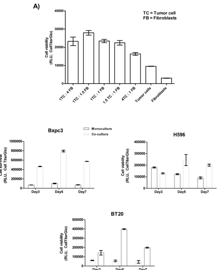
in the highest cell survival (Fig 1A). We further observed that cell survival values, increased from day 3 to day 5 and then decreased in most of the cell lines by day 7 (Fig 1B). Hence, we lected the 1:1.5 ratio and day 5 as a suitable time point to measure cell survival and cytokine se-cretion by the co-cultures in the screening experiments.
Using these conditions, we then compared the influence of 3D co-cultures on the survival of pancreatic cancer cells with that of 2D and trans-well co-cultures. The results of this compari-son indicated that 3D co-culture indeed induced differential cell survival in comparicompari-son to 2D co-culture and trans-well co-culture (Fig 2).
Three dimensional co-culture supports cell survival in a tumor
type-specific manner
To determine if the direct 3D co-culture of fibroblasts and tumor cells influences the survival of tumor cells from different indications (Table 1), we co-cultured a panel of pancreatic, lung and breast cancer cells with MRC5 fibroblasts and compared the tumor cell viability between the tumor cell mono-cultures and the co-cultures. For each cancer type, we identified cell lines that exhibited increased survival in co-culture with fibroblasts and other cell lines that did not exhibit this increase in survival.
In the pancreatic cancer cell panel, 7 out of the 9 cell lines exhibited a significant increase in cell survival when co-cultured with fibroblasts. In the lung and breast cancer panels, only 2 out of the 7 and 9 tested cell lines, respectively, exhibited increased survival when co-cultured with fibroblasts (Fig 3).
To validate this observation using primary TAFs, we selected one cell line that exhibited in-creased survival in co-culture and one that did not exhibit inin-creased survival from each of the cancer panels and co-cultured these cells with organ-specific fibroblasts or primary TAFs. Our data indicated that, similar to the data for the MRC5 fibroblasts, the cell lines that exhibited in-creased survival in co-culture also exhibited inin-creased survival in the presence of the corre-sponding primary TAFs or organ-specific fibroblasts (Fig 4).
Differential secretion of cytokines and growth factors by 3D co-culture
spheroids
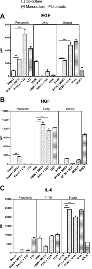
To investigate the potential role of soluble growth factors and cytokines in the increased prolif-eration observed in the co-cultures, we analyzed the mono- and co-culture supernatants, on day 5, for the presence of approximately 42 different cytokines. Several cytokines were found to be differentially secreted upon co-culture with MRC5 fibroblasts in the co-culture model (S4 Fig). We focused on the secretion of EGF, HGF and IL6, because these soluble factors have been associated with an important role in tumor progression, resistance and inflammation. EGF was secreted by breast TAFs and pancreatic fibroblast monocultures, by the Bxpc3 cells and BT20 cells in co-culture with fibroblasts (Fig 5A). High level of HGF was detected in the monoculture supernatants from the MRC5 fibroblasts and the primary lung TAFs and in the co-culture supernatant of lung cancer cells (H596) with MRC% fibroblasts or the primary lung TAFs (Fig 5B). IL6 was secreted at high levels by the primary breast TAFs and moderately by the primary lung TAFs and pancreatic fibroblasts. The breast cancer cells (BT20) secreted high levels of IL6 when co-cultured with MRC5 cells and primary TAFs whereas, the monocul-tures and co-culmonocul-tures of lung cancer cells (H596) secreted only moderate levels of IL6 (Fig 5C).
Cancer cell-fibroblast co-culture influences the response to therapeutic
agents
We further investigated whether the elevated levels of soluble factors in the co-cultures contributed to the increase in cell survival. To this end, cancer cells that were identified to se-crete increased levels of the aforementioned soluble factors were allowed to grow as either fibroblasts ratio of 1:1.5) on day 5.B) Optimization of the co-culture duration.Cancer cells and fibroblasts (MRC5) were cultured in 96-well plates as monocultures or co- cultures as described in the cell viability assay in the Materials and Methods section. The tumor cells and fibroblasts were co-cultured at a ratio of (1:1.5), and cell viability was measured on days 3, 5 and 7. The viability in of the co- cultured cells increased from day 3 to day 5 and then decreased slightly on day 7.
doi:10.1371/journal.pone.0127948.g001
Fig 2. Comparison of the Boyden chamber, 2D co-culture and 3D co-culture systems.To compare the 3D co-culture system to the 2D co-culture and trans- well co-culture systems, tumor cells and fibroblasts were cultured as either as mono-cultures or co-cultures for 5 days as described in the Materials and Methods section. Cell viability was measured on day 5. We observed that 3D co-culture of the tumor cells with fibroblasts induced differential proliferation in co-cultures compared to the Boyden chamber or the 2D co-culture system.
monocultures or were co-cultured with MRC5 fibroblasts or the corresponding TAFs for 5 days in the presence of inhibitory antibodies against EGFR (Erbitux), mAb cMet (monoclonal antibody cMet), mAb IL6, mAb IGF1R (R1507). Cell survival was measured using CellTiter-Glo. The percentage of surviving cells (% survival) was calculated for each treatment relative to the corresponding isotype controls.
The pancreatic cancer cells (Bxpc3), in monoculture were sensitive (approximately 50% sur-vival) to treatment with Erbitux. However, in co-culture with either MRC5 cells or the pancre-atic fibroblasts (LT2), these cells were less sensitive or were partially resistant (approximately 75% survival) to the same treatment. Additionally, Bxpc3 in co-culture responded to mAb IGF1R treatment (approximately 30% inhibition of proliferation) (Fig 6A).
Table 1. Cell line panel.
Catalog # Tumor cell line Source
Lung cancer
CCL-185 A549 LGC
CRL-5908 NCI-H1975 LGC
CRL-5909 NCI-H1993 LGC
CRL-5800 NCI-H23 LGC
CRL-5807 NCI-H358 LGC
HTB-177 NCI-H460 LGC
CRL-5810 NCI-H522 LGC
HTB-178 NCI-H596 LGC
Breast cancer
HTB-19 BT20 LGC
HTB-20 BT474 LGC
HTB-22 MCF7 LGC
HTB-26 MDAMB231 LGC
HTB-131 MDAMB453 LGC
HTB-132 MDAMB468 LGC
HTB-30 SKBR3 LGC
JIMT1 DSMZ
KPL4 DSMZ
Pancreatic cancer
CRL-1687 BxPc3 LGC
CRL-1469 Panc1 LGC
HTB-79 Capan1 LGC
HTB-80 Capan2 LGC
CRL-2119 HPAC LGC
CRL-1682 AsPc1 LGC
PK45P Oncotherapy Science, Inc.
Suit2 Oncotherapy Science, Inc.
PancTu-1 DSMZ
Fibroblasts
PC60161A (primary breast TAFs) 161A Asterand, PLC.
PC60129A1(primary lung TAFs) 129A Asterand, PLC.
CCL-171 MRC5 ATCC
SCR013 LT2 Millipore corporation
The lung cancer cells (H596), exhibited no significant reduction in survival when treated with Erbitux, mAb IL6 or mAb IGF1R in monoculture. In co-culture with MRC5 cells or pri-mary lung TAFs (129A) a significant reduction in survival (approximately 40%) was observed upon treatment with mAb cMet (Fig 6B), but not with the other therapeutic agents used.
The survival of the breast cancer cells (BT20) in co-culture with MRC5 cells was significant-ly reduced (approximatesignificant-ly 50%) upon treatment with by mAb IL6. This effect was not observed when the BT20 cells were in monoculture (Fig 6C). Treatment with Erbitux, mAb cMet or mAb IGF1R did not influence the survival of BT20 cells in monoculture or co-culture with corresponding fibroblasts.
Discussion
Although it is evident that tumor-stroma crosstalk appears to play a critical role in tumor pro-gression, and resistance to therapeutic agents, few suitablein vitrotools/models are available to examine these interactions. Most of thein vitrodata regarding the efficacy of therapeutic agents have been obtained from 2D mono-cultures of cancer cells in which the stromal component is lacking or from trans-well systems in which the tumor cells and stromal cells are physically separated. Alternatively,in vivodata have been obtained from xenograft models in which human tumor cells interact with mouse stromal cells. However, this microenvironment, if at all, is a poor substitute for the human TME. Thesein vitroandin vivomethods may overesti-mate the effects of therapeutic agents, in contrast to co-culture models in which human tumors cells and fibroblasts of human origin directly interact with each other. The co-culture model we described in this study involves culturing tumor cells and fibroblasts in a 3D setting that mimics thein vivomicro-environment. This model enables the monitoring of the effects of co-culturing and the contribution of the crosstalk between tumor cells and fibroblastsin vitroin the absence of exogenous factors, such as serum, growth factors or hormones, on cell survival. Our data from the experiment comparing trans-well based co-cultures and 2D co-cultures to 3D co-culture model clearly indicated that 3D co-culture exerts a differential impact on cell survival. Using this model, we revealed for the first time that different cancer cell types elicit distinct sets of secreted factors from stromal fibroblasts and, thus, can uniquely influence cell survival and therapeutic responses to therapeutic agents.
We used cancer cells from different tumor types and FAP-positive fibroblasts (S1 Fig) from different origins, including primary TAFs, for the co-culture experiments. Upon dissociation of spheroids on day 5 to identify the proliferating population, we found that the predominant proportion of the proliferating cells in the co-cultures was cancer cells (EpCAM-positive) (S2 Fig). However, co- culturing with fibroblasts did not induce enhanced proliferation of all can-cer cell lines tested. In fact, there were some cell lines that proliferated either equally well or better as mono-cultures indicating that there may be other factors influencing cell survival in co-cultures. The interaction between tumor cells and fibroblasts has been reported to induce the secretion of a variety of growth factors and cytokines by fibroblasts or cancer cells [18]. The expression of various soluble factors is associated with poor prognosis and may be of predictive value. It is known that the growth factor /cytokine (GC) profiles of cancer patients vary Fig 3. Co- culturing the tumor cells with MRC5 fibroblasts influences cell survival.Tumor cells and MRC5 fibroblasts were cultured as either co-cultures or monoco-cultures as described. Cell viability was measured based on the total ATP content on day 5 after cell seeding using CellTiterGlo.A)Seven of the 9 pancreatic cancer cell lines showed exhibited a significant increase in cell survival upon co- culturing with MRC5 cells. Bxpc3 cells exhibited the greatest fold-change in proliferation among these cell lines upon co-culturing.B)Two out of the 7 of the lung cancer cell lines exhibited a significant increase in cell survival upon co- culturing with MRC5 cells; out of which the H596 cells exhibited the greatest fold-change in proliferation upon co-culturing.C)Of the two breast cancer cell lines that exhibited an increase in proliferation upon co-culturing with MRC5 fibroblasts, only the BT20 cells exhibited a significant increase in cell survival.
Fig 4. Co- culturing the tumor cells with primary tumor associated fibroblasts (TAFs) influences cell survival similar to MRC5 fibroblasts.One tumor cell line that exhibited the greatest fold-change in cell survival due to co-culturing (Bxpc3, H596 and BT20) and one cell line that did not exhibit an increase in survival upon co-culture with MRC5 cells from each cancer type (Suit2, H1993 and SKBR3) were co-cultured with corresponding primary TAFs (129A, lung TAFs and or 161A, breast TAFs) or organs-specific fibroblasts (LT2, pancreatic fibroblasts) for 5 days followed by measurement of cell viability on day 5 using CellTiterGlo. All three cell lines that exhibited a significant increase in cell survival upon co-culturing with MRC5 fibroblasts (Bxpc3, H596 and BT20) also exhibited increased survival in co-culture with TAFs, whereas the cell lines that did not exhibit increased survival in co-culture with MRC5 (Suit2, H1993 and SKBR3) retained their
proliferative properties even upon co-culturing with TAFs.
depending on the cancer type and stage. In accordance with this understanding, the GC profile of our co-culture models varied depending on the cancer type. The GC profile of mono-cul-tured fibroblasts showed that although certain soluble factors were commonly produced by all fibroblasts, clear differences were observed between fibroblasts from different cancer types. For example IL-6 and IL-8 were secreted by all the fibroblasts we tested whereas HGF was specifi-cally secreted by lung fibroblasts (S3 Fig). Further, we observed that EGF is primarily secreted by co-cultures of pancreatic and breast cancer cells whereas HGF is primarily secreted by lung cancer cells and fibroblasts, and that IL6 is primarily secreted by the breast cancer co-cultures (Fig 5) indicating a cancer specific pattern in cytokine secretion. However, we also observed that some cell lines, such as Suit2 and H1993, both of which did not exhibit increased survival upon co-culture with fibroblasts, secreted growth factors (PDGF and TGFα, respectively),
al-ready in monoculture (S6 Fig). It would be interesting to evaluate whether these growth factors play a key role in attributing this fibroblast independent cell survival in this setting.
Our results from the pancreatic cancer cell panel indicated that most of the cell lines de-pended on fibroblasts for survival or are at least interdependent under these conditions. These data reflect the clinical situation, in which a desmoplastic stromal reaction containing fibro-blasts is considered as a hallmark of pancreatic cancer [5,12]. Pancreatic cancer cells are known to depend on EGFR signaling, and therapies targeting this signaling pathway are under evaluation in the clinic. However, the efficacy of drugs targeting EGFR is limited [17]. The pan-creatic cancer cell line, Bxpc3, exhibited reduced sensitivity to Erbitux in co-culture compared to mono-culture. One reason for this change in the response to Erbitux treatment could be the differential expression of EGFR between the mono-culture and the co-culture. However, we did not detect a significant difference in the EGFR levels between the mono- and co-cultures (S5A Fig), indicating that the resistance of these cells to treatment with Erbitux occursde novo and is potentially mediated by co-culturing with fibroblasts. Considering the recent findings that have implicated a role of the IGF1R pathway and the EGFR in pancreatic cancer progres-sion and therapeutic responses [16,19], we treated the Bxpc3 cells with mAb IGF1R to deter-mine whether the IGF1R influences the survival of these co-cultures In agreement with the clinical data, the Bxpc3 cells responded to IGF1R inhibition, suggesting that a combination therapy blocking the EGFR and IGF1R pathways may provide synergistic value in the clinic.
The resistance of lung cancer cell line, H596, to Erbitux in co-culture with fibroblasts and a corresponding increase in cMet expression and activation compared to the mono-cultures (S5B Fig), indicate that these cells have become resistant to EGFR therapy and depend on HGF produced by co-cultured fibroblasts for survival in co-cultures (Fig 6B). These results are in agreement with the data from other groups demonstrating that HGF produced by fibroblasts promotes tumor progression and induces resistance to EGFR inhibitors in lung cancer [20]. These observations further reflect the situation in non-small cell lung cancer patients, where treatment with inhibitors of the HGF pathway in combination with EGFR inhibitors has been suggested to serve as a better treatment strategy than treatment with either inhibitor alone[21]. factors are plotted in the graphs as the mean fluorescence intensities (MFI). The error bars represent the standard deviation of three replicates.A)The pancreatic and breast cancer co-culture supernatants contained increased EGF levels compared to the lung cancer co-culture supernatants.B)Increased HGF levels were detected in the supernatants from the lung cancer co-cultures but not in those from the corresponding mono-cultures. The Lung fibroblast cell line MRC5 and the TAFs, 129A, also secreted high levels of HGF into the supernatants in monoculture.C)High levels of IL6 were detected in the supernatants from BT20 cells co-cultured with MRC5 or 161A primary breast TAFs and in the supernatants from the H596 cell line mono- and co-cultures. The fibroblasts cell lines MRC5, and LT2 and the primary TAFs, 129A also produced IL6 in mono-culture.
The BT20 breast cancer cells used in these experiments were isolated from a breast tumor that was triple negative for the expression of Her2, estrogen receptor and progesterone recep-tor. Such triple negative tumors often express EGFR [22]. Triple negative breast cancer is also associated with increased stromal reaction with inflammatory component and a poor progno-sis. In our co-culture model, BT20 cells secreted increased amounts of various inflammatory cytokines, including the pro-inflammatory cytokine IL6 (S4 Fig). IL6, has been reported to in-duce cell proliferation in various cancers [23]. We also observed the activation of STAT3, a transcription factor that is downstream of the IL6 receptor, in the co-cultured BT20 cells, indi-cating the activation of this pathway (S5C Fig). Blocking IL6 resulted in a significant decrease in the survival of BT20 cells that were co-cultured with fibroblasts, indicating that the activa-tion of STAT3 and the subsequent increase in cell proliferaactiva-tion are mediated by IL6. Impor-tantly, the BT20 co-cultures also secreted EGF but did not respond to Erbitux treatment. In this context, it is important to note that the activation of the IL6 pathway has been implicated in the resistance to Trastuzumab (anti-Her2) in PTEN-deficient tumor cells [24]. Thus, IL6 se-cretion in BT20 co-cultures may be responsible for the resistance to Erbitux in our model. These data suggest that tumor cells can acquire resistance to therapeutic agents by utilizing al-ternative pathways depending on the availability of the corresponding ligands and the compo-sition of the TME.
Considering the complexity and the dynamics of tumor cell—fibroblast interactions in the TMEin vivo, our 3D co-culture system has also potential limitations. The ratio of tumor cells to fibroblasts and the time point of the measurements, for e.g. may need to be optimized based on the indication and the cell type examined. Other factors, such as mutations in certain genes that influence proliferation, were not taken into account in our system and may also contribute to the differential survival of the co-cultures with fibroblast. Nevertheless, this co-culture model can be used as a tool to elucidate the efficacy of potential therapies and/or the mecha-nisms underlying the resistance to these therapiesin vitro. This 3D co-culture system can be re-liably used as a method forin vitropre-clinical studies to understand tumor-stroma
interactions. Furthermore, the use of patient-derived primary cells could further increase the predictive value of this method. The possibility to extend this system to other cells of in the TME, including immune cells, is very attractive, and this advancement will be of great value once established.
Supporting Information
S1 Fig. The expression of fibroblast activation protein (FAP) by MRC5 and LT2 fibroblasts and primary TAFs.The cell surface expression of FAP, a fibroblast activation marker, was measured on fibroblast cells (MRC5 and LT2) and primary TAFs (129A and 161A) via flow cy-tometry. We observed that all the fibroblasts used expressed FAP on their cell surface.
(TIF)
measured on day 5 using the CellTiterGlo as described for the cell viability assay. The percentage of surviving cells (% survival) was calculated relative to the respective IgG control.A)The mono-cultured BxPc3 cells treated with Erbitux exhibited a significant reduction in cell survival, whereas in the BxPc3 cells co-cultured with MRC5 or LT2 cells were not strongly affected by Erbitux (approximately 20%). Upon treatment with the IGF1R antibody, the survival of Bxpc3 cells was moderately reduced in the co-cultures compared to the monocultures. Treatment with the IL6 and or cMet antibodies induced a slight reduction in cell survival.B) Upon treatment with the anti-cMet antibody lung cancer cell line, H596, did not exhibit a significant reduction in cell survival in monoculture. In co-culture with MRC5 or 129A TAFs, treatment with the anti-cMet antibody induced a significant reduction of in cell survival.C)The breast cancer cell line, BT20 responded to treatment with the anti-IL6 antibody in co-culture exhibiting a significant reduction of cell survival. The mono-cultured BT20 cells treated with the anti- IL6 antibody exhibited no reduction in cell survival.
S2 Fig. Tumor cell fibroblast co-culture induces cell proliferation and spheroid formation.
Cells were cultured either in monoculture or co-culture as indicated for the cell viability assay. Phase contrast images of mono and co-cultures were taken on day 5 using an inverted micro-scope with 20x magnification. All the cell lines investigated showed no or minimal formation of spheroids in monoculture. Upon co-culture with the MRC5 cells all three cell lines formed multicellular spheroids by day 5. Confocal imaging was performed on day 5 as described in M&M section with pre-labeled tumor cells and fibroblasts. The distribution of fibroblasts in spheroids varied between cell lines. The Bxpc3 and BT20 cells formed tight spheroids and the fibroblasts were mostly outside the spheroid unlike H596 which formed loose spheroids the fi-broblasts were found within the spheroid as well. FACS analysis of cell populations in co-cul-ture spheroids was performed on day 5. Cells were culco-cul-tured as indicated earlier. Spheroids were collected and treated with cell dissociation reagent to get single cells for the analysis. Cell sus-pensions were incubated with FAP antibody (activated fibroblast/ marker) or with anti-EpCAM antibody (Epithelial cell marker). Tumor cells expressed anti-EpCAM and could be de-tected in monoculture as well as co-culture with all the cell lines. However, few or no fibroblasts could be detected on day 5 indicating that even though initially more fibroblasts were added than tumor cells, the co-culture conditions favored tumor cell proliferation.
(TIF)
S3 Fig. GC profiles of the MRC5 and LT2 fibroblasts and the primary TAFs.The superna-tants from mono-cultured fibroblast spheroids were collected on day 5, and 42 different growth factors and cytokines were measured using Luminex multiplex technology. The growth factors and cytokines that were produced at detectable levels are depicted in the graph. Among these growth factors, the lung fibroblast cell lines MRC5 and 129A produced higher levels of HGF and VEGF than the pancreatic fibroblast cell line LT2 and the primary breast TAF cell line 161A. The LT2 cells secreted higher levels of PDGF than the other fibroblast cell types. Regard-ing the cytokines, all of the fibroblasts secreted high levels of IL6 and IL8. The expression of MCSF was higher in 161A breast TAFs than in the other fibroblast cell types. The LT2 pancre-atic fibroblasts produced higher levels of G-CSF and GM-CSF than the other fibroblast cell types.
(TIF)
S4 Fig. GC profiles of the tumor cell-MRC5 fibroblast co-cultures.The supernatants from co-culture spheroids were collected on day 5, and 42 different growth factors and cytokines were measured using Luminex multiplex technology. The growth factors and cytokines that were produced at detectable levels are depicted in the graph.
(TIF)
was also detectable in the monocultured fibroblasts. (TIF)
S6 Fig. Growth factor secretion by cell lines that were not dependent on fibroblast co-cul-ture for survival.The supernatants from co-culture spheroids of cell lines that were not depen-dent on fibroblast co-culture for survival were collected on day 5, and 42 different growth factors and cytokines were measured using Luminex multiplex technology. The growth factors and cytokines that were produced at detectable levels are depicted in the graph.
(TIF)
Acknowledgments
We would like thank Janina Findeis, for her support in with the cell culture and FACS experi-ments during the revision of this manuscript.
Author Contributions
Conceived and designed the experiments: MM. Performed the experiments: MM LPP MG. An-alyzed the data: MM. Wrote the paper: MM CHR.
References
1. Pietras K, Ostman A (2010) Hallmarks of cancer: interactions with the tumor stroma. Experimental cell research 316: 1324–1331. doi:10.1016/j.yexcr.2010.02.045PMID:20211171
2. de Kruijf EM, van Nes JG, van de Velde CJ, Putter H, Smit VT, Liefers GJ, et al. (2011) Tumor-stroma ratio in the primary tumor is a prognostic factor in early breast cancer patients, especially in triple-nega-tive carcinoma patients. Breast cancer research and treatment 125: 687–696. doi:
10.1007/s10549-010-0855-6PMID:20361254
3. Farmer P, Bonnefoi H, Anderle P, Cameron D, Wirapati P, Becette V, et al. (2009) A stroma-related gene signature predicts resistance to neoadjuvant chemotherapy in breast cancer. Nature medicine 15: 68–74. doi:10.1038/nm.1908PMID:19122658
4. Neesse A, Michl P, Frese KK, Feig C, Cook N, Jacobetz MA, et al. (2011) Stromal biology and therapy in pancreatic cancer. Gut 60: 861–868. doi:10.1136/gut.2010.226092PMID:20966025
5. Bremnes RM, Donnem T, Al-Saad S, Al-Shibli K, Andersen S, Sirera R, et al. (2011) The role of tumor stroma in cancer progression and prognosis: emphasis on carcinoma-associated fibroblasts and non-small cell lung cancer. Journal of thoracic oncology: official publication of the International Association for the Study of Lung Cancer 6: 209–217.
6. Ronnov-Jessen L, Petersen OW, Bissell MJ (1996) Cellular changes involved in conversion of normal to malignant breast: importance of the stromal reaction. Physiological reviews 76: 69–125. PMID:
8592733
7. Angeli F, Koumakis G, Chen MC, Kumar S, Delinassios JG (2009) Role of stromal fibroblasts in cancer: promoting or impeding? Tumour biology: the journal of the International Society for Oncodevelopmental Biology and Medicine 30: 109–120. doi:10.1159/000218708PMID:19440007
8. Mishra P, Banerjee D, Ben-Baruch A (2011) Chemokines at the crossroads of tumor-fibroblast interac-tions that promote malignancy. Journal of leukocyte biology 89: 31–39. doi:10.1189/jlb.0310182
PMID:20628066
9. Tyan SW, Kuo WH, Huang CK, Pan CC, Shew JY, Chang KJ, et al. (2011) Breast cancer cells induce cancer-associated fibroblasts to secrete hepatocyte growth factor to enhance breast tumorigenesis. PloS one 6: e15313. doi:10.1371/journal.pone.0015313PMID:21249190
10. Micke P, Ostman A (2004) Tumour-stroma interaction: cancer-associated fibroblasts as novel targets in anti-cancer therapy? Lung cancer 45 Suppl 2: S163–175. PMID:15552797
11. Samoszuk M, Tan J, Chorn G (2005) Clonogenic growth of human breast cancer cells co-cultured in di-rect contact with serum-activated fibroblasts. Breast cancer research: BCR 7: R274–283. PMID:
15987422
12. Camp JT, Elloumi F, Roman-Perez E, Rein J, Stewart DA, Harrell JC, et al. (2011) Interactions with fi-broblasts are distinct in Basal-like and luminal breast cancers. Molecular cancer research: MCR 9: 3–
13. Fujita H, Ohuchida K, Mizumoto K, Egami T, Miyoshi K, Moriyama T, et al. (2009) Tumor-stromal inter-actions with direct cell contacts enhance proliferation of human pancreatic carcinoma cells. Cancer sci-ence 100: 2309–2317. doi:10.1111/j.1349-7006.2009.01317.xPMID:19735487
14. Daniel VC, Marchionni L, Hierman JS, Rhodes JT, Devereux WL, Rudin CM, et al. (2009) A primary xe-nograft model of small-cell lung cancer reveals irreversible changes in gene expression imposed by cul-ture in vitro. Cancer research 69: 3364–3373. doi:10.1158/0008-5472.CAN-08-4210PMID:19351829
15. Shay JW, Wright WE (2007) Tissue culture as a hostile environment: identifying conditions for breast cancer progression studies. Cancer cell 12: 100–101. PMID:17692800
16. Li Q, Wang W, Yamada T, Matsumoto K, Sakai K, Bando Y, et al. (2011) Pleural mesothelioma insti-gates tumor-associated fibroblasts to promote progression via a malignant cytokine network. The American journal of pathology 179: 1483–1493. doi:10.1016/j.ajpath.2011.05.060PMID:21763682
17. Chong CR, Janne PA (2013) The quest to overcome resistance to EGFR-targeted therapies in cancer. Nature medicine 19: 1389–1400. doi:10.1038/nm.3388PMID:24202392
18. Bergmann U, Funatomi H, Yokoyama M, Beger HG, Korc M (1995) Insulin-like growth factor I overex-pression in human pancreatic cancer: evidence for autocrine and paracrine roles. Cancer research 55: 2007–2011. PMID:7743492
19. Castoldi R, Jucknischke U, Pradel LP, Arnold E, Klein C, Scheiblich S, et al. (2012) Molecular charac-terization of novel trispecific ErbB-cMet-IGF1R antibodies and their antigen-binding properties. Protein engineering, design & selection: PEDS 25: 551–559.
20. Yamada T, Matsumoto K, Wang W, Li Q, Nishioka Y, Sekido Y, et al. (2010) Hepatocyte growth factor reduces susceptibility to an irreversible epidermal growth factor receptor inhibitor in EGFR-T790M mu-tant lung cancer. Clinical cancer research: an official journal of the American Association for Cancer Re-search 16: 174–183.
21. Zucali PA, Ruiz MG, Giovannetti E, Destro A, Varella-Garcia M, Floor K, et al. (2008) Role of cMET ex-pression in non-small-cell lung cancer patients treated with EGFR tyrosine kinase inhibitors. Annals of oncology: official journal of the European Society for Medical Oncology / ESMO 19: 1605–1612.
22. Ryden L, Jirstrom K, Haglund M, Stal O, Ferno M (2010) Epidermal growth factor receptor and vascular endothelial growth factor receptor 2 are specific biomarkers in triple-negative breast cancer. Results from a controlled randomized trial with long-term follow-up. Breast cancer research and treatment 120: 491–498. doi:10.1007/s10549-010-0758-6PMID:20135347
23. Weidle UH, Klostermann S, Eggle D, Kruger A (2010) Interleukin 6/interleukin 6 receptor interaction and its role as a therapeutic target for treatment of cachexia and cancer. Cancer genomics & proteo-mics 7: 287–302.