Article
Dissecting Tumor-Stromal Interactions in Breast Cancer
Bone Metastasis
Yibin Kang
Department of Molecular Biology, Princeton University, Princeton, NJ, USA
Bone metastasis is a frequent occurrence in breast cancer, affecting more than 70% of late stage cancer patients with severe com-plications such as fracture, bone pain, and hypercalcemia. The pathogenesis of osteolytic bone metastasis depends on cross-com-munications between tumor cells and various stromal cells residing in the bone microenvironment. Several growth factor signal-ing pathways, secreted micro RNAs (miRNAs) and exosomes are functional mediators of tumor-stromal interactions in bone me-tastasis. We developed a functional genomic approach to systemically identified molecular pathways utilized by breast cancer cells to engage the bone stroma in order to generate osteolytic bone metastasis. We showed that elevated expression of vascular cell adhesion molecule 1 (VCAM1) in disseminated breast tumor cells mediates the recruitment of pre-osteoclasts and promotes
their differentiation to mature osteoclasts during the bone metastasis formation. Transforming growth factor β (TGF-β) is released
from bone matrix upon bone destruction, and signals to breast cancer to further enhance their malignancy in developing bone
me-tastasis. We furthered identified Jagged1 as a TGF-β target genes in tumor cells that engaged bone stromal cells through the acti -vation of Notch signaling to provide a positive feedback to promote tumor growth and to activate osteoclast differentiation. Sub-stantially change in miRNA expression was observed in osteoclasts during their differentiation and maturation, which can be
ex-ploited as circulating biomarkers of emerging bone metastasis and therapeutic targets for the treatment of bone metastasis. Fur -ther research in this direction may lead to improved diagnosis and treatment strategies for bone metastasis.
Keywords: Breast neoplasms; Bone metastasis; Tumor-stromal interaction; Osteoblasts; Osteoclasts; Transforming growth factor beta
INTRODUCTION
Breast cancer is the most common female malignancy and the second leading cause of cancer-related death in the United States. Among patients who die from breast cancer, more than 70% suffer from bone metastasis, which is often accompanied by severe bone pain, fracture, and potentially lethal complica-tions such as hypercalcemia [1-4]. Currently, although
anti-os-teolytic agents (such as bisphosphonate and receptor activator
of nuclear factor kappa-β ligand [RANKL] antibody denosum -ab) [5-7], radiotherapy, and chemotherapy can reduce morbidi-ty associated with bone metastasis, these treatments often do not significantly extend the survival time of the patients or pro-vide a cure [8,9], as metastatic cancers often acquire resistance to these treatments. More effective therapies are needed to im-prove the clinical outcome for stage IV breast cancer patients
Received: 18 April 2016, Revised: 26 April 2016, Accepted: 3 May 2016 Corresponding author: Yibin Kang
Department of Molecular Biology, Princeton University, Princeton, NJ 08544, USA
Tel: +1-609-258-8834, Fax: +1-609-258-2340, E-mail: [email protected]
Copyright © 2016 Korean Endocrine Society
This is an Open Access article distributed under the terms of the Creative
Com-mons Attribution Non-Commercial License (http://creativecomCom-mons.org/
with bone metastasis. Tumor-stromal interaction plays a major role in promoting bone metastasis of breast cancer [4]. The bone microenvironment mainly includes osteoblast lineage cells, osteoclasts, hematopoietic cells, and additional stromal
cell types residing in the bone marrow (BM). Formation of
bone metastasis is the result of complicated interactions be-tween tumor cells and various stromal cells in bone, leading to the initial survival of cancer cells in the bone microenviron-ment, activation from dormancy or indolent growth, and ex-pansion of overt osteolytic lesions. Understanding the molecu-lar mechanism of tumor-stromal interactions in bone metastasis is crucial for the improved early detection of bone metastasis, as well as more effective therapies to prevent or slow down the development of bone metastasis.
STROMAL CELLS IN THE BONE
MICROENVIRONMENT
As a crucial organ to foster hematopoiesis and osteogenesis in healthy individuals, bone represents a biologically highly ac-tive microenvironment containing various stromal niches to regulate the dynamic balance of stem, progenitor and mature cells of different lineages [10,11]. Recent studies from the field of hematopoiesis indicate the existence of two major niches in the BM: the osteoblastic niche and the perivascular niche. Within these niches, two major cell lineages derived from he-matopoietic stem cells (HSCs) and mesenchymal stem/stromal cells (MSCs) have complex interactions with each other to
sus-tain normal hematopoiesis and osteogenesis [11-13]. Located
at the inner surface of the bone cavity, the osteoblastic niche has been previously reported to mainly accommodate long-term quiescent HSCs, although recent findings revealed it as the primary site for early B-cell progenitors and certain lym-phoid progenitors [12,14-16]. In contrast, recent studies sug-gested that HSCs are mostly localized in the perivascular niche where the endothelial cells, C-X-C motif chemokine 12
(CXCL12)-abundant reticular (CAR) cells and MSCs regulate
the HSCs through a series of growth factors, cytokines and
chemokines such as stem cell factor (SCF), CXCL12, and an -giopoietin-1 [12]. In particular, BM-MSCs are capable of gen-erating osteoprogenitor cells to form the osteoblastic niche, and
releasing CXCL12, thrombopoietin and other factors to main -tain HSC self-renewal and proliferation. In addition to main-taining healthy bone development, the bone niche supplies im-mune cells and tissue progenitor cells reconstitute the peripher-al immune system and contribute to tissue repair and
regenera-tion [17-19]. The osteoblastic and the perivascular niches have both been reported to impact metastatic survival and tumor cell proliferation during bone metastasis. Direct competition for the osteoblastic niche has been observed between HSCs and meta-static cancer cells [20]. The perivascular niche has been char-acterized as an alternate site for bone metastatic colonization [21-23].
Osteoblasts are differentiated from MSCs [24]. Together with osteocytes that have been terminally differentiated from osteo-blasts, these cells are the major cell types with bone building functions. Through depositing cross-linked collagen, calcium and other mineral substrates, osteoblast cells are responsible for building up the hard yet elastic bone matrix [25]. Osteoblasts also play a significant role in maintaining bone homeostasis, as
osteoblasts secrete cytokine RANKL to promote the maturation
of the bone degrading osteoclasts [26]. Recent studies have demonstrated that disseminated tumor cells (DTCs) in the bone microenvironment usually engage osteoblasts to survive and form proliferating colonies. DTCs have been shown to compete with HSCs for occupancy in the osteoblast niches [20]. In breast cancer bone metastasis, tumor cells use heterotypic cadherin in-teractions with osteogenic cells to activate prosurvival mamma-lian target of rapamycin-Akt signaling [27].
The bone resorbing osteoclast is another major stromal cell type in bone that play an important role in physiological bone remodeling [28,29], and in pathological conditions such as Paget’s disease and lytic bone metastasis [4]. Osteoclast differ-entiation is crucially dependent on macrophage
colony-stimu-lating factor (M-CSF) and RANKL [28,29] and are additional -ly controlled by other growth factors and cytokines [28,29]. As the only cell type in human body that is capable of bone degra-dation, osteoclasts has been the focus in the study of tumor-stromal interactions in bone metastasis, and the development of osteoclast-targeting treatments for bone metastasis [30].
Other bone stromal cells that have been implicated in the de-velopment of bone metastasis include CD4+
T cells, myeloid-derived suppressor cells (MDSCs), Tregs, and Dendritic cells. Contrary to the common perception of the anti-tumor effects of T cells, CD4+
T cells have been demonstrated to be part of the pre-metastatic niche in bone, and activate bone remodeling by
secreting RANKL [31]. Inflammatory molecule prostaglandin
E2 released from breast tumor cells have also been reported to recruit Tregs to establish a pro-metastatic niche in bone [32].
Furthermore, plasmocytoid dendritic cells have been shown to
recruit MDSCs and Tregs and inhibit the cytotoxicity of CD8+
TUMOR-STROMAL INTERACTIONS IN
BONE METASTASIS
Bone tissue constantly undergoes dynamic remodeling mediat-ed by the balancmediat-ed activity of osteoclasts and osteoblasts. Met-astatic cancer cells often exploit the normal bone homeostatic process and tip the equilibrium toward either hyperactive bone lysis or bone growth to facilitate the formation of bone metas-tasis. The proclivity of breast cancer in forming osteolytic bone metastasis has been frequently cited as the classic example of “seed and soil” interactions between tumor and stroma in me-tastasis. A “vicious cycle” of molecular crosstalk between tu-mor cells and the bone microenvironment often takes place in osteolytic bone metastasis whereby tumor cell-produced fac-tors stimulate osteoclast maturation or activity, leading to ex-tensive degradation of the bone matrix and the release of bone-derived factors that further enhance tumor growth. A major ef-fort of our research is devoted to identifying molecular media-tors used by tumor cells to engage various stromal cell types in the bone microenvironment to promote the initiation and pro-gression of bone metastasis.
Most bone relapses of breast cancer occur many years after the initial treatment of primary tumors. The molecular basis for the activation of dormant bone micrometastasis to
life-threat-ening overt metastasis remains largely unknown, in large part due to the lack of appropriate animal models that closely reca-pitulate the process. We developed a novel mouse model to mimic the activation of indolent micrometastases to aggressive lesions. In this model, the single cell progeny clone number 6 (SCP6) single-cell derived clone of the MDA-MB-231 breast cancer cell line was known to have no basal bone metastatic ability after intracardiac injection into nude mice recipients [34]. Interestingly, a small number of mice eventually devel-oped osteolytic bone lesions after more than 6 months of ap-parent bone metastasis free survival, suggesting that certain ge-netic/epigenetic changes occurs in the DTCs that endowed them with bone metastatic ability. Careful gene expression pro-filing analysis and functional studies led to the identification and validation of vascular cell adhesion molecule 1 (VCAM1) as a crucial functional driver of conversion from indolent mi-crometastases to overt osteolytic bone metastasis [35]. Mecha-nistically, we determined that tumor-derived soluble VCAM1 serves as a chemoattractant to recruit circulating monocytic precursors of osteoclasts. By interacting with its cognate
recep-tor, α4β1 integrin, VCAM1 also promotes the adhesion of
pre-osteoclasts to the surface of tumor cells, leading to cell fusion and differentiation of pre-osteoclasts and initiation of bone
de-struction (Fig. 1).
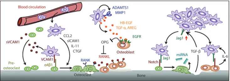
Fig. 1. Tumor-stromal interactions in bone metastasis. Key pathways uncovered by our lab are highlighted, including: (1) vascular cell adhesion molecule 1 (VCAM1) activated osteolytic expansion of indolent bone micrometastasis (left); (2) osteolytic paracrine signaling cascade initiated by matrix metalloproteinase (MMPs, middle); (3) a positive feedback loop in bone metastasis mediated by Jagged1/
Notch and transforming growth factor β (TGF-β) signaling pathways (right). See text for details. sVCAM1, soluble vascular cell adhe
-sion molecule 1; CCL2, chemokine (C-C motif) ligand 2; sICAM1, soluble intercellular adhe-sion molecule 1; IL-11, interleukin 11; CTGF, connective tissue growth factor; ADAMTS1, ADAM metallopeptidase with thrombospondin type 1 motif, 1; HB-EGF, heparin-binding epidermal growth factor-like growth factor; AREG, amphiregulin; EGFR, epidermal growth factor receptor; OPG, osteoprote
-gerin; RANK, receptor activator of nuclear factor kappa-β; RANKL, receptor activator of nuclear factor kappa-β ligand; Jag1, Jagged1; miRNA, micro RNA; IL-6, interleukin 6.
TGF-β Jag1
EGFR HB-EGF TGF-α, AREG
IL-6
Bone Osteoclast
CTGF IL-11 sICAM1 CCL2
Jag1 miRNA Notch
Osteoblast
ADAMTS1 MMP1
OPG
RANKL RANK
Blood circulation
sVCAM1
Pre-osteoclast α4β1
To systematically identify tumor-derived factors that pro-mote bone metastasis, we developed an in vivo selection strate-gy to isolate bone-metastatic breast cancer variants [34]. The MDA-MB-231 cell line contains a heterogeneous population of cancer cells based on morphological and gene expression analysis. When the parental cell line was injected into nude mice via the left cardiac ventricle to form bone metastasis, about 20% to 30% of mice developed osteolytic bone lesions. More than half of the sublines of cancer cells isolated from these lesions displayed dramatically increased ability to metas-tasize to bone, while some sublines displayed mildly or no in-crease of bone metastatic ability. These isogenic sublines with differential bone metastatic ability provided an ideal cohort to identify candidate bone metastasis genes based on gene
expres-sion profiling. Genes in the bone metastasis expresexpres-sion signa -ture included previously reported bone metastasis genes, such as C-X-C chemokine receptor type 4 (CXCR4) [36], but also contains many novel candidate metastasis genes that were sub-sequently validated in follow-up studies, including interleukin
11 (IL-11), osteopontin, connective tissue growth factor (CTGF), Jagged1, matrix metalloproteinase-1 (MMP1), ADAM
metallopeptidase with thrombospondin type 1 motif, 1
(AD-AMTS1), and chemokine (C-C motif) ligand 2 (CCL2) [34,37-39]. Functional characterization of candidate bone metastasis
genes revealed novel mechanisms of tumor-stromal
interac-tions. For example, we showed that two metalloproteases,
MMP1 and ADAMTS1, perform important signaling functions in osteoclast differentiation through activating a paracrine cas-cade mediated by three different cell types [38]. MMP1 and ADAMTS1 proteolytically cleave the membrane-bound
epi-dermal growth factor (EGF) family ligands, including heparin-binding epidermal growth factor-like growth factor (HB-EGF)
and amphiregulin, which activate epidermal growth factor
re-ceptor (EGFR) signaling in osteoblasts, leading to reduced ex -pression of osteoprotegerin, the decoy receptor and antagonist
of RANKL. Increased RANKL activity promotes osteoclast differentiation and osteolytic bone metastasis (Fig. 1).
It is believed that growth factors embedded in bone matrix are released during bone destruction and further stimulate the malignancy of cancer cells, forming a “vicious cycle” in bone metastasis. Among the bone-derived growth factors, we are particularly interested in the role of transforming growth factor
β (TGF-β) since it is one of the most abundant bone-embedded growth factors. Furthermore, many of the bone metastasis genes are direct transcriptional targets of TGF-β. We first used
genetic, pharmacological and advanced imaging approaches to
demonstrate that TGF-β is released from the bone during bone
destruction and further promotes tumor malignancy [40]. Us-ing a MDA-MB-231 cell line engineered to have conditional Smad4 expression and also contain a dual luciferase report
sys-tem for imaging TGF-β signaling activity (using firefly lucifer -ase driven by Smad binding elements) and tumor burden (using cytomegalovirus promoter driven Renilla luciferase), we ex-plored the temporal-spatial dynamics and requirement of
TGF-β signaling in bone metastasis. We showed that TGF-β
signaling activity was dramatically elevated in osteolytic bone lesions, and such activation was inhibited when the mice are treated with bisphosphonates to reduce bone lysis. This result
indicated that bone is indeed a major source of TGF-β during
bone metastasis. Importantly, both genetic (using Tet-off
ex-pression control of Smad4) and pharmacological (using TGF-β receptor kinase inhibitor treatment) inhibition of TGF-β signal -ing in mice dramatically reduce the development of bone me-tastasis [40].
We subsequently identified Jagged1 as an important TGF-β
downstream target with a crucial role in engaging bone stromal cells in osteolytic metastasis [39]. Tumor-derived Jagged1 acti-vates Notch signaling in osteoblasts to increase the expression
of IL-6, which feeds back to tumor cells to stimulate their
growth. In parallel, Jagged1 also directly activates osteoclast
differentiation for bone destruction (Fig. 1). Thus, two devel
-opmentally conserved pathways, TGF-β and Notch, converge
to constitute a vicious cycle that may account for the frequent bone metastasis of breast cancer. This research has led to our current collaborative effort with Amgen (Thousand Oaks, CA, USA) to develop humanized, Jagged1-blocking antibodies, which are showing excellent efficacy in pre-clinical testing.
Turning our attention to bone stromal cells, we delineated a micro RNA (miRNA) regulatory network [41] that controls the
activation of osteoclasts by RANKL and tumor-derived factors,
such as soluble intercellular adhesion molecule 1 (ICAM1)
[41] and CCL2 [37]. Osteoclast miRNAs down-regulated dur -ing osteoclastogenesis are potent inhibitors of bone resorption and osteolytic bone metastasis, while up-regulated miRNAs, such as miR-16 and miR-378, can be detected in circulation as
CONCLUSIONS AND FUTURE
PERSPECTIVES
The study of bone metastasis has produced substantial new in-sights into the intricate cross-talk between metastatic cancer cells and bone stromal cells, particularly osteoblasts and osteo-clasts. These findings have resulted in the successful
develop-ment of bisphosphonates and denosumab (RANKL-neutraliz
-ing antibody) as some of the first U.S. Food and Drug Admin -istration-approved stroma-targeting treatments for metastatic cancer. However, such treatments are generally not curative, and simply reduce skeletal-related events (bone fracture, bone pain, etc.) without improving the overall survival of patients. Interestingly, adjuvant treatment of bisphosphonates improved the survival of post-menopausal breast cancer patients [42], suggesting early application of anti-metastasis treatments may have better outcome than in late-stage diseases. With rapid ad-vance in the field of bone metastasis research, it is likely that several new bone metastasis targeting agents such as Jagged1 neutralizing antibody may become available soon. It will be imperative to test the potential synergistic effect of combing different agents with distinct targeting mechanisms. It is possi-ble that stroma-targeting treatment may enhance the efficacy of traditional chemotherapy and radiation therapy, which are com-monly used to control bone metastasis, as well as the newly de-veloped immune checkpoint therapy. As bone metastases are usually more refractory to various cancer treatments than the primary tumor, understanding the molecular mechanism of treatment resistance in bone metastasis may further provide a promising avenue of further investigation that may significantly improve the outcome of patients with bone metastasis.
CONFLICTS OF INTEREST
No potential conflict of interest relevant to this article was re-ported.
ACKNOWLEDGMENTS
I thank members of our laboratories for helpful discussions.
The work in our laboratory was supported by a Susan G. Ko
-men Fellowship to T. C-T (PDF15332075), and grants from the Brewster Foundation, the Breast Cancer Research Foundation,
Department of Defense (BC123187), and the National Insti-tutes of Health (R01CA141062).
ORCID
Yibin Kang http://orcid.org/0000-0002-1626-6730
REFERENCES
1. Guise TA, Kozlow WM, Heras-Herzig A, Padalecki SS,
Yin JJ, Chirgwin JM. Molecular mechanisms of breast can-cer metastases to bone. Clin Breast Cancan-cer 2005;5 Suppl: S46-53.
2. Mundy GR. Metastasis to bone: causes, consequences and
therapeutic opportunities. Nat Rev Cancer 2002;2:584-93.
3. Nguyen DX, Bos PD, Massague J. Metastasis: from dis-semination to organ-specific colonization. Nat Rev Cancer 2009;9:274-84.
4. Weilbaecher KN, Guise TA, McCauley LK. Cancer to
bone: a fatal attraction. Nat Rev Cancer 2011;11:411-25.
5. Kennedy MJ. Metastatic breast cancer. Curr Opin Oncol 1996;8:485-90.
6. Fizazi K, Lipton A, Mariette X, Body JJ, Rahim Y, Gralow
JR, et al. Randomized phase II trial of denosumab in pa-tients with bone metastases from prostate cancer, breast cancer, or other neoplasms after intravenous bisphospho-nates. J Clin Oncol 2009;27:1564-71.
7. Fizazi K, Carducci M, Smith M, Damiao R, Brown J, Karsh
L, et al. Denosumab versus zoledronic acid for treatment of
bone metastases in men with castration-resistant prostate
cancer: a randomised, double-blind study. Lancet 2011;377:
813-22.
8. Coleman RE, Guise TA, Lipton A, Roodman GD, Berenson
JR, Body JJ, et al. Advancing treatment for metastatic bone cancer: consensus recommendations from the Second Cam-bridge Conference. Clin Cancer Res 2008;14:6387-95.
9. Mackiewicz-Wysocka M, Pankowska M, Wysocki PJ. Progress in the treatment of bone metastases in cancer pa-tients. Expert Opin Investig Drugs 2012;21:785-95.
10. Shen Y, Nilsson SK. Bone, microenvironment and hemato-poiesis. Curr Opin Hematol 2012;19:250-5.
11. Frenette PS, Pinho S, Lucas D, Scheiermann C. Mesenchy -mal stem cell: keystone of the hematopoietic stem cell niche and a stepping-stone for regenerative medicine. Annu Rev Immunol 2013;31:285-316.
12. Morrison SJ, Scadden DT. The bone marrow niche for hae-matopoietic stem cells. Nature 2014;505:327-34.
13. Mendez-Ferrer S, Michurina TV, Ferraro F, Mazloom AR,
-poietic stem cells form a unique bone marrow niche. Nature 2010;466:829-34.
14. Ding L, Morrison SJ. Haematopoietic stem cells and early
lymphoid progenitors occupy distinct bone marrow niches. Nature 2013;495:231-5.
15. Ding L, Saunders TL, Enikolopov G, Morrison SJ. Endo -thelial and perivascular cells maintain haematopoietic stem cells. Nature 2012;481:457-62.
16. Greenbaum A, Hsu YM, Day RB, Schuettpelz LG, Christo
-pher MJ, Borgerding JN, et al. CXCL12 in early mesenchy -mal progenitors is required for haematopoietic stem-cell maintenance. Nature 2013;495:227-30.
17. Hess D, Li L, Martin M, Sakano S, Hill D, Strutt B, et al.
Bone marrow-derived stem cells initiate pancreatic regen-eration. Nat Biotechnol 2003;21:763-70.
18. Orlic D, Kajstura J, Chimenti S, Jakoniuk I, Anderson SM,
Li B, et al. Bone marrow cells regenerate infarcted myocar -dium. Nature 2001;410:701-5.
19. Mercier FE, Ragu C, Scadden DT. The bone marrow at the
crossroads of blood and immunity. Nat Rev Immunol 2011; 12:49-60.
20. Shiozawa Y, Pedersen EA, Havens AM, Jung Y, Mishra A, Joseph J, et al. Human prostate cancer metastases target the hematopoietic stem cell niche to establish footholds in mouse bone marrow. J Clin Invest 2011;121:1298-312.
21. Kaplan RN, Psaila B, Lyden D. Bone marrow cells in the
‘pre-metastatic niche’: within bone and beyond. Cancer Metastasis Rev 2006;25:521-9.
22. Kuhn NZ, Tuan RS. Regulation of stemness and stem cell niche of mesenchymal stem cells: implications in tumori-genesis and metastasis. J Cell Physiol 2010;222:268-77.
23. Colmone A, Amorim M, Pontier AL, Wang S, Jablonski E,
Sipkins DA. Leukemic cells create bone marrow niches
that disrupt the behavior of normal hematopoietic progeni-tor cells. Science 2008;322:1861-5.
24. Prockop DJ. Marrow stromal cells as stem cells for nonhe-matopoietic tissues. Science 1997;276:71-4.
25. Sims NA, Martin TJ. Coupling the activities of bone forma-tion and resorpforma-tion: a multitude of signals within the basic multicellular unit. Bonekey Rep 2014;3:481.
26. Sethi N, Kang Y. Dysregulation of developmental pathways in bone metastasis. Bone 2011;48:16-22.
27. Wang H, Yu C, Gao X, Welte T, Muscarella AM, Tian L, et
al. The osteogenic niche promotes early-stage bone coloni-zation of disseminated breast cancer cells. Cancer Cell 2015; 27:193-210.
28. Boyle WJ, Simonet WS, Lacey DL. Osteoclast differentia -tion and activa-tion. Nature 2003;423:337-42.
29. Teitelbaum SL, Ross FP. Genetic regulation of osteoclast
development and function. Nat Rev Genet 2003;4:638-49.
30. Clezardin P. Therapeutic targets for bone metastases in breast cancer. Breast Cancer Res 2011;13:207.
31. Monteiro AC, Leal AC, Goncalves-Silva T, Mercadante
AC, Kestelman F, Chaves SB, et al. T cells induce pre-met -astatic osteolytic disease and help bone metastases estab-lishment in a mouse model of metastatic breast cancer.
PLoS One 2013;8:e68171.
32. Karavitis J, Hix LM, Shi YH, Schultz RF, Khazaie K,
Zhang M. Regulation of COX2 expression in mouse
mam-mary tumor cells controls bone metastasis and PGE2-in
-duction of regulatory T cell migration. PLoS One 2012;7:
e46342.
33. Sawant A, Hensel JA, Chanda D, Harris BA, Siegal GP,
Maheshwari A, et al. Depletion of plasmacytoid dendritic cells inhibits tumor growth and prevents bone metastasis of breast cancer cells. J Immunol 2012;189:4258-65.
34. Kang Y, Siegel PM, Shu W, Drobnjak M, Kakonen SM, Cordon-Cardo C, et al. A multigenic program mediating breast cancer metastasis to bone. Cancer Cell 2003;3:537-49.
35. Lu X, Mu E, Wei Y, Riethdorf S, Yang Q, Yuan M, et al.
VCAM-1 promotes osteolytic expansion of indolent bone
micrometastasis of breast cancer by engaging α4β1-positive
osteoclast progenitors. Cancer Cell 2011;20:701-14.
36. Muller A, Homey B, Soto H, Ge N, Catron D, Buchanan
ME, et al. Involvement of chemokine receptors in breast cancer metastasis. Nature 2001;410:50-6.
37. Lu X, Kang Y. Chemokine (C-C motif) ligand 2 engages
CCR2+ stromal cells of monocytic origin to promote breast cancer metastasis to lung and bone. J Biol Chem 2009;284: 29087-96.
38. Lu X, Wang Q, Hu G, Van Poznak C, Fleisher M, Reiss M,
et al. ADAMTS1 and MMP1 proteolytically engage
EGF-like ligands in an osteolytic signaling cascade for bone
me-tastasis. Genes Dev 2009;23:1882-94.
39. Sethi N, Dai X, Winter CG, Kang Y. Tumor-derived JAG
-GED1 promotes osteolytic bone metastasis of breast cancer
by engaging notch signaling in bone cells. Cancer Cell 2011;19:192-205.
40. Korpal M, Yan J, Lu X, Xu S, Lerit DA, Kang Y. Imaging
Med 2009;15:960-6.
41. Ell B, Mercatali L, Ibrahim T, Campbell N, Schwarzenbach
H, Pantel K, et al. Tumor-induced osteoclast miRNA changes as regulators and biomarkers of osteolytic bone metastasis. Cancer Cell 2013;24:542-56.
42. Early Breast Cancer Trialists’ Collaborative Group (EBCTCG),
Coleman R, Powles T, Paterson A, Gnant M, Anderson S, et
al. Adjuvant bisphosphonate treatment in early breast cancer: meta-analyses of individual patient data from randomised