1
UNIVERSIDADE FEDERAL DE SÃO CARLOS
CENTRO DE CIÊNCIAS BIOLÓGICAS E DA SAÚDE
PROGRAMA DE PÓS-GRADUAÇÃO EM ECOLOGIA E
RECURSOS NATURAIS
BIOTIC FACTORS DRIVE BACTERIOPLANKTON
COMMUNITY IN A TROPICAL COASTAL SITE
OF THE EQUATORIAL ATLANTIC OCEAN
Vinicius Silva Kavagutti
São Carlos
–
SP
2
UNIVERSIDADE FEDERAL DE SÃO CARLOS
CENTRO DE CIÊNCIAS BIOLÓGICAS E DA SAÚDE
PROGRAMA DE PÓS-GRADUAÇÃO EM ECOLOGIA E RECURSOS
NATURAIS
BIOTIC FACTORS DRIVE BACTERIOPLANKTON COMMUNITY
IN A TROPICAL COASTAL SITE OF THE EQUATORIAL
ATLANTIC OCEAN
Vinicius Silva Kavagutti
Dissertação apresentada ao Programa de Pós-Graduação em Ecologia e Recursos Naturais da Universidade Federal de São Carlos, como parte dos requisitos para obtenção do título de MESTRE em ECOLOGIA, área de concentração: Ecologia e Recursos Naturais.
Orientador: Prof Dr. Hugo Miguel Preto de Morais Sarmento
Ficha catalográfica elaborada pelo DePT da Biblioteca Comunitária UFSCar Processamento Técnico
com os dados fornecidos pelo(a) autor(a)
K21b Kavagutti, Vinicius Silva Biotic factors drive bacterioplankton community in a tropical coastal site of the equatorial atlantic ocean / Vinicius Silva Kavagutti. -- São Carlos : UFSCar, 2016.
73 p.
Dissertação (Mestrado) -- Universidade Federal de São Carlos, 2016.
4
5
Agradecimentos
Ao Prof. Dr. Hugo Sarmento, minha imensa gratidão por tudo. Aos anos de “se
vira”, “é Windows, não Mac, não é?! ”, “não aceito Excel”, broncas, conselhos, dedicação, paciência, exemplo e amizade, MUITO OBRIGADO.
À Maiara Menezes, Janaina Rigonato e Inessa Lacativa, muito obrigado! Sem a contribuição e paciência de vocês, esse trabalho não teria sido possível.
Agradeço aos Profs. Drs. Armando Vieira, Marli Fiore, Maria da Graça Gama Melão, Flávio Henrique Silva, André Megali Amado e a todos aqueles que cederam seus laboratórios nos quais pude trabalhar.
Muito obrigado aos membros da banca Prof. Dr. Hugo Sarmento, Prof. Dr. Josep Gasol e Profa. Dra. Odete Rocha pelas contribuições durante a defesa da dissertação.
Ao pessoal de Natal-RN em especial a Iagê Terra pela hospedagem. E a Eliana Ribeiro por toda atenção e cuidado durante minhas idas a Piracicaba. Obrigado.
Agradeço ainda aos meus companheiros de laboratório Erick, Roberta, Renan, Mariana, Michaela e Aline pela amizade, cafés, risadas, infinitas brigas, procrastinações, correções e discussões (produtivas ou não). Além da turma do lab do
café: Julia, Erika, Ângela por todos os bólos e risadas. Obrigado.
À minha família, obrigado. Não fiz mais que a minha obrigação, eu sei, mas obrigado por existirem.
Às amizades de longa data e distância, muito dos quais não leram e nem lerão esse trabalho, muito obrigado por existirem e me suportarem durante todo esse tempo. Minha gratidão a Lais, Fábio, Livia, Cachoni, Paulo, Vivi, Pam, Ana Rúbia, Fê, Thaísa, Jaguarinha, Brunão, Erick, Lexandre, Fernanda (Chicó), Mailane, Jé, Andreza, Tonho, Fer e Rê Muy.
7
Resumo
A relação entre a latitude e diversidade microbiana no oceano é controversa. Modelos de nicho preveem maior riqueza em altas latitudes no inverno, enquanto amostragens pontuais indicam uma maior riqueza em latitudes intermediárias, com valores mais baixos para regiões equatoriais e polares. No entanto, dada a natureza dinâmica do ecossistema oceânico, é difícil explicar variações temporais da biodiversidade microbiana nas avaliações empíricas. Nesse trabalho comparamos os componentes da diversidade (riqueza e equitabilidade) e estabilidade das populações microbianas (coeficiente de variação) em dois observatórios oceânicos costeiros com estados tróficos semelhantes, localizados em latitudes contrastantes: um localizado no Oceano Atlântico Equatorial e um em clima temperado localizado no noroeste do Mar Mediterrâneo, a fim de avaliar quais fatores estruturam a dinâmica das comunidades microbianas em cada local. Observamos que tal como animais e plantas, as comunidades microbianas exibem maior (ou pelo menos similar) riqueza no equador pelo menos em águas costeiras. Também encontramos evidências de aumento da estabilidade com o aumento da uniformidade nas comunidades microbianas tropicais, quando comparadas com as de clima temperado. De modo geral, temperatura e silicatos foram as variáveis que condicionaram as comunidades procariotas de vida livre no observatório da região temperada, enquanto que no observatório tropical, fatores estocásticos tais como interações bióticas com eucariotos, foram os fatores que mais influenciaram as comunidades bacterianas. Assim, propomos um quadro conceitual onde a composição da comunidade microbiana seria impulsionada por fatores determinísticos em latitudes mais elevadas, enquanto que em latitudes menores, seriam determinados por fatores mais estocásticos, como interações bióticas. Nosso estudo destaca a importância de estudos comparativos utilizando series temporais Eulerianas em diferentes latitudes para entender os padrões de diversidade das comunidades microbianas no oceano.
Palavras-chave
8
Abstract
The relationship between latitude and microbial diversity in the ocean is controversial. Niche models predict higher richness at high latitudes in winter, while snapshot field-sampling point towards higher richness at intermediate latitudes, with lower values both towards equatorial and Polar Regions. However, given the dynamic
nature of ocean’s ecosystem it is difficult to account for temporal variations in empirical
assessments of microbial biodiversity. Here, we compared the components of diversity (richness and evenness) and microbial population stability (coefficient of variation) in two coastal ocean observatories with similar trophic state located in contrasting latitudes, one located in the Equatorial Atlantic Ocean, and one temperate located in the Northwestern Mediterranean Sea, to evaluate which factors drive the dynamics of microbial communities in each site. Our observations support the view that, as animals and plants, microbial communities exhibit higher (or at least similar) richness towards the equator, at least in the coastal ocean. We also found evidence of increasing stability with increasing evenness in tropical microbial communities when compared to the temperate ones. Temperature and silicates drove temperate free-living prokaryotic communities, while tropical ones were driven by stochastic factors such as biotic interactions with eukaryotes. We propose a conceptual framework where microbial community composition would be driven by deterministic factors in higher latitudes and once the factor temperature is removed moving towards the equator, more stochastic factors such as biotic interactions would emerge as the main factors shaping microbial communities. This study highlights the importance of comparative studies on Eulerian time-series distributed at different latitudes to fully understand the diversity patterns of microbial communities in the ocean.
Keywords
9
Lista de Abreviações e Siglas
BBMO - Blanes Bay Microbial Observatory CV – Coefficient of Variation
DNA - Deoxyribonucleic Acid
EAMO - Equatorial Atlantic Microbial Observatory Euk – Eukaryotes
FL – Free-Living (< 3 µm) FL3 – Fluorescence Type 3 (Red)
H’ – Shannon Index HNA - High Nucleic Acid
NGS – Next-Generation Sequencing OTU – Operational Taxonomic Units PA – Particle-Attached (> 3 µm) PCR - Polymerase Chain Reaction Prok – Prokaryotes
rRNA - Ribosomal Ribonucleic Acid SSC – Side-Scatter
10
Lista de figuras
11 Figure 2. 1. Location of sampling stations (Equatorial Atlantic Microbial Observatory - EAMO and Mediterranean Sea in the Blanes Bay Microbial Observatory - BBMO), coordinates in the text. The plots on the sides illustrate the accumulated precipitation, air temperature (minimum and maximum) and wind speed during the period of this study in the tropical site (EAMO) on the right, and for the temperate site (BBMO) on the left. Arrows indicate the months sampled in each site. ... 36 Figure 2. 2. Rank abundance of the free-living (< 3 μm) and particle-attached (> 3 μm)
prokaryotes. Dashed lines represent the tropical site; solid lines represent the temperate site. ... 42
Figure 2. 3. Rank abundance of the small (< 3 μm) and large (> 3 μm) size fraction
13
Material suplementar
Supplementary Material 1. Free-living (FL) prokaryotic groups that contribute with at least 1% of the community at the different sampling dates at EAMO. ... 62 Supplementary Material 2. Free-living (FL) prokaryotic OTUs that contribute with at
least 1% (sum of all dates) of the community at the different sampling dates at EAMO. ... 63 Supplementary Material 3. Particle-attached (PA) prokaryotic groups that contribute
with at least 1% of the community at the different sampling dates at EAMO. ... 64 Supplementary Material 4. Particle-attached (PA) prokaryotic OTUs that contribute
with at least 1% (sum of all dates) of the community at the different sampling dates at EAMO. ... 65 Supplementary Material 5. Free-living (FL) eukaryotic groups that contribute with at
least 1% of the community at the different sampling dates at EAMO. ... 66 Supplementary Material 6. Free-living (FL) eukaryotic OTUs that contribute with at
least 1% (sum of all dates) of the community at the different sampling dates at EAMO. ... 67 Supplementary Material 7. Larger fraction (PA) eukaryotic groups that contribute with
at least 1% of the community at the different sampling dates at EAMO. ... 68 Supplementary Material 8. Larger fraction (PA) eukaryotic OTUs that contribute with at
least 1% (sum of all dates) of the community at the different sampling dates at EAMO. ... 69
Supplementary Material 9. Prokaryote’s Venn diagram of tropical site by size fraction.
Free-living = < 3 µm and Particle-attached = > 3 µm. ... 70
Supplementary Material 10. Eukaryote’s Venn diagram of tropical site by size fraction.
Smaller fraction = < 3 µm and larger fraction = > 3 µm. ... 70 Supplementary Material 11. Prokaryote’s Venn diagram of free-living OTUs between
sites, BBMO = Blanes Bay Microbial Observatory and EAMO = Equatorial
Atlantic Microbial Observatory. Free-living = < 3 µm. ... 71 Supplementary Material 12. Prokaryote’s Venn diagram of particle-attached OTUs
between sites, BBMO = Blanes Bay Microbial Observatory and EAMO =
14
Supplementary Material 13. Eukaryote’s Venn diagram of smaller fraction OTUs
between sites, BBMO = Blanes Bay Microbial Observatory and EAMO =
Equatorial Atlantic Microbial Observatory. Smaller fraction = < 3 µm. ... 72
Supplementary Material 14. Eukaryote’s Venn diagram of larger fraction OTUs
between sites, BBMO = Blanes Bay Microbial Observatory and EAMO =
Equatorial Atlantic Microbial Observatory. Larger fraction = > 3 µm. ... 72
Lista de tabelas
15
Sumário
Capítulo I ... 16
Introdução Geral ... 16
Introdução geral ... 17
Referências ... 24
Capítulo II ... 31
Introduction ... 32
Hypotheses ... 34
Objective ... 34
Material and methods ... 35
Sampling and study sites ... 35
Chemical Analyses ... 37
Bacterial abundance and biomass ... 37
Community composition ... 37
Extraction, PCR and bioinformatics ... 38
Statistical treatment ... 38
Results ... 40
Discussion ... 49
Acknowledgements ... 54
Conclusões ... 55
References ... 56
Supplementary Material ... 61
PCR conditions ... 61
16
Capítulo I
Introdução Geral
17
Introdução geral
Os microrganismos marinhos foram os primeiros organismos vivos na Terra e únicos durante milhões de anos de evolução. Cada litro de água do mar está repleta de milhões de vírus, bactérias e protistas, ultrapassando todos os metazoários multicelulares em abundância, biomassa, atividade metabólica e diversidade genética e bioquímica (Falkowski et al., 2008; Kirchman, 2008; Hug et al., 2016). Na visão mais
atual da árvore da vida (Figura 1.1), os o conjunto de todas as formas de vida eucariótica (como animais e plantas, por exemplo) caberiam em uma pequena ramificação dessa árvore, sendo a esmagadora maioria da diversidade filogenética constituída por bactérias e arqueas (procariotos).
Ao contrário dos animais e plantas, morfologicamente diferenciáveis em sua maioria, apenas uma parte da diversidade microbiana pode ser identificada com base na morfologia. Isso é válido especialmente no caso de protistas e picoeucariotos, dentre os quais a existência de espécies crípticas é uma característica generalizada (de Vargas et al., 2015b). Justamente estes microrganismos constituem o maior conjunto de
diversidade genômica e metabólica (funcional) ainda a ser explorada (Yooseph et al.,
2007, 2010). Até há bem pouco tempo, as técnicas de biologia molecular disponíveis para estudar a identidade filogenética dos microrganismos se limitavam aos organismos mais abundantes ou a alguns (poucos) cultiváveis, geralmente pouco representativos das comunidades presentes no ambiente (Yooseph et al., 2007, 2010; Pedrós-Alió, 2006)
(Figura 1.2). Entretanto, nos últimos anos a ecologia microbiana passou por uma revolução impulsionada pelas tecnologias de sequenciamento de nova geração (next generation sequencing - NGS) (Sogin et al., 2006). Esta nova abordagem independente
18 bioativas. Recentemente, estas técnicas de NGS combinadas com ferramentas bioinformáticas permitiram a caracterização de comunidades microbianas diretamente de amostras ambientais (Logares et al., 2013; Sunagawa et al., 2015; Venter et al.,
2004).
Figura 1. 1. Visão atual da árvore da vida, adaptado de Hug et al. (2016), considerando
19 Figura 1. 2. Relação entre a abundância de indivíduos e os taxa ranqueados do mais abundante para o menos abundante (rank-abundance) adaptado de Pedrós-Alió (2006). A área vermelha representa organismos mais abundantes, enquanto a área azul representa a biosfera rara (organismos menos abundantes). A seta em vermelho indica os organismos que contribuem para o fluxo de carbono, em rosa, os que contribuem para o fluxo de outros elementos, em azul, os que contribuem para a real diversidade microbiana e que têm potencial para contribuir para o fluxo de carbono e outros elementos, ao se tornarem abundantes.
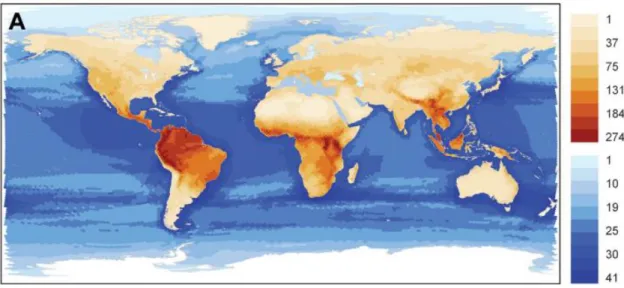
Modelos macro-ecológicos predizem maior riqueza em regiões tropicais, onde a estabilidade do ambiente favorece a complexidade e um aumento da riqueza (e.g. Schipper et al., 2008; Olson et al., 2001; Kreft and Jetz, 2007) (Figuras 1.3 e 1.4). Em
contrapartida, no que se refere a microrganismos não existe ainda um consenso. Observações empíricas realizadas em expedições oceanográficas que cobrem largas escalas espaciais indicam maior diversidade em latitudes intermediárias (Sunagawa et al., 2015) (Figura 1.5), enquanto modelos de nicho predizem uma maior riqueza em
20 Figura 1. 3. Padrões globais de distribuição da riqueza de mamíferos, em ambientes terrestres e de água doce (em marrom) e no oceano (em azul), adaptado de Schipper, et al (2008).
Figura 1. 4. Padrão de distribuição geográfica da riqueza de plantas vasculares, adaptado de Barthlott et al (2007).
O movimento das massas de água no oceano dificultam o rastreamento das mesmas por longos períodos (Fuhrman et al., 2015). Assim, a maioria dos estudos de
21 em transectos (e.g. Milici et al., 2016), que tentam seguir as correntes e massas de água
tanto quanto possível (amostragem Lagrangiana). No entanto, esta estratégia de amostragem não tem uma boa cobertura das variações temporais que ocorrem ao longo do ano. Deste modo, estudos de séries temporais, coletadas em um mesmo ponto durante um intervalo de tempo determinado (amostragem Euleriana), deveriam ser mais comuns para a compreensão da dinâmica microbiana em ecossistemas marinhos (Fuhrman et al., 2015). No entanto, existem poucos observatórios microbianos que
monitoram comunidades microbianas marinhas ao longo do tempo, e os que existem estão predominantemente concentrados em regiões temperadas (Fuhrman et al., 2015).
Vários trabalhos abordando diferentes aspetos da diversidade microbiana têm sido publicados em diversos ambientes marinhos, por exemplo, zonas costeiras (Thompson
et al., 2011), gradientes costa - oceano aberto (Pommier et al., 2010), oceano aberto
(Delong, 2005), oceano profundo (Delong et al., 2006; Salazar et al., 2015; Sogin et al.,
2006) e até mesmo em regiões polares (Ghiglione et al., 2012; Logares et al., 2013). Na
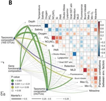
maioria dos casos, os fatores ambientais que determinam a estrutura microbiana são a temperatura (Sunagawa et al., 2015) ou a duração do dia (horas de luz) (Gilbert et al.,
22 Figura 1. 5. Fatores ambientais que determinam a composição da comunidade microbiana de superfície, adaptado de Sunagawa et al (2015). O gradiente de cor nas
comparações de variáveis ambientais par a par indicam os coeficientes de correlação de Spearman.
Figura 1. 6. Modelo da diversidade bacteriana global, adaptado de Ladau et al (2013).
23 Porém, estudos deste tipo em oceano tropical onde variações da temperatura e das horas de luz ao longo do ano são pouco significativas, são ainda incipientes. Considerando que a maior parte da superfície do oceano global é constituída por regiões quentes de oceano tropical, esta poderia ser uma lacuna importante no esclarecimento dos mecanismos que determinam os padrões de diversidade microbiana no oceano. Essas regiões tropicais do oceano são geralmente oligotróficas, e a produção orgânica
concentra-se na subsuperfície, sendo dominada por procariotos unicelulares dos gêneros
Synechoccocus e Prochloroccocus e eucariotos de tamanho inferior a 2μm
(picoplâncton). A área do oceano com estas características (estratificado e dominadas por picoplâncton) representa cerca de 74% da superfície total do oceano global (Behrenfeld et al., 2006). Por cobrir uma área tão grande do oceano e por apresentar um
funcionamento ecológico diferenciado, o estudo das comunidades planctônicas que ocorrem no oceano quente e oligotrófico são de particular interesse para a interpretação de fenômenos de impacto global. Apesar dos esforços e avanços recentes, a diversidade microbiana e sua dinâmica no oceano tropical ainda representa uma lacuna importante.
Dessa forma, esse estudo realizou uma análise comparativa da estabilidade e dos componentes da diversidade (riqueza e equitabilidade) das comunidades microbianas em dois observatórios microbianos situados em latitudes contrastantes, além de determinar quais fatores (ambientais, biológicos ou neutros) dirigem a estrutura dessas comunidades em cada local. Para isso, aplicamos técnicas de NGS, sequenciando
amplicons do gene rRNA, 16S (para procariotos) e 18S (para eucariotos) para descrever
24
Referências
Acinas SG, Antón J, Rodríguez-Valera F. (1999). Diversity of free-living and attached bacteria in offshore western Mediterranean waters as depicted by analysis of genes encoding 16S rRNA. Appl Environ Microbiol65: 514–522.
Alvain S, Moulin C, Dandonneau Y, Loisel H. (2008). Seasonal distribution and succession of dominant phytoplankton groups in the global ocean: A satellite view. Global Biogeochem Cycles22: 1–15.
Apprill a, McNally S, Parsons R, Weber L. (2015). Minor revision to V4 region SSU rRNA 806R gene primer greatly increases detection of SAR11 bacterioplankton.
Aquat Microb Ecol75: 129–137.
Barthlott W, Hostert A, Kier G, Küper W, Mutke J, Rafiqpoor MD, et al. (2007).
Patterns of Vascular Plant Diversity. Erdkunde61: 305–315.
Behrenfeld MJ, O’Malley RT, Siegel D a, McClain CR, Sarmiento JL, Feldman GC, et
al. (2006). Climate-driven trends in contemporary ocean productivity. Nature444:
752–755.
Boyce DG, Frank KT, Leggett WC. (2015). From mice to elephants: Overturning the
‘one size fits all’ paradigm in marine plankton food chains. Ecol Lett 18: 504–
515.
Connolly SR, MacNeil MA, Caley MJ, Knowlton N, Cripps E, Hisano M, et al. (2014).
Commonness and rarity in the marine biosphere. Proc Natl Acad Sci 111: 8524–
8529.
Delong EF. (2005). Microbial community genomics in the ocean. Nat Rev Microbiol3:
459–69.
Delong EF, Preston CM, Mincer T, Rich V, Hallam SJ, Frigaard N, et al. (2006).
25
Science (80- )311: 496–503.
Doney SC, Ruckelshaus M, J. ED, Barry JP, Chan F, English CA, et al. (2002). Climate
Change Impacts on Marine Ecosystems. Estuaries25: 149–164.
Edgar RC. (2010). Search and clustering orders of magnitude faster than BLAST. doi:10.1093/bioinformatics/btq461.
Edgar RC. (2013). UPARSE: Highly accurate OTU sequences from microbial amplicon reads. doi:dx.doi.org/10.1038/nmeth.2604.
Falkowski PG, Fenchel T, Delong EF. (2008). The Microbial Engines That Drive Earth
’s Biogeochemical Cycles. Science (80- )320: 1034–1039.
Fuhrman JA, Cram JA, Needham DM. (2015). Marine microbial community dynamics and their ecological interpretation. Nat Rev Microbiol13: 133–146.
Fuhrman JA, Hewson I, Schwalbach MS, Steele JA, Brown M V, Naeem S. (2006). Annually reoccurring bacterial communities are predictable from ocean conditions. Proc Natl Acad Sci U S A103: 13104–9.
Galand PE, Casamayor EO, Kirchman DL, Lovejoy C. (2009). Ecology of the rare microbial biosphere of the Arctic Ocean. Proc Natl Acad Sci U S A 106: 22427–
32.
Gasol JM, Giorgio P a DEL, del Giorgio P a. (2000). Using flow cytometry for counting natural planktonic bacteria and understanding the structure of planktonic bacterial communities. Sci Mar64: 197–224.
Ghiglione JF, Conan P, Pujo-Pay M. (2009). Diversity of total and active free-living vs. particle-attached bacteria in the euphotic zone of the NW Mediterranean Sea.
FEMS Microbiol Lett299: 9–21.
Ghiglione J-F, Galand PE, Pommier T, Pedros-Alio C, Maas EW, Bakker K, et al.
26 communities. Proc Natl Acad Sci109: 17633–17638.
Gilbert JA, Steele JA, Caporaso JG, Steinbrück L, Reeder J, Temperton B, et al. (2012).
Defining seasonal marine microbial community dynamics. ISME J6: 298–308.
Giovannoni SJ, Vergin KL. (2012). Seasonality in Ocean Microbial Communities.
Science (80- )335: 671–676.
Grasshoff K, Kremling K, Ehrhardt M. (1999). Methods of seawater analysis. doi:10.1016/0304-4203(78)90045-2.
Herlemann DP, Labrenz M, Jürgens K, Bertilsson S, Waniek JJ, Andersson AF. (2011). Transitions in bacterial communities along the 2000 km salinity gradient of the Baltic Sea. ISME J5: 1571–9.
Hug LA, Baker BJ, Anantharaman K, Brown CT, Probst AJ, Castelle CJ, et al. (2016).
A new view of the tree and life’s diversity. Nat Microbiol 1: Manuscript
submitted for publication.
Kindt R, Coe R. (2005). Tree diversity analysis. A manual and software for common statistical methods for ecological and biodiversity studies.
Kirchman DL. (2008). Microbial Ecology of the Oceans. 2nd ed. ISBN: 978-0-470-04344-8: Cambridge doi:10.1017/CBO9781107415324.004.
Kreft H, Jetz W. (2007). Global patterns and determinants of vascular plant diversity.
Proc Natl Acad Sci104: 5925–5930.
Ladau J, Sharpton TJ, Finucane MM, Jospin G, Kembel SW, O’Dwyer J, et al. (2013).
Global marine bacterial diversity peaks at high latitudes in winter. ISME J 7:
1669–77.
Lima-Mendez G, Faust K, Henry N, Decelle J, Colin S, Carcillo F, et al. (2015).
27 Logares R, Audic S, Bass D, Bittner L, Boutte C, Christen R, et al. (2014). Patterns of
Rare and Abundant Marine Microbial Eukaryotes. Curr Biol24: 813–821.
Logares R, Lindström ES, Langenheder S, Logue JB, Paterson H, Laybourn-Parry J, et al. (2013). Biogeography of bacterial communities exposed to progressive
long-term environmental change. ISME J7: 937–948.
Logares R, Sunagawa S, Salazar G, Cornejo-Castillo FM, Ferrera I, Sarmento H, et al.
(2014). Metagenomic 16S rDNA Illumina tags are a powerful alternative to amplicon sequencing to explore diversity and structure of microbial communities.
Environ Microbiol16: 2659–2671.
Margalef R. (1978). Life-forms of phytoplankton as survival alternatives in an unstable environment. Oceanol Acta1: 493–509.
McCann KS. (2000). The diversity–stability debate. Nature405: 228–233.
Milici M, Tomasch J, Wos-Oxley ML, Wang H, Jáuregui R, Camarinha-Silva A, et al.
(2016). Low diversity of planktonic bacteria in the tropical ocean. Sci Rep 6:
19054.
Morana C, Sarmento H, Descy J-P, Gasol JM, Borges A V., Bouillon S, et al. (2014).
Production of dissolved organic matter by phytoplankton and its uptake by heterotrophic prokaryotes in large tropical lakes. Limnol Oceanogr 59: 1364–
1375.
Norland S. (1993). The relationship between biomass and volume of bacteria. Kemp P, Sherr BF, Sherr E, Cole JJ, Eds Handb methods Aquat Microb Ecol 303–307.
Oksanen J, Blanchet F, Kindt R, et al. (2015). vegan: Community Ecology Package. Olson DM, Dinerstein E, Wikramanayake ED, Burgess ND, Powell GVN, Underwood
EC, et al. (2001). Terrestrial Ecoregions of the World: A New Map of Life on
28 Pedrós-Alió C. (2006). Marine microbial diversity: can it be determined? Trends
Microbiol14: 257–263.
Pommier T, Neal P, Gasol J, Coll M, Acinas S, Pedrós-Alió C. (2010). Spatial patterns of bacterial richness and evenness in the NW Mediterranean Sea explored by pyrosequencing of the 16S rRNA. Aquat Microb Ecol61: 221–233.
Quast C, Pruesse E, Yilmaz P, Gerken J, Schweer T, Yarza P, et al. (2013). The SILVA
ribosomal RNA gene database project: Improved data processing and web-based tools. Nucleic Acids Res41: 590–596.
R Core Team. (2011). R: A Language and Environment for Statistical Computing. Vienna, Austria : the R Foundation for Statistical Computing. GNU General
Public License (GPL) - http://www.gnu.org/licenses/old-licenses/gpl-2.0.html. Salazar G, Cornejo-Castillo FM, Borrull E, Díez-Vives C, Lara E, Vaqué D, et al.
(2015). Particle-association lifestyle is a phylogenetically conserved trait in bathypelagic prokaryotes. Mol Ecol 5692–5706.
Sarmento H, Gasol JM. (2012). Use of phytoplankton-derived dissolved organic carbon by different types of bacterioplankton. Environ Microbiol14: 2348–2360.
Sarmento H, Montoya JM, Vázquez-Domínguez E, Vaqué D, Gasol JM. (2010). Warming effects on marine microbial food web processes: how far can we go when it comes to predictions? Philos Trans R Soc Lond B Biol Sci365: 2137–49.
Schipper J, Chanson JS, Chiozza F, Cox NA, Hoffmann M, Katariya V, et al. (2008).
The Status of the World’s Land and Marine Mammals: Diversity, Threat, and
Knowledge. Science (80- )322: 225–230.
Sogin ML, Morrison HG, Huber JA, Welch, David Mark, Huse SM, Neal PR, et al.
(2006). Microbial Diversity in the Deep Sea and the Underexplored ‘Rare
29 Soininen J, Passy S, Hillebrand H. (2012). The relationship between species richness and evenness: A meta-analysis of studies across aquatic ecosystems. Oecologia 169: 803–809.
Stoeck T, Bass D, Nebel M, Christen R, Jones MDM, Breiner HW, et al. (2010).
Multiple marker parallel tag environmental DNA sequencing reveals a highly complex eukaryotic community in marine anoxic water. Mol Ecol19: 21–31.
Sunagawa S, Coelho LP, Chaffron S, Kultima JR, Labadie K, Salazar G, et al. (2015).
Structure and function of the global ocean microbiome. Science348: 1261359.
Thompson FL, Bruce T, Gonzalez A, Cardoso A, Clementino M, Costagliola M, et al.
(2011). Coastal bacterioplankton community diversity along a latitudinal gradient in Latin America by means of V6 tag pyrosequencing. Arch Microbiol 193: 105–
114.
de Vargas C, Audic S, Henry N, Decelle J, Mahé F, Logares R, et al. (2015a).
Eukaryotic plankton diversity in the sunlit ocean. Science348: 1261605.
de Vargas C, Audic S, Henry N, Decelle J, Mahé F, Logares R, et al. (2015b). Ocean
plankton. Eukaryotic plankton diversity in the sunlit ocean. Science348: 1261605.
Venables WN, Ripley BD. (2002). Modern Applied Statistics with S. Fourth Edition. Venter JC, Remington K, Heidelberg JF, Halpern AL, Rusch D, Eisen J a, et al. (2004).
Environmental genome shotgun sequencing of the Sargasso Sea. Science304: 66–
74.
Yooseph S, Nealson KH, Rusch DB, McCrow JP, Dupont CL, Kim M, et al. (2010).
Genomic and functional adaptation in surface ocean planktonic prokaryotes.
Nature468: 60–66.
Yooseph S, Sutton G, Rusch DB, Halpern AL, Williamson SJ, Remington K, et al.
30 of protein families. PLoS Biol5: 0432–0466.
Zhang J, Kobert K, Flouri T, Stamatakis A. (2014). PEAR: a fast and accurate Illumina Paired-End reAd mergeR. Bioinformatics30: 614–620.
Zhang Z, Schwartz S, Wagner L, Miller W. (2000). A greedy algorithm for aligning DNA sequences.
31
Capítulo II
Biotic Factors Drive
Bacterioplankton Community in a
Tropical Coastal Site of the
Equatorial Atlantic Ocean
“Tenho a impressão de ter sido uma criança brincando à beira-mar, divertindo-me em descobrir uma pedrinha mais lisa ou uma concha mais bonita que as outras,
enquanto o imenso oceano da verdade continua misterioso diante de meus olhos”.
32
Introduction
Marine microbial communities play a crucial role in most global biogeochemical cycles (Falkowski et al., 2008) and a bear an important share of Earth’s biodiversity
(Sunagawa et al., 2015). Understanding the factors that drive the dynamics of marine
microbial communities at a global scale is an important step to predict the consequences of the growing impacts (Doney et al., 2002). In recent years, aquatic microbial ecology
has suffered an authentic revolution driven by high throughput sequencing technologies (Venter et al., 2004). Combined with bioinformatics tools, these new sequencing
technologies allowed a characterization of microbial populations directly from environmental samples, and brought a new perspective about the structure, evolution and ecology of the microbial world (e.g. de Vargas et al., 2015; Sunagawa et al., 2015).
Microbial diversity surveys showed that communities are dominated by a relatively small number of some OTUs (operational taxonomic units), but thousands of other less abundant populations (the rare biosphere) are responsible for most of the phylogenetic diversity observed ( e. g. Galand et al., 2009; Connolly et al., 2014;
Logares, Audic, et al., 2014). Recent large-scale oceanographic cruises focused on how
OTU (operational taxonomic units) richness is distributed across latitudinal gradients suggesting that, differently from animals and plants, microorganisms present maximal richness at intermediate latitudes (Milici et al., 2016; Sunagawa et al., 2015). However,
ocean’s ecosystem is extremely dynamic and seasonal variations are difficult to capture
in such cruises covering large spatial scales (Fuhrman et al., 2015). Niche models, a
33 In a different approach, microbial observatories provided valuable information on microbial community dynamics, where different populations responded to environmental changes revealing recurrent patterns year after year (Fuhrman et al.,
2015; Giovannoni and Vergin, 2012; Gilbert et al., 2012). However, the existing
microbial observatories have a restricted distribution in mid-latitudes (~22º to 50º) northern hemisphere (Fuhrman et al., 2015) and we lack of comprehensive comparative
studies in ocean time-series using the same methods to access microbial diversity, and covering a wide range of latitudes.
Most of the world’s ocean surface consists of warm tropical conditions. These regions are usually oligotrophic, where primary production is usually dominated by single-celled prokaryotes Synechococcus, Prochlorococcus and small eukaryotes
(picoplankton) (Alvain et al., 2008). Considering that warm oligotrophic tropical waters
represent about 74% of the total area of the global ocean (Behrenfeld et al., 2006), we can not aim to fully understand the global dynamics of microbial communities without
studying this part of the world’s ocean in detail.
A critical point to decipher large-scale biodiversity patterns is which proxy of diversity one should look at. Most studies have focused on richness and this may provide a restricted view on this question. Other proxies of diversity, independent from richness, such as evenness (Soininen et al., 2012) may provide new insights into this
topic. When time-series are available, one can also evaluate stability, and determine which factors (environmental, biological or neutral) drive microbial communities through time (Giovannoni and Vergin, 2012), thus, understand the mechanisms of macro-ecological patterns.
34 ability of the system to resist or recover from changes (resistance and resilience) and those based on the system’s dynamic stability (McCann, 2000). Here, we used the definition of dynamic stability, expressed as the variance of population relative abundance through time (McCann, 2000).
Hypotheses
Given that temperature is the most important factor shaping bacterial community composition (Sunagawa et al., 2015) and the lack of knowledge on the dynamics of marine bacterial communities in oligotrophic tropical regions, we wondered which factors would act in places where temperature remains nearly constant throughout the year, such as the equatorial Atlantic Ocean. We hypothesize that tropical microbial communities are more stable through time, and when the factor temperature is removed, biological factors arise as the structuring factor of microbial communities.
Objective
35
Material and methods
Sampling and study sites
37 Chemical Analyses
Water transparency was determined with a Secchi disc and water temperature, conductivity, dissolved oxygen were measured in situ with multi-parameter probes. To determine the Chlorophyll a concentration we filtered 2 to 4 liters of seawater, extracted
pigments with acetone (90% v/v) in the dark and measured fluorescence with Turner fluorimeter.
The concentrations of dissolved organic nutrients were determined by spectrophotometry following standard procedures in 0.2 µm filtered seawater (Grasshoff et al., 1999).
Bacterial abundance and biomass
The samples reserved for flow cytometry were preserved with 1% paraformaldehyde + 0.05% glutaraldehyde (final conc.). Bacterial abundance was analyzed by flow cytometry (BD FACSCalibur flow cytometer) with SybrGreen, according to Gasol and del Giorgio (2000). Picocyanobacteria were subtracted in independent counts in non-stained samples in a plot of SSC (90º side-scatter) versus FL3 (red fluorescence). Bacterial biomass was calculated using the volume-to-carbon relationship where pgC cell-1 = 0.12 pg (µm3 cell-1)0.7 (Norland, 1993).
Community composition
38 Extraction, PCR and bioinformatics
DNA extraction was carried out with a phenol-chloroform protocol, cutting the filters into small pieces (Logares, Sunagawa, et al., 2014), and subsequent purification
using Amicon column (Millipore® 100KDa/100.000MWCO).
PCR amplification was performed using the primers 341F (5'- CCTACGGGNGGCWGCAG-3') (Herlemann et al., 2011) and 805R (5'-GACT
ACHVGGGTATCTAATCC-3') (Apprill et al., 2015) for the 16S rRNA gene V3-V4
region. For eukaryotes the primers used were TAReukFWD1 (5'-CCAGC ASCYGCGGTAATTCC-3') and TAReukREV3 (TTTCGTTCTTGATYRA 5'-CA-3') of the 18S rRNA gene V4 region (Stoeck et al., 2010).
Samples were sequenced in an Illumina MiSeq platform and processed in a UPARSE (Edgar, 2013) based pipeline implemented internally (available in: https://github.com/ramalok/amplicon_processing). In summary, we obtained the pairing of sequences using PEAR (Zhang et al., 2014). All sequences shorter than 100
nucleotides were discarded. Quality dereplication checking, OTU clustering (UPARSE
algorithm, similarity ≥ 99%) and filtering of chimeras (using SILVA v.119 (Quast et al.,
2013) as a reference database), were made via USEARCH (Edgar, 2010). The taxonomic classification was made by comparison with the database through the BLASTn 119.1 SILVA (Zhang et al., 2000) (at least 75% similarity). The final result of
this pipeline is a clean OTU table that was rarefied, obtaining a relative abundance OTU table.
Statistical treatment
We used the software R version 3.2.3 (R Development CoreTeam, 2011) with the packages vegan (Oksanen et al., 2015), MASS (Venables and Ripley, 2002),
40
Results
As expected, seawater temperatures were significantly lower in the temperate coastal site, and more constant (warmer) values in the tropical site (Tab. 1). Nutrient concentrations were also different between sites, excepting silica and nitrate concentrations. Phosphate and nitrite concentrations were higher in the temperate site, while ammonium was significantly higher in the tropical costal site. Water transparency was higher in the temperate Mediterranean coastal site, up to 20 m, while the maximum Secchi disk depth in the tropical coastal site was only 7 m (Tab. 1).
Average chlorophyll a did not differed among sites, but the smaller fraction
(chlorophyll a < 3m) was significantly higher in the tropical coastal site. Concerning
picophytoplankton abundance, Prochlorococcus and picoeukaryotes given, by flow
cytometry, were not significantly different between sites. In contrast, Synechococcus
41 Table 1. Minimum, mean and maximum values of environmental and biological parameters collected in coastal waters of the Mediterranean Sea (temperate) and tropical Atlantic Ocean (asterisks indicate statistical significant differences (*p < 0.05, **p < 0.01).
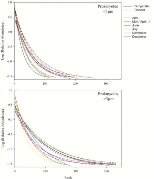
Concerning the sequencing results, a total of 4.665.345 reads were obtained for the 16S amplicon and 6.631.839 reads for the 18S amplicon, at least 13.281 (up to 727.129) reads per sample for the 16S and at least 10.748 (up to 840.077) reads per sample for the 18S amplicon. The prokaryotic community rank-abundance curves point towards a greater dominance of fewer OTUs in the temperate site for both size fractions
Temperate Tropical
Min. Avg. Max. Min. Avg. Max.
Temperature (ᵒC) 14.4 16.7 21.6 27.3 28.5 29.6**
NH4+ (µM) 0.32 0.81 1.96 1.20 1.79 2.19*
NO2- (µM) 0.11 0.19 0.38 0.02 0.03 0.05*
NO3- (µM) 0.13 0.72 1.53 0.31 1.43 5.87
PO43- (µM) 0.07 0.10 0.17 0.04 0.05 0.08*
SiO42- (µM) 0.46 1.20 1.88 0.88 1.70 3.04
Secchi disk (m) 8.5 15.1 20.0 4.0 5.3 7.0*
Chlorophyll a (mg/m³) 0.31 0.51 0.75 0.17 0.31 0.79
Chlorophyll a < 3m (%) 21.3 41.6 59.2 49.2 59.7 67.9*
Synechococcus abundance (104 cell/ml) 1.1 3.1 7.9 4.0 9.0 11.0* Prochlorococcus abundance (102 cell/ml) 0.4 36.9 118.5 18.6 34.7 65.7
Picoeukaryots abundance (103 cell/ml) 0.7 2.5 5.1 0.4 1.4 2.6
Bacterial abundance (105 cell/ml) 7.5 9.8 144.0 4.5 6.1 7.9
% of HNA 47.0 52.8 63.7 24.0 37.1 54.9*
42 (Fig. 2.2.). A similar and even more pronounced pattern was observed for eukaryotic communities throughout the year (Fig. 2.3.).
Figure 2. 2. Rank abundance of the free-living (< 3 μm) and particle-attached (> 3 μm)
43 Figure 2. 3. Rank abundance of the small (< 3 μm) and large (> 3 μm) size fraction
eukaryotes. Dashed lines represent the tropical site; solid lines represent the temperate site.
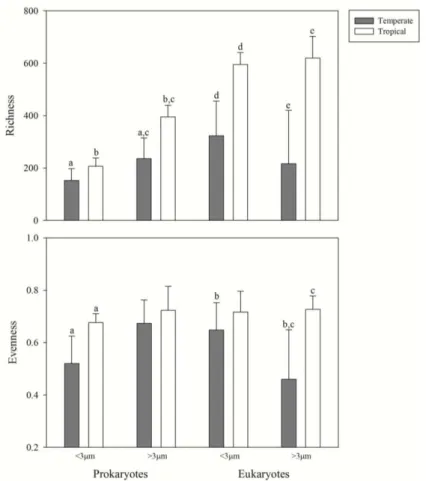
Prokaryotes of the larger size fractions (> 3 μm) had higher richness than smaller fractions (< 3 μm), differently from eukaryotes, where no significant differences could be pointed out in both sites (Fig. 2.4.). No statistical significant differences among sites
44 m). Significant differences could also be observed for eukaryote richness between
temperate and tropical sites for both size fractions.
The patterns observed for evenness were strikingly different. There were significant differences between the two sites for prokaryotes < 3 μm and for eukaryotes > 3 μm. Furthermore, analyzing the differences within the same site, evenness was higher in eukaryotes < 3 μm in the tropical site (Fig. 2.4.). However, pairwise comparisons of the Shannon diversity index, evenness and richness indicate higher values for the tropical site (Fig. 2.5.).
46 lower-right side of the graphs, while above-left side of the line indicate higher values in the tropical site.
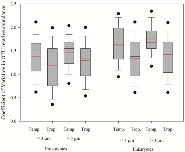
In order to evaluate microbial population stability, we calculated the coefficient of variation of each OTU relative abundance in all samples. We found significant differences (p <0.001) between the average coefficients of variation between sites with constantly higher values in the temperate coastal ocean (Fig. 2.5.).
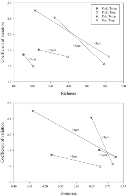
47 Plotting the average coefficient of variation against average richness for both prokaryotes and eukaryotes, we observe that all points tend to go towards the lower right side of the graph, indicating more stable and richer communities in the tropical coastal ocean (Fig 2.7. upper panel). That trend was even stronger in the plot of the coefficient of variation against evenness (Fig 2.7. lower panel).
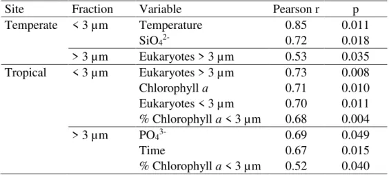
48 Then we tried to determine if the prokaryotic communities were responding to the same variables in contrasting latitudes, using Mantel test correlation between distance matrices. We divided the variables measured (Tab. 1) in environmental (temperature, nutrients, water transparency), potential biotic interactions (eukaryotic communities and chlorophyll a) or neutral factors (time). The dissimilarity matrix (Bray-Curtis distance)
of the temperate free-living (< 3 m) prokaryotic community was correlated with environmental factors such as temperature and silica. In contrast, in the tropical site, the prokaryotic community distance matrix was correlated with biological variables such as eukaryotic community (Bray-Curtis dissimilarity matrix) > 3 μm and < 3 μm and chlorophyll a (Tab. 2).
The attached prokaryotes (> 3 m) in the temperate site were correlated with the larger eukaryotes (> 3 m), while in the tropical site, the best explanatory variables were phosphate, time between samples (Euclidean distance), and percentage of chlorophyll a < 3 µm (Tab. 2).
Table 2. Mantel test significant relationships between dissimilarity (Bray Curtis distance) matrices of the free-living (< 3 μm) and particle-attached (> 3 μm) prokaryotes and environmental, biological and temporal (Euclidean) distance matrices.
Site Fraction Variable Pearson r p
Temperate < 3 µm Temperature 0.85 0.011
SiO42- 0.72 0.018
> 3 µm Eukaryotes > 3 µm 0.53 0.035
Tropical < 3 µm Eukaryotes > 3 µm 0.73 0.008
Chlorophyll a 0.71 0.010
Eukaryotes < 3 µm 0.70 0.011
% Chlorophyll a < 3 µm 0.68 0.004
> 3 µm PO43- 0.69 0.049
Time 0.67 0.015
49
Discussion
We found evidence of increasing stability with increasing richness and evenness in tropical marine microbial communities when compared to the temperate ones (Fig. 2.7). With the exception of the free-living bacteria (< 3 m fraction), all fractions had higher richness in the tropical site. These observations on samples taken at different times in the same location contradicts those from snapshot-sampling approaches covering a wide latitude range (Sunagawa et al., 2015; Milici et al., 2016). Given that
environmental conditions and microbial communities in the ocean are extremely dynamic (Fuhrman et al., 2015), snapshot-sampling strategies may miss a lot of that
variability, hampering the detection of consistent large-scale ecological patterns. Our observations support the view that microbial communities actually follow a similar macro-ecological pattern as animals and plants, exhibiting higher (or at least similar) richness towards the equator, at least in the coastal ocean. It is possible that these observations do not hold for the open ocean, as coastal systems have different dynamics, but our results highlight the importance of Eulerian time-series to fully understand microbial diversity patterns in the ocean.
Nutrient concentration and Chlorophyll a indicated that both sites had comparable
trophic state, rather oligotrophic. Still, the tropical site had a higher proportion of the smaller fraction of phytoplankton biomass (Chlorophyll a < 3 m) and higher Synechococcus abundance (Tab. 1). This is probably due to higher amplitude of
50 The higher richness of particle-attached prokaryotes already observed in the Mediterranean Sea (Ghiglione et al., 2009; Acinas et al., 1999) was also confirmed for
tropical waters. Contrastingly, this pattern could not be observed for eukaryotes. The richness of the two size fractions were different between sites, but not between size fractions in the tropical site. Again, this could reflect the lack of true phytoplankton succession cycle in constantly warm tropical waters, usually dominated by picoplankton (Alvain et al., 2008; Boyce et al., 2015).
The rank-abundance plots and evenness values evidenced a higher dominance of few OTUs in the Mediterranean Sea contrasting with a more even distribution in the tropical ocean. This pattern was particularly clear for eukaryotes indicating “bloom”
events. Curiously, rank-abundance curves of winter months in the Mediterranean Sea were those that resembled the most to the tropical ones, with a more even distribution for both prokaryotes and eukaryotes, corroborating the niche model predictions of higher microbial richness in winter in temperate regions (Ladau et al., 2013) and also
previous observations in the Western English Channel Observatory (Gilbert et al.,
2012). It worth noticing that taking the free-living prokaryotes alone, OTU richness was not significantly different between sites, but this fraction was less stable in the temperate site, as reflected by the lower evenness. These observations are also compatible with the niche model predictions (Ladau et al., 2013) where the higher
winter richness in mid and higher latitudes compensate the lower richness during summer, compared to the always moderate richness predicted for tropical regions.
Temperature was the main factor driving the free-living prokaryotic communities (< 3 µm) in the temperate site, which is a deterministic factor (Fuhrman et al., 2006),
51 prokaryote community (> 3 µm) was better explained by the concomitant “living particles” (eukaryotes > 3 µm), indicating strong associations between the prokaryotes and the particles they are sitting on (Lima-Mendez et al., 2015).
Unlike the temperate site, the structure of coastal tropical ocean prokaryotic community was clearly driven by more stochastic factors such as biotic interactions (Tab. 2). We may infer that, once the deterministic factors such as day length and temperature removed, stochastic features such as the interactions between organisms emerge as drivers of free-living prokaryotic community dynamics. In one hand, being more evenly distributed and stable throughout the year, the amplitude of fluctuations in tropical plankton populations is low, with no true succession or phenology. On the other hand, biological processes occur at higher rates because they are not constrained by temperature (e.g. Sarmento et al., 2010). Furthermore, high light and low nutrient
conditions increase the release of photosynthates by primary producers (Zlotnik and Dubinsky, 1989; Morana et al., 2014), which might increase mutualistic interactions
54 We propose a conceptual framework where microbial community composition would be driven by different proportions deterministic and stochastic factors along a latitudinal gradient: in higher latitudes most of the variation would be explained by deterministic factors (day length, for example, Gilbert et al., 2012) dictating a true
phenology throughout seasons. In intermediate latitudes (BBMO) where the annual temperature amplitude in maximal, microbial communities would mainly dictated by temperature (Figure 2.9.). Finally, in low latitudes (EAMO), where the factor temperature is removed, more stochastic biotic interactions would be the main factors shaping microbial communities.
This study demonstrates the importance of comparative studies on Eulerian time-series distributed at different latitudes to fully understand the macro-ecological patterns of diversity of microbial communities in the ocean.
Acknowledgements
55
Conclusões
1. Encontramos um aumento significativo da estabilidade nas comunidades marinhas microbianas tropicais costeiras, juntamente com um aumento na riqueza e equabilidade, quando comparado às comunidades marinhas microbianas temperadas costeiras.
2. Comunidades microbianas tropicais costeiras parecem seguir os mesmos padrões de riqueza e diversidade macroecológicos mundiais, demonstrando maior riqueza de espécies em baixas latitudes em comparação com regiões de maior latitude.
3. Amostras coletadas no inverno no mar mediterrâneo possuem riquezas semelhantes as amostras de verão dos trópicos, corroborando a ideia de que a riqueza é maior em regiões temperadas no inverno.
4. A temperatura (fator determinístico) foi preponderante na definição da estrutura das comunidades microbianas temperadas costeiras, enquanto fatores mais estocásticos como as interações biológicas parecem determinar a estrutura das comunidades microbianas tropicais costeiras
5. Finalmente, este trabalho demonstra a importância de estudos comparativos
56
References
Acinas SG, Antón J, Rodríguez-Valera F. (1999). Diversity of free-living and attached bacteria in offshore western Mediterranean waters as depicted by analysis of genes encoding 16S rRNA. Appl Environ Microbiol65: 514–522.
Alvain S, Moulin C, Dandonneau Y, Loisel H. (2008). Seasonal distribution and succession of dominant phytoplankton groups in the global ocean: A satellite view. Global Biogeochem Cycles22: 1–15.
Apprill a, McNally S, Parsons R, Weber L. (2015). Minor revision to V4 region SSU rRNA 806R gene primer greatly increases detection of SAR11 bacterioplankton.
Aquat Microb Ecol75: 129–137.
Behrenfeld MJ, O’Malley RT, Siegel D a, McClain CR, Sarmiento JL, Feldman GC, et
al. (2006). Climate-driven trends in contemporary ocean productivity. Nature444:
752–755.
Boyce DG, Frank KT, Leggett WC. (2015). From mice to elephants: Overturning the
‘one size fits all’ paradigm in marine plankton food chains. Ecol Lett 18: 504–
515.
Connolly SR, MacNeil MA, Caley MJ, Knowlton N, Cripps E, Hisano M, et al. (2014).
Commonness and rarity in the marine biosphere. Proc Natl Acad Sci 111: 8524–
8529.
Doney SC, Ruckelshaus M, J. ED, Barry JP, Chan F, English CA, et al. (2002). Climate
Change Impacts on Marine Ecosystems. Estuaries25: 149–164.
57 Edgar RC. (2013). UPARSE: Highly accurate OTU sequences from microbial amplicon
reads. doi:dx.doi.org/10.1038/nmeth.2604.
Falkowski PG, Fenchel T, Delong EF. (2008). The Microbial Engines That Drive Earth
’s Biogeochemical Cycles. Science (80- )320: 1034–1039.
Fuhrman JA, Cram JA, Needham DM. (2015). Marine microbial community dynamics and their ecological interpretation. Nat Rev Microbiol13: 133–146.
Fuhrman JA, Hewson I, Schwalbach MS, Steele JA, Brown M V, Naeem S. (2006). Annually reoccurring bacterial communities are predictable from ocean conditions. Proc Natl Acad Sci U S A103: 13104–9.
Galand PE, Casamayor EO, Kirchman DL, Lovejoy C. (2009). Ecology of the rare microbial biosphere of the Arctic Ocean. Proc Natl Acad Sci U S A 106: 22427–
32.
Gasol JM, Giorgio P a DEL, del Giorgio P a. (2000). Using flow cytometry for counting natural planktonic bacteria and understanding the structure of planktonic bacterial communities. Sci Mar64: 197–224.
Ghiglione JF, Conan P, Pujo-Pay M. (2009). Diversity of total and active free-living vs. particle-attached bacteria in the euphotic zone of the NW Mediterranean Sea.
FEMS Microbiol Lett299: 9–21.
Gilbert JA, Steele JA, Caporaso JG, Steinbrück L, Reeder J, Temperton B, et al. (2012).
Defining seasonal marine microbial community dynamics. ISME J6: 298–308.
Giovannoni SJ, Vergin KL. (2012). Seasonality in Ocean Microbial Communities.
Science (80- )335: 671–676.
58 doi:10.1016/0304-4203(78)90045-2.
Herlemann DP, Labrenz M, Jürgens K, Bertilsson S, Waniek JJ, Andersson AF. (2011).
Transitions in bacterial communities along the 2000 km salinity gradient of the
Baltic Sea. ISME J5: 1571–9.
Kindt R, Coe R. (2005). Tree diversity analysis. A manual and software for common statistical methods for ecological and biodiversity studies.
Ladau J, Sharpton TJ, Finucane MM, Jospin G, Kembel SW, O’Dwyer J, et al. (2013).
Global marine bacterial diversity peaks at high latitudes in winter. ISME J 7:
1669–77.
Lima-Mendez G, Faust K, Henry N, Decelle J, Colin S, Carcillo F, et al. (2015).
Determinants of community structure in the global plankton interactome.pdf. 348: 1–10.
Logares R, Audic S, Bass D, Bittner L, Boutte C, Christen R, et al. (2014). Patterns of
Rare and Abundant Marine Microbial Eukaryotes. Curr Biol24: 813–821.
Logares R, Sunagawa S, Salazar G, Cornejo-Castillo FM, Ferrera I, Sarmento H, et al.
(2014). Metagenomic 16S rDNA Illumina tags are a powerful alternative to amplicon sequencing to explore diversity and structure of microbial communities.
Environ Microbiol16: 2659–2671.
Margalef R. (1978). Life-forms of phytoplankton as survival alternatives in an unstable environment. Oceanol Acta1: 493–509.
McCann KS. (2000). The diversity–stability debate. Nature405: 228–233.
Milici M, Tomasch J, Wos-Oxley ML, Wang H, Jáuregui R, Camarinha-Silva A, et al.
59 19054.
Morana C, Sarmento H, Descy J-P, Gasol JM, Borges A V., Bouillon S, et al. (2014).
Production of dissolved organic matter by phytoplankton and its uptake by heterotrophic prokaryotes in large tropical lakes. Limnol Oceanogr 59: 1364–
1375.
Norland S. (1993). The relationship between biomass and volume of bacteria. Kemp P, Sherr BF, Sherr E, Cole JJ, Eds Handb methods Aquat Microb Ecol 303–307.
Oksanen J, Blanchet F, Kindt R, et al. (2015). vegan: Community Ecology Package. Quast C, Pruesse E, Yilmaz P, Gerken J, Schweer T, Yarza P, et al. (2013). The SILVA
ribosomal RNA gene database project: Improved data processing and web-based tools. Nucleic Acids Res41: 590–596.
R Core Team. (2011). R: A Language and Environment for Statistical Computing. Vienna, Austria : the R Foundation for Statistical Computing. GNU General
Public License (GPL) - http://www.gnu.org/licenses/old-licenses/gpl-2.0.html. Sarmento H, Gasol JM. (2012). Use of phytoplankton-derived dissolved organic carbon
by different types of bacterioplankton. Environ Microbiol14: 2348–2360.
Sarmento H, Montoya JM, Vázquez-Domínguez E, Vaqué D, Gasol JM. (2010). Warming effects on marine microbial food web processes: how far can we go when it comes to predictions? Philos Trans R Soc Lond B Biol Sci365: 2137–49.
Soininen J, Passy S, Hillebrand H. (2012). The relationship between species richness and evenness: A meta-analysis of studies across aquatic ecosystems. Oecologia 169: 803–809.
60 Multiple marker parallel tag environmental DNA sequencing reveals a highly complex eukaryotic community in marine anoxic water. Mol Ecol19: 21–31.
Sunagawa S, Coelho LP, Chaffron S, Kultima JR, Labadie K, Salazar G, et al. (2015).
Structure and function of the global ocean microbiome. Science348: 1261359.
de Vargas C, Audic S, Henry N, Decelle J, Mahé F, Logares R, et al. (2015). Eukaryotic
plankton diversity in the sunlit ocean. Science348: 1261605.
Venables WN, Ripley BD. (2002). Modern Applied Statistics with S. Fourth Edition. Venter JC, Remington K, Heidelberg JF, Halpern AL, Rusch D, Eisen J a, et al. (2004).
Environmental genome shotgun sequencing of the Sargasso Sea. Science304: 66–
74.
Zhang J, Kobert K, Flouri T, Stamatakis A. (2014). PEAR: a fast and accurate Illumina Paired-End reAd mergeR. Bioinformatics30: 614–620.
Zhang Z, Schwartz S, Wagner L, Miller W. (2000). A greedy algorithm for aligning DNA sequences.
61
Supplementary Material
PCR conditions
For each sample, triplicate PCR reactions were performed in 25 l and consisted of 0.4 U of Phusion high-fidelity DNA polymerase (Finnzymes, Vantaa, Finland), 1X Phusion HF reaction buffer (Finnzymes), 200 M of each dNTP (Invitrogen, imported by Sinapse, São Paulo, SP, Brazil), 0.5 µM of each primer (DNA Express, São Paulo, SP, Brazil), 0.4 mg ml-1 of BSA (Invitrogen, imported by Sinapse, São Paulo, SP,
Brazil) and 5 – 10 ng of template DNA.
PCR incubations for 16S rRNA were comprised an initial denaturation step at 95 C for 5 min, followed by 25 cycles of denaturation at 95 C for 40 sec, annealing at 53 C for 40 C and extension of 72 C for 1 min and finalized with a 10 min extension step at 72 C. And PCR incubations for 18S rRNA were comprised 95°C during 2 min, followed by 10 cycles of 30 sec at 95°C, 60 sec at 50°C and 40s at 72°C, and afterwards by 20 cycles of 30 sec at 95°C, 30 sec at 45 °C and 40 sec at 72°C, including a final elongation step at 72°C for 10 minutes.
After pooling amplicons from the triplicate reactions, PCR-products were cleaned using the AMPure XP purification kit (Beckman Coulter Inc., Brea, CA, USA) following the instructions from the manufacturer.
Microbial diversity at the Equatorial Atlantic Microbial Observatory
(EAMO)
62 Supplementary Material 1. Free-living (FL) prokaryotic groups that contribute with at least 1% of the community at
64 Supplementary Material 3. Particle-attached (PA) prokaryotic groups that contribute with at least 1% of the community at
68 Supplementary Material 7. Larger fraction (PA) eukaryotic groups that contribute with at least 1% of the community at the
70 Supplementary Material 9. Prokaryote’s Venn diagram of tropical site by size fraction. Free-living = < 3 µm and Particle-attached = > 3 µm.
71 Supplementary Material 11. Prokaryote’s Venn diagram of free-living OTUs between sites, BBMO = Blanes Bay Microbial Observatory and EAMO = Equatorial Atlantic Microbial Observatory. Free-living = < 3 µm.
72 Supplementary Material 13. Eukaryote’s Venn diagram of smaller fraction OTUs between sites, BBMO = Blanes Bay Microbial Observatory and EAMO = Equatorial Atlantic Microbial Observatory. Smaller fraction = < 3 µm.
Supplementary Material 14. Eukaryote’s Venn diagram of larger fraction OTUs between sites, BBMO = Blanes Bay Microbial Observatory and EAMO = Equatorial Atlantic Microbial Observatory. Larger fraction = > 3 µm.

