Epiphytic marine pigmented bacteria: A prospective source of natural antioxidants
Ravindra Pawar
1, Chellandi Mohandass
1, Elakkiya Sivaperumal
2, Elaine Sabu
1,
Raju Rajasabapathy
1, Tanaji Jagtap
1,31
Biological Oceanography Division, National Institute of Oceanography, Panaji, India.
2
Nallamuthu Gounder Mahalingam Centre for Research and Development, Nallamuthu Gounder Mahalingam College, Bharathiar University, Tamilnadu, India.
3
Cholamandalam MS Risk Services Pvt Ltd., Panaji, India.
Submitted: April 1, 2013; Approved: June 6, 2014.
Abstract
Awareness on antioxidants and its significance in human healthcare has increased many folds in re-cent time. Increased demand requisite on welcoming newer and alternative resources for natural anti-oxidants. Seaweed associated pigmented bacteria screened for its antioxidant potentials reveals 55.5% of the organisms were able to synthesize antioxidant compounds. DPPH assay showed 20% of the organisms to reach a antioxidant zone of 1 cm and 8.3% of the strains more than 3 cm. Pseudomo-nas koreensis(JX915782) aSargassumassociated yellowish brown pigmented bacteria have better activity than known commercial antioxidant butylated hydroxytoluene (BHT) against DPPH scav-enging.Serratia rubidaea(JX915783), an associate of Ulva sp. andPseudomonas argentinensis (JX915781) an epiphyte ofChaetomorpha media, were also contributed significantly towards ABTS (7.2%±0.03 to 15.2±0.09%; 1.8%±0.01 to 15.7±0.22%) and FRAP (1.81±0.01 to 9.35±0.98; 7.97±0.12 to 18.70±1.84mg/mL of AsA Eq.) respectively. 16S rRNA gene sequence analysis re-vealed bacteria that have higher antioxidant activity belongs to a bacterial class Gammaproteo-bacteria. Statistical analysis of phenolic contents in relation with other parameters like DPPH, ABTS, reducing power and FRAP are well correlated (p < 0.05). Results obtained from the current study in-ferred that the seaweed associated pigmented bacteria have enormous potential on antioxidant com-pounds and need to be extracted in a larger way for clinical applications.
Key words:antioxidant, epiphytic, pigmented bacteria, seaweeds.
Introduction
Oxidants are commonly known as free radicals that are chemically reactive and unstable species which need to be controlled to avoid a chain of unwanted reactions in the living system. The most important free radicals in the body are the reactive oxygen species (ROS). They try to react with the surrounding macromolecules like lipids, proteins, deoxyribonucleic acids (DNA) and certain carbohydrates in order to stabilise (Cheeseman and Slater, 1993; Dekkers et al., 1996), which leads to oxidative stress and finally re-sults in cell death (Halliwell and Gutteridge, 1999). How-ever, in order to cope up with oxidative stress the human body has an elaborate antioxidant defence system (Alscher
et al., 1997). Antioxidants are the bio-molecules manufac-tured within the body as well as extracted from the food that we consume where a variety of vitamins, minerals and en-zymes helps to protect the body by scavenging free radicals (Dekkerset al., 1996).
Coastal inhabitants like seaweeds and associated bac-teria are continuously exposed to various environmental stresses like desiccation, freezing, ultraviolet radiation, and heavy metal fluctuations which result in gradual production and accumulation of ROS (Dring, 2005). Overproduction of ROS hampers not only the process of photosynthesis and growth in plants (Dring, 2005) but also affects the cellular structures and physiology in microbes (Fridovich, 1986). In order to withstand such stress conditions, these organisms
Send correspondence to C. Mohandass. Biological Oceanography Division, CSIR-National Institute of Oceanography, Dona Paula, 403 004 Goa, Panaji, India. E-mail: cmohan@nio.org.
activate antioxidant defence mechanisms to neutralize ROS.
Seaweeds are well evaluated in the context of radical scavenging properties and are reported to possess natural antioxidants such as carotenoids, pigments, polyphenols, enzymes and diverse functional polysaccharides (Vinayak et al., 2011). Common seaweeds like,Sargassum, (Gar-cia-Casalet al., 2009) andChaetomorpha(Kelmanet al., 2012) species are found to possess strong radical scaveng-ing activities. Not only seaweeds, some of the marine pig-mented bacteria and their derivatives are also known to produce high antioxidant efficiency compounds (Dufosse, 2006; Konzenet al., 2006; Shindo et al., 2007; Correa-Llantenet al., 2012).
Seaweeds and the pigmented bacteria are independ-ently studied and reported to exhibit various antioxidant re-sponses towards excess of ROS. But pigmented bacteria and their symbiotic interactions to produce antioxidant compounds remain unexplored for quite a long time. Sea-weeds can host many heterotrophic bacteria which play an important role in maintaining the health of the host organ-ism (Bolincheset al., 1998) by producing unique bioactive secondary metabolites. Hence, epibiotic bacteria become an incredible source of new natural products (Holmstrom and Kjelleberg, 1999) for the biomedical and biopharma-ceutical industry. Thus, seaweed-associated bacteria may be useful in the development and production of bioactive compounds and biocontrol agent. The initial-step in the de-velopment of any bioactive compounds involves isolation
of appropriate bacteria from natural sources and optimiza-tion of its antagonistic activity. In this background, the present study is initiated to explore the antioxidant poten-tial of marine culturable pigmented bacteria associated with seaweeds from the central west coast of India (CWCI) as a suitable source for antioxidant compounds.
Materials and Methods
Chemicals
Sodium carbonate (Na2CO3), butylated hydroxy
tolu-ene (BHT), 2,2 diphenyl-1-picraylhydrozyl (DPPH), dimethyl sulfoxide (DMSO), ascorbic acid, trichloro acetic acid (TCA), 2,2 azinobis (3-ethylbenzothiozoline-6-sul-fonic acid) diammonium salt (ABTS), quercetin, potassium ferric cyanide, ferric chloride (FeCl3), thiobarbituric acid
(TBA), ethylenediaminetetraacetic acid (EDTA) and gallic acid were purchased from Sigma-Aldrich chemical Pvt. Ltd., Bangalore. For molecular analysis, the gel extraction and purification kit was purchased from QIAGEN India Pvt. Ltd., New Delhi. All other chemicals and reagents used for experimental work were of extra pure quality.
Sample collection and isolation of marine bacteria
The seaweed samples (Table 1) were picked with sterile forceps from the intertidal region along the central west coast of India during 2010-2011 (Figure 1). All the samples were stored in sterile plastic bottles and brought to the laboratory in ice box. Isolation of bacteria was carried
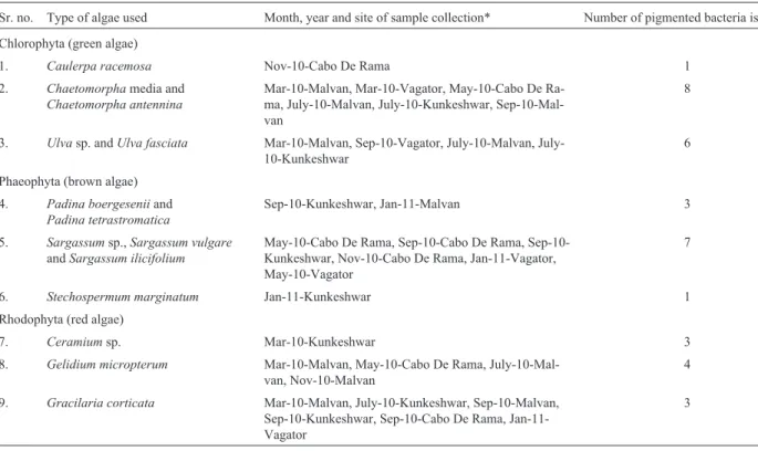
Table 1- Type of seaweeds used for isolation of epiphytic pigmented bacteria.
Sr. no. Type of algae used Month, year and site of sample collection* Number of pigmented bacteria isolated Chlorophyta (green algae)
1. Caulerpa racemosa Nov-10-Cabo De Rama 1
2. Chaetomorphamedia and Chaetomorpha antennina
Mar-10-Malvan, Mar-10-Vagator, May-10-Cabo De Ra-ma, July-10-Malvan, July-10-Kunkeshwar, Sep-10-Mal-van
8
3. Ulvasp. andUlva fasciata Mar-10-Malvan, Sep-10-Vagator, 10-Malvan, July-10-Kunkeshwar
6
Phaeophyta (brown algae) 4. Padina boergeseniiand
Padina tetrastromatica
Sep-10-Kunkeshwar, Jan-11-Malvan 3
5. Sargassumsp.,Sargassum vulgare andSargassum ilicifolium
May-10-Cabo De Rama, Cabo De Rama, Sep-10-Kunkeshwar, Nov-10-Cabo De Rama, Jan-11-Vagator, May-10-Vagator
7
6. Stechospermum marginatum Jan-11-Kunkeshwar 1
Rhodophyta (red algae)
7. Ceramiumsp. Mar-10-Kunkeshwar 3
8. Gelidium micropterum Mar-10-Malvan, May-10-Cabo De Rama, July-10-Mal-van, Nov-10-Malvan
4
9. Gracilaria corticata Mar-10-Malvan, July-10-Kunkeshwar, Sep-10-Malvan, Sep-10-Kunkeshwar, Sep-10-Cabo De Rama, Jan-11-Vagator
3
out by spread plate method (Lakshmanaperumalsamy and Purushothaman, 1982) using Zobell marine agar 2216 me-dia (Hi-Meme-dia, M384, Mumbai). Seaweed samples were rinsed with sterile seawater to remove debris and unat-tached bacteria. Approximately 10 g (wet weight) of sea-weed sample was suspended in 100 mL of sterile seawater in conical flask. This was kept in a rotary shaker at 150 rpm for 30 min. Then the water samples were serially diluted and plated in duplicates. Plates were incubated at 28±2 °C for 48-72 h. Distinct pigmented colonies were selected and purified. These cultures were stored in glycerol vials at -80 °C until further analysis.
Primary screening of antioxidant producing strains
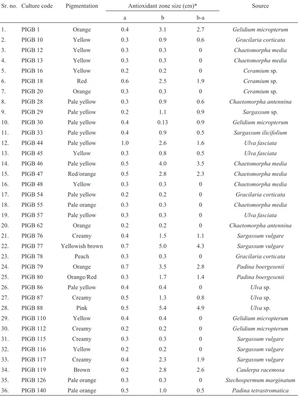
The simple primary DPPH-antioxidant screening method applied by Takaoet al.(1994) was followed for ini-tial screening. Pure culture suspension broth (2 mL) was drop inoculated on nutrient agar plates and incubated at 28 °C for 24 h. A sterilized filter paper (Whatman no. 1) was then placed on the agar plate, so that colonies and their extracellular metabolites get replicated on the paper. Incu-bation was further continued at 28 °C for another 24 h. Then the filter paper was taken out, dried and sprayed with a DPPH solution (80mg mL-1in methanol). Strains show-ing a white on purple spot were considered as antioxidant producing strains. Colony size and zones of decolorization were noted for each analysed culture (Table 2).
Mass culture, extraction and dry weight determination
An aliquot of 100mL of selected pure cultures (selec-tion based on highest zone of DPPH decoloriza(selec-tion) were inoculated in sterile nutrient broth (300 mL). It was incu-bated on shaker incubator at 28 °C with 120 rpm for 48 h. It
was then centrifuged (10 °C, 10 min at 6153 x g) and supernatant was collected in another sterile collection tube. The supernatant was filtered (0.45mm) to remove all the bacterial cells from broth and extracted with ethyl acetate (30% v/v). The extracts were evaporated and concentrated with rotary evaporator (Equitron, Roteva (63) R-V) until complete dryness and finally retained in minimum HPLC grade methanol. 0.5-1.0 mL of sample was taken in a pre-weighed aluminium foil and allowed to dry for weight determination (Ekanayakeet al., 2004). The known con-centrations of sample viz. 1.0, 5.0, 10 and 15 mg/mL were prepared in methanol, and used for quantitative antioxidant analysis.
Antioxidant analysis
DPPH radical scavenging activity
Radical scavenging activity of different bacterial ex-tracts against DPPH radicals was assessed according to the method of Larrauriet al.(1998). Briefly, 0.8 mL (0.05 mM) DPPH-methanol solution was mixed with different concen-tration of samples and final volume was made up to 1.0 mL. Reaction mixture was incubated for 30 min at 25 °C in dark and absorbance was measured at 517 nm. Control was maintained with DPPH and methanol instead of antioxidant solution while blanks contained only methanol instead of DPPH solution throughout the experiment. BHT was used as positive control. The inhibition of DPPH radicals by the samples was calculated according to the following equa-tion,
DPPH scavenging activity (%) = [1-(Absorbance of the sample/Absorbance of the control)] x 100.
Ferric reducing antioxidant power (FRAP) assay
FRAP analysis was done with the modified method of Benzie and Strain’s (1996). The working FRAP reagent was prepared by mixing 300 mM acetate buffer (pH 3.6), 10 mM TPTZ solution (10 mM TPTZ in 40 mM HCl) and 20 mM FeCl36H2O in a 10:1:1 ratio just before use and
heated to 37°C. A total of 150mL of working FRAP reagent was added to each well of a 96 well microtiter plate. A blank reading was then taken at 600 nm using an ELISA plate reader (Biorad 680 XR, Biosciences). A total of 20mL of sample was then added to each well. After addition of sample to the FRAP reagent, a second reading at 595 nm was performed after 8 min. Ascorbic acid was used as stan-dard and the antioxidant content of the extract was ex-pressed as ascorbic acid equivalents (AsA Eq.)mg/mL of crude sample extract.
ABTS assay
The ABTS radical cation decolourization assay (Reet al., 1999) was used to evaluate antioxidant power of sample extracts. ABTS was dissolved in water to a 7 mM concen-tration. ABTS radical cations were produced by reacting Figure 1- Sampling locations along the central west coast (CWC) of
ABTS stock solution with 2.45 mM potassium per sulphate (final concentration) and allowing the mixture to stand in the dark for 12-16 h before use. The stock solution was di-luted with ethanol (1:89 v/v) to an absorbance of 0.70 (±0.02) at 734 nm and equilibrated at 30°C exactly 6 min after initial mixing. One millilitre of diluted ABTS solution was mixed with 10 mL of sample extracts of different strength (1.0 to 15 mg/mL). The decrease of absorbance
was measured at 734 nm and the antioxidant capacity was expressed in percent inhibition (%I). Quercetin was used as a standard antioxidant and IC50value was calculated from
regression analysis.
Determination of reducing power
The reducing abilities of sample extracts were deter-mined according to the method of Ferreira (2007). Briefly, Table 2- Primary antioxidant screening results of DPPH decolorization assay.
Sr. no. Culture code Pigmentation Antioxidant zone size (cm)* Source
a b b-a
1. PIGB 1 Orange 0.4 3.1 2.7 Gelidium micropterum
2. PIGB 10 Yellow 0.3 0.9 0.6 Gracilaria corticata
3. PIGB 12 Yellow 0.3 0.3 0 Chaetomorpha media
4. PIGB 13 Yellow 0.3 0.3 0 Chaetomorpha media
5. PIGB 16 Yellow 0.2 0.2 0 Ceramiumsp.
6. PIGB 18 Red 0.6 2.5 1.9 Ceramiumsp.
7. PIGB 20 Orange 0.3 0.3 0 Ceramiumsp.
8. PIGB 28 Pale yellow 0.3 0.9 0.6 Chaetomorpha antennina
9. PIGB 29 Pale yellow 0.2 1.1 0.9 Sargassumsp.
10. PIGB 30 Pale yellow 0.4 0.13 0.9 Gelidium micropterum
11. PIGB 33 Pale yellow 0.4 0.9 0.5 Sargassum ilicifolium
12. PIGB 44 Pale yellow 1.0 2.6 1.6 Ulva fasciata
13. PIGB 45 Yellow 0.3 0.8 0.5 Ulva fasciata
14. PIGB 46 Pale yellow 0.5 4.0 3.5 Chaetomorpha media
15. PIGB 47 Red/orange 0.5 2.8 2.3 Chaetomorpha media
16. PIGB 48 Yellow 0.3 0.3 0 Chaetomorpha media
17. PIGB 54 Pale yellow 0.2 0.2 0 Gracilaria corticata
18. PIGB 55 Pale orange 0.3 0.3 0 Chaetomorpha media
19. PIGB 57 Pale yellow 0.3 0.3 0 Ulva fasciata
20. PIGB 62 Orange 0.2 0.2 0 Chaetomorpha antennina
21. PIGB 76 Creamy 0.4 1.5 1.1 Sargassum vulgare
22. PIGB 77 Yellowish brown 0.7 5.0 4.3 Sargassum vulgare
23. PIGB 78 Peach 0.3 0.3 0 Gracilaria corticata
24. PIGB 79 Orange 0.7 3.5 2.8 Padina boergesenii
25. PIGB 80 Orange/Red 0.3 1.7 1.4 Padina boergesenii
26. PIGB 86 Pale yellow 0.4 0.4 0 Ulvasp.
27. PIGB 87 Creamy 0.5 1.3 0.8 Ulvasp.
28. PIGB 88 Pink 0.5 5.4 4.9 Ulvasp.
29. PIGB 110 Yellow 0.4 0.4 0 Gelidium micropterum
30. PIGB 112 Creamy 0.2 0.2 0 Gelidium micropterum
31. PIGB 115 Creamy 0.3 0.3 0 Sargassum vulgare
32. PIGB 116 Yellow 0.2 0.2 0 Sargassum vulgare
33. PIGB 117 Creamy 0.4 2.3 1.9 Sargassum vulgare
34. PIGB 119 Brown 0.2 2.8 2.6 Caulerpa racemosa
35. PIGB 126 Pale orange 0.3 0.3 0 Stechospermum marginatum
36. PIGB 140 Pale orange 0.5 1.0 0.5 Padina tetrastromatica
0.25 mL aliquots of various concentrations of sample were mixed with 2.5 mL of 200 mM sodium phosphate buffer (pH 6.6) and 2.5 mL of 1% potassium ferricyanide. The mixture was then incubated at 50 °C for 20 min. Later, 2.5 mL of 10% TCA (w/v) was added to the reaction mix-ture and centrifuged at 4062 xgfor 10 min. Finally, 5 mL aliquot of the upper layer was mixed with 5 mL of distilled water and 1.0 mL of 0.1% ferric chloride. The absorbance was measured at 700 nm.
Determination of total phenolic contents
Total Phenolic compounds in extracellular extracts were quantified by using Folin-ciocalteu’s colorimetric method described by Shanet al.(2005). Different concen-trations of extracted samples (0.2 mL) were initially oxi-dized with 1 mL of 0.5 M of Folin-ciocalteu’s reagent for 4 min. The reaction was finally neutralized with 1 mL of saturated sodium carbonate (75 g/L) after 30 min of incuba-tion and absorbance of the resulting blue color was mea-sured at 760 nm. Quantification was done from standard of gallic acid curve and results were expressed as mg of gallic acid equivalents (GAE) per mL of sample extract. All tests were performed in triplicate.
Identification of antioxidant potent strains by 16S rRNA gene
Genomic DNA was extracted from all the potent iso-lates by using DNeasy Blood and Tissue kit (Qiagen) ac-cording to manufacturer’s instructions. The extracted DNA was subjected to polymerase chain reaction (PCR) with universal primers 27F and 1492R (Lane, 1991). DNA se-quencing was performed with an automatic sequencer (Ap-plied Biosystems 3130xl Genetic Analyzer) with the bacterial primers 27F, 518F and 1492R. The 16S rRNA gene sequences obtained were submitted to the GenBank and the accession numbers were assigned from JX915781 to JX915783.
Statistical analysis
Each sample was run in triplicate for statistical analy-sis. Results are expressed as mean ± standard deviation (SD). Changes in biochemical parameters were tested using one-way ANOVA and post hoc tests (Newman-Keuls) to discriminate between means of values. Differences were considered statistically significant when p < 0.05.
Results
Pigmented bacteria from the seaweeds
A total of 36 pigmented bacterial strains isolated from 14 different seaweeds are listed in Table 1. Highest num-bers of pigmented bacteria were associated with Chaetomorpha and Sargassum species. While, the sea-weeds like Caulerpa racemosa and Stechospermum marginatum indicated the poor association with the
pig-mented bacteria. These two species could associate only one pigmented bacterial colony throughout the year. More-over, high numbers of pigmented bacteria were retrieved from the algal surface which belongs to Chlorophyta (green algae) while, Phaeophyta (brown algae) and Rhodophyta (red algae) were seen to be loaded with near about equal numbers of bacteria. Seaweeds (Chaetomorpha, SargassumandUlva spp.) possessing highest number of pigmented bacteriai.e.58.34% of the total population were observed to be dominating during pre-monsoon and mon-soon seasons. Another interesting finding is that though the Gracilaria corticatawas dominating most of the sampling stations throughout the year it was able to give only limited number of pigment producers.
Primary screening of extracellular antioxidants
DPPH screening method indicates that 20 different pigmented bacteria were able to produce extracellular anti-oxidants. Cultures showing decolonization zone above 3 cm were considered as high potent strains and below this zone were excluded for further investigation. Bacterial as-sociates ofUlva,SargassumandChaetomorphaspp. ex-hibited high DPPH activity in terms of the hollow zones of 4.9, 4.3 and 3.5 cm respectively. Isolates obtained from Caulerpa racemosa, Gelidium micropterum and Padina boergeseniiexpressed second level of activity and exhib-ited the zones of 2.6-2.8 cm (Table 2).
Antioxidant potential of pigmented strains
Of the total pigmented isolates tested ~55.5% bacte-ria showed antioxidant properties. The bactebacte-ria coloured yellow, yellowish brown and pink were able to produce prominent antioxidant zones. The second level of activities were obtained with near about 20% of strains having or-ange, yellowish-brown, pink and brown pigmentation whereas creamy and pale yellow coloured colonies (44.5%) exhibited nil or negligible amount of activity. This clearly indicates variation in pigment coloration contributes signif-icantly towards radical scavenging activities. Table 2 shows that, among the total antioxidant producers 27% were associated with green algae (Chlorophyta) and 8.3% of the pigmented bacteria (PIGB 46, PIGB 77 and PIGB 88) exhibiting highest activities were selected for quantitative analysis.
DPPH radical scavenging assay on selected bacteria
syn-thetic BHT used in the study (Table 3).Serratia rubidaea andP. argentinensisalso exhibited 57.5±3.03 and 28.7± 0.92% of DPPH scavenging while increasing the sample concentrations (Table 3).
ABTS assay
P. argentinensis and S. rubidaea exhibited poor ABTS scavenging activity 1.8±0.01 to 15.7±0.22% and 7.2±0.03 to 15.2±0.09% respectively at 1 to 15 mg of sample concentration. Highest activities by P. koreensis (19.2±0.05 to 69.5± 1.00%) with lowest EC50value of
9.41 ± 0.09 mg/mL indicate its potential role towards extracellular antioxidant (Table 3). Other strains, though having different EC50 values (P. argentinensis,
EC50 = 46.14 ± 3.49 and S. rubidaea, EC50 = 88.34 ±
2.15 mg/mL) were able to exhibit the same ABTS scaveng-ing effects (Table 3). However, higher concentrations of the extracts were more effective in quenching free radicals in the system.
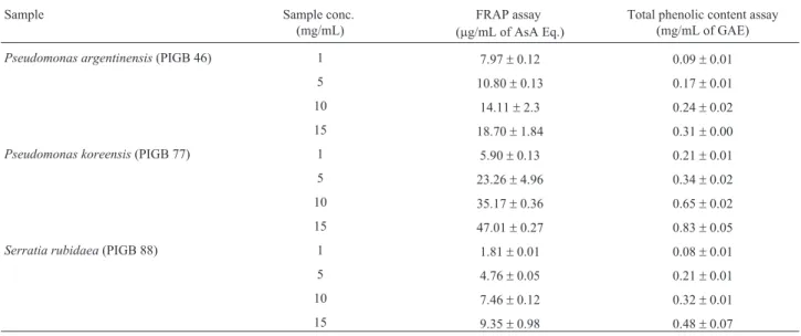
FRAP assay
Results of FRAP analysis indicated significant levels of AsA Eq. in P. koreensis which was followed by P. argentinensisandS. rubidaea. Increasing orders of FRAP values were observed in case ofP. koreensisfrom 5.90± 0.13 for 1 mg and 47.01±0.27mg AsA Eq./mL for 15 mg of samples (Table 4). Contrastingly,S. rubidaeawas found to possess low levels of AsA Eq. though it exhibited better DPPH and ABTS scavenging than that ofP.argentinensis. Moreover, correlation results of FRAP were significant with total phenolic contents.
Reducing power assay
The reducing capacities of ethyl acetate extracts were determined based on the change in absorbancei.e.in terms of increasing OD values and results were represented in Figure 2.P. koreensisshowed highest absorbance. The ac-tivity tested withP. koreensiswas much higher than that of standard compoundi.e., ascorbic acid. However, these re-ducing abilities were also shown linear with the increase in concentrations.
Table 3- Antioxidant activities of extracellular bacterial extracts by DPPH and ABTS radical scavenging assay. Values are mean±SD (n = 3). Sample conc. (mg/mL) DPPH scavenging assay
(%)
EC50
(mg/mL)
ABTS scavenging assay (%)
EC50
(mg/mL) Pseudomonas argentinensis(PIGB 46)
1 10.9±0.28 31.13±4.15 1.8±0.01 46.14±3.49
5 13.7±0.39 4.7±0.01
10 22.4±0.33 8.4±0.03
15 28.7±0.92 15.7±0.22
Pseudomonas koreensis(PIGB 77)
1 47.3±0.77 1.17±0.06 19.2±0.05 9.41±0.09
5 76.1±2.61 38.9±1.98
10 93.4±6.54 53.4±2.75
15 95.6±5.61 69.5±1.00
Serratia rubidaea(PIGB 46)
1 18.0±0.17 10.31±0.48 7.2±0.03 88.34±2.15
5 35.5±0.32 9.3±0.03
10 53.0±0.56 11.3±0.03
15 57.5±3.03 15.2±0.09
Standards
Sample conc. (mg/mL) DPPHiScavenging assay
(%)
EC50
(mg/mL)
*ABTSiiscavenging
assay (%)
Sample conc. (mM/mL)
40 56.6±1.63 24.33±1.82 78.0±1.14 5.74±1.26
80 80.3±0.61 97.6±1.25
120 82.8±0.72 99.6±1.18
160 84.3±1.34 99.5±1.06
Standards used for various assays:iDPPH scavenging assay - BHT,iiABTS scavenging assay -Quercetin. *Sample concentrations used for ABTS
Total phenolic content assay
The TPCs in all the tested samples showed less than 1 mg/mL of GAE. Analysis results clearly showed thatP. koreensisextracts had the highest phenolic content (0.83± 0.05 mg GAE/mL). While, other species like P. argentinensis(0.31±0.00 mg GAE/mL) andS. rubidaea (0.48±0.07 mg GAE/mL) were observed to possess low TPCs (Table 4). Results of correlation analysis between each antioxidant parameter and total phenolic content are shown (Figure 3). TPC exhibited significant r2values over all the other antioxidant tests performed in this investiga-tion (p < 0.005).
High potent strains and their source
Considering the source of the potent strains, the high-est antioxidant producing strain PIGB77 was found to be associated withSargassumspecies (Phaeophyta) whereas the other two strains belonged toChaetomorphaandUlva
spp. (Chlorophyta) (Table 5). This shows a bacterium liv-ing in the vicinity of seaweeds belongliv-ing to Phaeophyta and Chlorophyta supports more bioactive flora than Rhodophyta.
High potent strains and their molecular identity (16S rRNA)
The sequence data obtained was visualised by CromasPro 1.41. Closest matches assessed using BLASTn and accession numbers obtained from GenBank were given in Table 5. Out of 3 selected strains two belonged to Pseu-domonas genus while the other was from Serretia. Se-quence match for species identity revealed selected strains as, P. argentinensis (97%), P. koreensis (100%), and S. rubidaea(99%). Moreover, 16S rRNA results highlights Gammaproteobacteria group as a common origin for anti-oxidant potent bacterial flora (Table 5).
Figure 2- Reducing potentials of selected bacteria.*Concentrations of the sample: 1, 5, 10, 15 mg/mL, STD: standard compound usedi.e.ascorbic acid.
Table 4- FRAP and total phenolic contents in extracellular ethyl acetate extracts of selected bacterial strains.
Sample Sample conc.
(mg/mL)
FRAP assay (mg/mL of AsA Eq.)
Total phenolic content assay (mg/mL of GAE)
Pseudomonas argentinensis(PIGB 46) 1 7.97±0.12 0.09±0.01
5 10.80±0.13 0.17±0.01
10 14.11±2.3 0.24±0.02
15 18.70±1.84 0.31±0.00
Pseudomonas koreensis(PIGB 77) 1 5.90±0.13 0.21±0.01
5 23.26±4.96 0.34±0.02
10 35.17±0.36 0.65±0.02
15 47.01±0.27 0.83±0.05
Serratia rubidaea(PIGB 88) 1 1.81±0.01 0.08±0.01
5 4.76±0.05 0.21±0.01
10 7.46±0.12 0.32±0.01
15 9.35±0.98 0.48±0.07
Discussion
Bacteria are well known to be associated with sea-weed surfaces. Earlier studies had revealed distinct diversi-ties among the seaweed epiphytic bacterial flora (Oberbeckmann, 2007; Menezeset al., 2010). These bacte-ria have recently gained attention of the scientific commu-nity due to their source for novel compounds of industrial importance. Pigmented bacteria are of similar concern ob-served to have distinct physiological and biological roles. We retrieved prominent numbers of such bacteria from the surfaces of selected seaweeds (Table 1). Supporting growth for pigmented bacteria had been reported by Shibaet al. (1979), who found thalli of Enteromorpha linza and Sargassum horneriwere dominant with sixteen strains of aerobic pink or orange bacteria. Moreover, novel species of pigmented bacteria such as ArenibacterandFlavobacter are also found to be associated with seaweeds (Nedashkovskayaet al., 2004, 2006). This clearly shows that pigmented bacteria also form a major part of the epi-phytic bacterial flora. The reason behind their attached
life-style may be the utilization of various substances secreted by living or dead algae and sometimes vice versa; these as-sociations play an important role in host (algae) growth (Soria-Mercadoet al., 2012).
Takaoet al.(1994), first gave a simple and efficient method for screening antioxidants of bacterial origin using DPPH decolorization assay. Our investigation followed a similar method for primary screening and selection of anti-oxidant potent pigmented strains. Of the total strains tested, 55.5% showed positive results which may be due to the scavenging properties of extracellular compounds includ-ing pigments. Few of the recent studies on bacterial pig-ments of marine realme.g.violacein, astaxanthin, cantha-xanthin, zeacantha-xanthin, rubrolone and carotenoid derivatives evidence their efficient radical scavenging activities (Dufosse, 2006; Shindoet al., 2007). These studies clearly indicate that variation in pigment coloration contributes to-wards their radical scavenging activities.
The DPPH activity is a measure of the reactivity where decrease in absorbance is estimated as the reaction between DPPH and antioxidant progresses. All the bacte-Figure 3- Correlation analysis of antioxidant parameteres. PIGB 46; PIGB 77; PIGB 88. A. TPC Vs DPPH (PIGB 46; r2: 0.976, PIGB 77; r2: 0.741, PIGB
88; r2: 0.855) p = 0.004, B. TPC Vs ABTS (PIGB 46; r2: 0.872, PIGB 77; r2: 0.964, PIGB 88; r2: 0.790) p = 0.009, C. TPC Vs Reducing power (PIGB 46; r2: 0.979, PIGB 77; r2: 0.725, PIGB 88; r2: 0.943) p = 0.01, D. TPC Vs FRAP (PIGB 46; r2: 0.971, PIGB 77; r2: 0.962, PIGB 88; r2: 0.975) p = 0.01.
Table 5- 16S rRNA identity for high potent antioxidant strains.
Isolate Isolation source Closest match (accession number) Phylogenetic group Similarity (%) Pigmentation PIGB 46 Chaetomorpha media Pseudomonas argentinensis(JX915781) g-proteobacteria 97 Yellow PIGB 77 Sargassum vulgare Pseudomonas Koreensis(JX915782) g-proteobacteria 100 Yellow/brown
rial extracellular extracts under the study exhibited increas-ing DPPH scavengincreas-ing activities with increase in sample concentration (Table 2). These scavenging effects may be due to the donation of hydrogen atoms to quench free radi-cals by extracellularly released compounds like carotenoid pigments by selected bacterial strains. The same kind of quenching effects of hydrogen donation by an antioxidant sample were also observed by Kahkonenet al.(1999). Ca-rotenoids of bacterial origin are well reported to possess DPPH scavenging and or antioxidant properties (Kuoet al., 2011, Shindoet al., 2007). Mikiet al.(1994), showed the presence of carotenoid derived compounds in Pseudomo-nassp. isolated from marine environment. In addition, Ye et al.(2012) had recently reported strong DPPH scavenging by exopolysaccharides isolated fromPseudomonasPF-6. So the overall scavenging effects in the present study may be due to the combined quenching effect of carotenoids and polysaccharide like reductants. Moreover, positive correla-tion with reducing power and phenolic contents also indi-cates their responsible roles (Figure 3).
Scavenging of proton radicals is an important attrib-ute of natural antioxidants. ABTS is a synthetic protonated radical which exhibits decrease in absorbance with the scavenging of the proton radicals (Mathew and Abraham, 2004). Ethyl acetate extracts ofP. koreensiswere able to scavenge these radicals efficiently, indicating higher re-sponses towards their activity and comparable with stan-dard compound (Quercetin) used (Table 3). Various factors like stereo-selectivity of the radicals, the presence of func-tional groups in the bioactive compound, solubility of the extract in different testing systems and polarity of the sol-vent are reported to react and quench free radicals (Yuet al., 2002). Wanget al.(1998), had reported that some com-pounds having ABTS scavenging property may not show similar DPPH scavenging effects. In our study Serratia rubidaea extracts also showed increased DPPH and de-creased ABTS scavenging (Table 3). Whereas,P. koreensis showed strong scavenging effects towards both DPPH and ABTS radicals. These results indicate that the radical scav-enging activities may be in relation to properties of a com-pound excreted by the bacteria.
Increased FRAP value represents higher content of antioxidants in the sample extract responsible for reduction of ferric ion to the ferrous ion (Smetet al., 2006). We ob-served similar kind of expression in FRAP with P. koreensis and P. argentinensis (Table 4). Although S. rubidaeaexhibited good radical scavenging activities, the FRAP values were found to be lower in this case. Synergis-tic effects of the substances like carotenoids, vitamins and minerals are responsible in antioxidant activity variations (Ratnamet al., 2006) and this type of material might have contributed radical scavenging activity of the above spe-cies.
All the sample extracts showed increase in absor-bance with increasing sample concentration (Figure 2). The
reducing potential of the sample involves various reactions such as prevention of chain initiation, binding of transition metal ion catalysts, decomposition of peroxides, prevention of continued hydrogen abstraction and radical scavenging (Diplock, 1997). Higher reducing powers withSargassum extract were reported at 10 mg/mL (Choet al., 2007) and until now there are no reports in associated bacteria which can yield similar results. FurtherP. koreensis, associated withSargassumin our study could show the highest reduc-ing abilities within a concentration of 5 mg/mL (Figure 3). This result suggests that, reducing capabilities of samples could be enhanced by the associated bacteria and it can be used as an alternative source for antioxidative compounds.
Generally, plants are well known source of phenolic and flavonoid compounds and till now limited amount of information is available on their presence in bacterial spe-cies. The current investigation on seaweed associated bac-teria shows a positive indication of producing significant levels of TPCs (Table 4). Polyphenolics and their deriva-tives appear to function as good electron and hydrogen atom donors and therefore, cause termination of radical chain reaction by converting free radicals to more stable products (Shahidi and Wanasundara, 1992). Earlier studies on TPCs were strongly correlated with antioxidant capaci-ties (Kaltet al., 1999). Our studies also showed positive correlations of TPC (p < 0.05) towards the other assessed antioxidant activities. As the prominent levels of phenolic contents were observed in theP. koreensisextracts, it may be assumed that this species may possess one or more bioactivities of similar kind.
In the current study we have come across one of the bacterial species Pseudomonas koreensis (JX915782) a Sargassumassociated yellowish brown pigmented bacte-rium with high antioxidant activity while comparing with a known commercial antioxidant butylated hydroxytoluene (BHT) against DPPH scavenging. BHT is primarily used as an antioxidant additive in food, cosmetics, pharmaceu-ticals, rubber electrical transformer oil, and embalming fluid (Malhotra, 2010). BHT also finds uses in hydraulic fluids, turbine and gear oils, jet fuels (Michael and Irene, 2004). Keeping the various application of BHT listed above by many researchers we could suggest here our material on the above isolatePseudomonas koreensis(JX915782) will definitely form a better natural resource in terms of biotech-nological prospects and may be looked for its future appli-cations.
com-pounds. All the three high potent strains worked out in the present study are expected to have similar or related genes in their nucleotide sequences for their synthesis; however analysis of such specific genes for quantifying these com-pounds remains the objective of future study.
Acknowledgments
Authors like to thank the Director of NIO, Dr. P. Vethamony, Dr. N. Ramaiah and Mr. Ram Murti Meena for their support in this study. The authors declare that there are no conflicts of interest. This is NIO contribution number 5592.
References
Alscher RG, Donahue JL, Cramer CL (1997) Reactive oxygen species and antioxidants: relationships in green cells. Phy-siol Plant 100:224-233.
Benzie IFF, Strain JJ (1996) The ferric reducing ability of plasma (FRAP) as a measure of `antioxidant power’: the FRAP as-say. Anal Biochem 239:70-76.
Bolinches J, Lemos ML, Barja JL (1988) Population dynamics of heterotrophic bacterial communities associated withFucus vesiculosus and Ulva rigida in an estuary. Microb Ecol 15:345-357.
Cheeseman KH, Slater TF (1993) An introduction to free radical biochemistry. Bri Med Bull 49:481-493.
Cho SH, Kang SE, Cho JY, Kim AR, Park SM, Hong YK, Ahn DH (2007) The antioxidant properties of brown seaweed (Sargassum siliquastrum) extracts. J Med Food 10:479-485. Correa-Llanten DN, Amenabar MJ, Blamey JM (2012)
Antioxi-dant capacity of novel pigments from an Antarctic bacte-rium. J Microbiol 50:374-379.
Dekkers JC, Van Doornen LJPH, Kemper CG (1996) The role of antioxidant vitamins and enzymes in the prevention of exer-cise-induced muscle damage. Sports Med 21:213-238. Diplock AT (1997) Will the `good fairies’ please proves to us that
vitamin E lessens human degenerative of disease? Free Rad Res 27:511-532.
Dring MJ (2005) Stress resistance and disease resistance in sea-weeds: the role of reactive oxygen metabolism. Adv Bot Res 43:175-207.
Dufosse L (2006) Microbial production of food grade pigments. Food Technol Biotechnol 44:313-321.
Ekanayake P, Lee YD, Lee J (2004) Antioxidant activity of flesh and skin of Eptatretus burgeri(Hag Fish) and Enedrias nebulosus(White Spotted Eel). Food Sci Tech Int 10:171-177.
Ferreira ICFR, Baptista P, Vilas-Boas M, Barros L (2007) Free-radical scavenging capacity and reducing power of wild ed-ible mushrooms from northeast Portugal: individual cap and stipe activity. Food Chem 100:1511-1516.
Fridovich I (1986) Biological effects of the superoxide radical. Arch Biochem Biophys 247:1-11.
Garcia-Casal MN, Ramirez J, Leets I, Pereira AC, Quiroga MF (2009) Antioxidant capacity, polyphenol content and iron bioavailability from algae (Ulva sp., Sargassum sp. and
Porphyrasp.) in human subjects. Br J Nutr 101:79-85. Halliwell B, Gutteridge JM (1999) Free Radicals in Biology and
Medicine. 3rd ed. Oxford University Press, Oxford.
Holmstrom C, Kjelleberg S (1999) MarinePseudoalteromonas
species are associated with higher organisms and produce biologically active extracellular agents. FEMS Microbiol Ecol 30:285-293.
Kahkonen MP, Hopia AI, Vuorela HJ, Rauha JP, Pihlaja K, Kujala TS, Heinonen M (1999) Antioxidant activity of plant extracts containing phenolic compounds. J Agric Food Chem 47:3954-3962.
Kalt W, Forney CF, Martin A, Prior RL (1999) Antioxidant ca-pacity, vita-min C, phenolics, and anthocyanins after fresh storage of small fruits. J Agr Food Chem 47:4638-4644. Kelman D, Posner EK, McDermid KJ, Tabandera NK, Wright
PR, Wright AD (2012) Antioxidant activity of Hawaiian marine algae. Mar Drugs 10:403-416.
Konzen M, De Marco D, Cordova CA, Vieira TO, Antonio RV, Creczynski-Pasa TB (2006) Antioxidant properties of viola-cein: possible relation on its biological function. Bioorg Med Chem 14:8307-8313.
Kuo YH, Liang TW, Liu KC, Hsu YW, Hsu HC, Wang SL (2011) Isolation and identification of a novel antioxidant with antitumor activity fromSerratia ureilyticausing squid pen as fermentation substrate. Marine Biotechnol 13:451-461. Lakshmanaperumalsamy P, Purushothaman A (1982)
Heterotro-phic bacteria associated with seaweed. Proc Indian Acad Sci 91:487-493.
Lane DJ (1991) 16S/23S rRNA sequencing.In: Stackebrandt, E., Goodfellow, M. (eds). Nucleic Acid Techniques in Bacterial Systematic. John Wiley and Sons Incorporation, New York, pp 115-175.
Larrauri JA, Sanchez-Moreno C, Saura-Calixo F (1998) Effect of temperature on the free radical scavenging capacity of ex-tracts from red and white grape pomace peels. J Agri Food Chem 46:2694-2697.
Malhotra GK (2010) Chemical Process Simplification: Improving Productivity and Sustainability. Wiley-Blackwell (John Wiley & Sons Ltd), Chicester.
Mathew S, Abraham TE (2006) In-vitro antioxidant activity and scavenging effect of Cinnamomum verumleaf extract as-sayed by different methodologies. Food Chem Toxicol 44:198-206.
Menezes CB, Bonugli-Santos RC, Miqueletto PB, Passarini MR, Silva CH, Justo MR, Leal RR, Fantinatti-Garboggini F, Oliveira VM, Berlinck RG, Sette LD (2010) Microbial di-versity associated with algae, ascidians and sponges from the north coast of Sao Paulo state. Brazil Microbiol Res 165:466-482.
Michael A, Irene A (2004) Handbook of Preservatives. Synapse Information Resources Inc., Endicott, NY.
Miki W, Otaki N, Yokoyama A, Izumida H, Shimidzu N (1994) Okadaxanthin, a novel C50-carotenoid from a bacterium,
Pseudomonas sp. KK10206C associated with marine sponge,Halichondria okadai. Experientia 50:684-686. Nedashkovskaya OI, Kim SB, Han SK, Lysenko A, Mikhailov
VV, Bae KS, Swings J (2006)Arenibacter palladensissp. nov., a novel marine bacterium isolated from the green alga
Ulva fenestrata, and emended description of the genus
Arenibacter. Int J Syst Evol Microbiol 56:155-160. Nedashkovskaya OI, Kim SB, Han SK, Rhee MS, Lysenko AM,
Falsen E, Frolova GM, Mikhailov VV, Bae KS (2004)
the familyFlavobacteriaceaeisolated from the green alga
Ulva fenestrata. Int J Syst Evol Microbiol 54:119-123. Oberbeckmann S, Gerdts G, Wichels A, Laatsch H, Schumann G
(2007) Diversity of epiphytic marine bacteria associated with fronds of the brown algaeLaminaria. Annual Confer-ence of the Association for General and Applied Microbiol-ogy, Osnabruck, Germany.
Ratnam DV, Ankola DD, Bhardwaj V, Sahana DK, Kumar MN (2006) Role of antioxidants in prophylaxis and therapy: A pharmaceutical prospective. J Control Release 113:189-207. Re R, Pellegrini N, Proteggente A, Pannala A, Yang M, Rice-Evans C (1999) Antioxidant activity applying an improved ABTS radical cation decolorization assay. Free Radic Biol Med 26:1231-1237.
Shahidi F, Wanasundara PKJPD (1992) Phenolic antioxidants. Crit Rev Food Sci Nutr 32:67-103.
Shan B, Cai YZ, Brooks JD, Corke H (2007) The in vitro antibac-terial activity of dietary spice and medicinal herb extracts. Int J Food Microbiol 117:112-119.
Shiba T, Simidu U, Taga N (1979) Distribution of aerobic bacteria which contain bacteriochlorophylla. Appl Environ Micro-biol 38:43-45.
Shindo K, Kikuta K, Suzuki A, Katsuta A, Kasai H, Yasumoto-Hirose M, Matsuo Y, Misawa N, Takaichi S (2007) Rare ca-rotenoids, (3R)-saproxanthin and (3R,2’S)-myxol, isolated from novel marine bacteria (Flavobacteriaceae) and their antioxidative activities. Appl Microbiol Biotechnol 74:1350-1357.
Smet K, Raes K, De Smet S (2006) Novel approaches in measur-ing the antioxidative potential of animal feeds: the FRAP and DPPH methods. J Sci Food Agric 86:2412-2416. Soria-Mercado IE, Villarreal-Gomez LJ, Rivas GG, Sanchez
NEA (2012) Bioactive compounds from bacteria associated to marine algae.In: Sammour, R.H. (ed). Biotechnology -Molecular Studies and Novel Applications for Improved Quality of Human Life. Croatia, pp. 25-44.
Takao T, Kitatani F, Watanabe N, Yagi A, Sakata K (1994) A sim-ple screening method for antioxidants and isolation of sev-eral antioxidant produced by marine bacteria from fish and shellfish. Biosci Biotech Biochem 58:1780-1783.
Vinayak RC, Sabu AS, Chatterji A (2011) Bio-prospecting of a few brown seaweeds for their cytotoxic and antioxidant ac-tivities. Evid Based Complement Alternat Med 7:1-9. Wang M, Li J, Rangarajan M, Shao Y, La Voie EJ, Huang TC, Ho
CT (1998) Antioxidative phenolic compounds from sage (Salvia officinalis). J Agric Food Chem 46:4869-4873. Ye S, Liu F, Wang J, Wang H, Zhang M (2012) Antioxidant
activ-ities of an exopolysaccharide isolated and purified from ma-rine Pseudomonas PF-6. Carbohydrate Polymers 87:764-770.
Yu L, Haley S, Perret J, Harris M, Wilson J, Qian M (2002) Free radical scavenging properties of wheat extracts. J Agric Food Chem 50:1619-1624.
Associate Editor: Lara Durães Sette