EXPRESSION OF MOUSE BETA DEFENSIN 2 IN ESCHERICHIA COLI AND ITS BROAD-SPECTRUM
ANTIMICROBIAL ACTIVITY
TianxiangGong1, Wanyi Li2, Yueling Wang2, Yan Jiang2, QiangZhang2, Wei Feng1, Zhonghua Jiang2, Mingyuan Li2, 3*
¹Chengdu Blood Centre, 610041 Chengdu, China; ²Department of Microbiology, West China School of Preclinical and Forensic Medicine, Sichuan University, 610041 Chengdu, China; ³State Key Laboratory of Oral Diseases, Sichuan University, 610041
Chengdu, China.
Submitted: October 23, 2009; Approved: March 14, 2011.
ABSTRACT
Mature mouse beta defensin 2 (mBD2) is a small cationic peptide with antimicrobial activity. Here we established a prokaryotic expression vector containing the cDNA of mature mBD2 fused with thioredoxin (TrxA), pET32a-mBD2. The vector was transformed into Escherichia Coli (E. coli) Rosseta-gami (2) for expression fusion protein. Under the optimization of fermentation parameters: induce with 0.6 mM isopropylthiogalactoside (IPTG) at 34ºC in 2×YT medium and harvest at 6 h postinduction, fusion protein TrxA-mBD2 was high expressed in the soluble fraction (>95%). After cleaved fusion protein by enterokinase, soluble mature mBD2 was achieved 6 mg/L with a volumetric productivity. Purified recombinant mBD2 demonstrated clear broad-spectrum antimicrobial activity for fungi, bacteria and virus. The MIC of antibacterial activity of against Staphylococcus aureus was 50 µg/ml. The MIC of against
Candida albicans (C. albicans) and Cryptococcus neoformans (C. neoformans) was 12.5µg/ml and 25µg/ml, respectively. Also, the antimicrobial activity of mBD2 was effected by NaCl concentration. Additionally, mBD2 showed antiviral activity against influenza A virus (IAV), the protective rate for Madin-Darby canine kidney cells (MDCK) was 93.86% at the mBD2 concentration of 100 µg/ml. These works might provide a foundation for the following research on the mBD2 as therapeutic agent for medical microbes.
Key words: Mouse beta defensin 2, Expression, Purification, Antimicrobial activity
INTRODUCTION
Defensins are cationic antimicrobial peptides that exert a direct antimicrobial effect on invading microbes and are expressed by many different organisms (1). They have broad-spectrum antimicrobial activity against a variety of organisms
including bacteria, fungi, and enveloped viruses (2). Based on the spatial distribution of their six-cysteine residues and the connectivity of the disulfide bonds, defensins can be classified into three categories: -, -, and -defensins (3). -defensins are predominantly produced by barrier epithelial cells, they not only attack invading microbes, but may also modulate the
host’s immune response and cell-mediated immunity via cytokine expression, providing an interface between innate and adaptive immune response (4). With the growing problem of pathogenic organisms which are resistant to conventional antibiotics, there is increased interest in the pharmacological application of antimicrobial peptides to treat infection.
Especially, with the increasing of occurrence of immunocompromised patients and increasing use of catheters and implants, the incidence of invasive fungal infections in humans has increased considerably. Candida bloodstream infections have steadily increased since the 1980s and account for 8-15% of all bloodstream infections (5). It had initiated a search for innate peptide antibiotics as alternative drug therapies because of the presence of fungal strains with multi-drug resistance. Mouse -defensin 2 was firstly reported by Morrison et al. in 1999 (6), its gene sequence is similarly to another mouse and human airway beta defensins. In the past decade, the information on mBD2 function was very scarce, mBD2 was only reported to show activity against
Staphylococcus aureus in vivo (7). The effects of mBD2 on pathogenic bacteria, fungi and virus had been poorly studied.
In the present paper, we established a prokaryotic expression system for producing recombinant mBD2 (rmBD2). The conditions of cultivation and induction were optimized systematically for further improve mBD2 productivity. Purified rmBD2 showed not only antibacterial activity against
Staphylococcus aureus and antifungal activity against C.
albicans and C. neoformans, but also antiviral activity against IAV. It might be as a therapeutic agent for the inhibition of microbe infection and avoiding the problems of acquired resistance.
MATERIALS AND METHODS
Strain, medium, and enzyme
E. coli JM109 was used as the host strain for gene manipulation. E. coli Rosseta-gami (2) (Novagen, Shanghai,
China) were used as host strains for fusion protein TrxA-mBD2 expression. Staphylococcus aureus (ATCC 25923),
Escherichia coli ATCC 25922, Pseudomonas aeruginosa (clinical isolate) were used for antibacterial assay. C. albicans (ATCC 10231) and C. neoformans (clinical isolate) were used for antifungal assay. IAV A/PuertoRico/8/34 (PR-8, H1N1) titer was determined by the 50% tissue culture infective does (TCID50) analysis in MDCK and evaluated by the method of Reed and Muench.
Luria-Bertani (LB) medium (w/v), containing 0.5% yeast extract, 1% tryptone, and 1% NaCl, was used for manipulation of molecular clone, simple recombinant expression, and seed culture. 2×YT medium (w/v): 1.6% tryptone, 1% yeast extract, 0.5% NaCl, was used for fermentation. Mueller-Hinton broth (M-H medium) containing (w/v) 0.5% beef extract, 1.75% casamino acid, 0.15% starch was used for antibacterial assay. Sabouraud’s medium (w/v), containing 1% peptone, 4% glucose and YPG medium (w/v) containing 1% yeast extract, 1% peptone, 2% glucose were used for antifungal assay.
Taq DNA polymerase, restriction enzymes, and T4 DNA ligase were purchased from Takara Biotech Co.Ltd (Dalian, China).
techniques were used in vector construction. The PCR conditions
were following: 94ºC, 4 min; 30 cycles of 94ºC, 30 s; 58ºC, 30 s; 72ºC, 30 s; 72ºC, 5 min. PCR product was cleaved by Xho I and
Kpn I, and the mBD2 fragment was inserted into similarly digested pET32a(+) (Novagen, Shanghai, China) to construct the
expression vector pET32a-mBD2. Vector pET32a-mBD2 was
transformed into JM109 and the correct sequence was confirmed through DNA sequencing at the DNA sequencing laboratory of
Takara Biotech Co. Ltd (Dalian, China). Vector pET32a-mBD2 was transformed into E.coli Rosseta-gami (2) for fusion protein
expression. Isolated colonies were used to inoculate LB medium (containing 100 µg/ml ampicillin) overnight with shaking at 37°C.
Then, the overnight cell suspension was added to medium (with
100 µg/ml ampicillin) with the ratio of 1% (v/v) at 37ºC and cultured following the optimized condition: induce with 0.6 mM
IPTG at 34ºC in 2×YT medium, and harvest at 6 h post-induction. Each gram of cell paste was suspended in 10 ml of binding buffer
(20 mM sodium phosphate, 500 mM NaCl, 40 mM imidazole, pH
7.4), which contains 1mM PMSF, and lyzed by sonication and subsequent centrifugation 12,000 rpm for 25 min at 4ºC. The
fusion protein was analyzed by 15% SDS-PAGE. The expression yield was analyzed using Quantity One Quantitation software
(Bio-Rad) according to the relative band intensities of Coomassie blue staining.
Product purification and analysis
Purification was performed on the ÄKTA Purifer system (Amersham Pharmacia Biotech) with His TrapTM FF crude (GE,
Healthcare), which was prepacked with the affinity medium Ni Sepharose 6 Fast Flow. The purified fusion protein (TrxA-mBD2)
was desalted by Amicon® Ultra-15 10K Centrifugal Filter Devices (Millipore, USA) and subjected to recombinant
Enterokinase-His (rEK-His, Zhongda Nanhai Marine Biotech, Guangdong, China) digestion at 25ºC for 16 h in recommended
buffer (0.2 mM Tris-HCl, 100 mM NaCl, pH 8.0). The mixture
buffer was further purified by His TrapTM HP crude. Released mature mBD2 peptide (theoretical molecular weight of 4.5 kDa)
was obtained by the effluent of loading sample of digestion mixture and further desalted by Amicon® Ultra-15 3K Centrifugal
Filter Devices. Finally, the released mature mBD2 was examined
by Tricine-SDS-PAGE (8) and Western blot with anti-mBD2
antibody (Santa Cruz, CA, USA). The Bradford assay was used for quantitative analysis of protein.
Antibacterial assay
The antibacterial activity of rmBD2 was evaluated using a
broth dilution assay (9). Bacterial organisms were grown
overnight at 37ºC with shaking in M-H medium. The grown bacteria were inoculated into fresh broth and grown to mid-log
phage. The grown bacteria were washed three times in 10 mM sodium phosphate buffer (PB: Na2HPO4/NaH2PO4, pH7.5) and the
final concentration was adjusted to 104 cfu/ml. 50 µl grown
bacteria and serial dilutions of rmBD2 in PB was added to each well of a microtiter plate, followed by incubation for 3 h at 37ºC.
100 µl of 2×M-H medium was added to each well, and after 24 h of incubation at 37ºC, the MIC was determined by visual
inspection and by measurement of the OD600. Various concentration of NaCl (from 0-l50 mM) was used in the culture
with 50 µg/ml rmBD2 to analyze the effect of NaCl concentration
on the antibacterial activity of against Staphylococcus aureus.
Antifungal activity assays
For MIC testing (10), fungal cells were grown in Sabouraud’s medium and resuspended in the fresh yeast complete medium
YPG with diluted 100-fold. The medium was dispensed by 90 µl
aliquots into the wells of a microplate containing 10 µl of either 0.2 mM NaHCO3 or the twofold serial dilutions of rmBD2 or
TrxA-mBD2 (0.88-200 µg/ml) in 0.2 mM NaHCO3. Growth of fungi was evaluated by visual analysis after 24 h at 30ºC.
Colony counting assay, concentration killing curve of rmBD2 was
determined using colony counting assay (11) with slight modifications. Briefly, fungal cells were grown overnight in YPG
medium. On the following day, the cells were washed three times in buffer 10 mM PB and the final concentration was adjusted to
1×10⁶ cells/ml. The cell suspensions were mixed with serial dilutions of rmBD2 in 10 mM PB and incubated for 3 h at 37ºC.
Cell suspensions were diluted 200-fold in 10 mM PB, followed by spread onto YPG agar plates and incubation at 25ºC. Control
culture was incubated with 10 mM PB alone. After 48 h, fungal
0-l50 mM) was used in the culture with 25 µg/ml rmBD2 to analyze the effect of NaCl concentration on the antifungal activity
of against C. albicans and C. neoformans.
Antiviral activity assay
MTT assay was employed for evaluating the activity of rmBD2 against IAV [12]. Briefly, The PR-8 (10TCID50) were
preincubated with serial double diluted rmBD2 or TrxA-mBD2 (100 3.125 µg /ml) in 10 mM PB for 1 h. The regular medium of
confluent monolayer MDCK cells was aspirated and washed with PBS, followed by added the 100 µl of the virus-peptide mixture to
MDCK cells for 1 h at 37 . Cells were washed with PBS and
overlaid with 100 µl fresh DMEM medium. Tray was incubated at
37 in 5% CO2 for 48 h and cytotoxicity was measured with the
MTT [3-(4,5-dimethylthiazolyl-2)-2,5-diphenyl tetrazolium
bromide] kit (GMS10039, Genmed Scientifics Inc) following the manufacture’s instructions.
RESULTS AND DISCUSSION
Protein expression and purification
Because of its well-established expression systems, fast growth rate and low cost, the E. coli is used commonly as a host
cell to express proteins. In our present study, the employed expression system is the E. coli pET/Origami, in which the E. coli
Origami host strains have mutations in both the thioredoxin
reductase and the glutathione reductase genes to enhance greatly cytoplasmic disulfide bond formation (13). Additionally, the
Rosseta strains supply rare tRNAs to enhance the expression of eukaryotics proteins. In the expression vector pET32a–mBD2
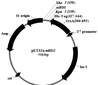
(Figure 1), mBD2 was fused with TrxA, this fusion expression
alleviates the obstacles in using E. coli as the host cell for antibiotic peptides expression, including the host-killing activity
and the susceptibility to degradation of product (14). Between the TrxA and mBD2 coding sequence, there is a His·Tag, which
serves as the detection and purification tag in later steps. This expression vector was transformed into E. coli Rosetta-gami (2)
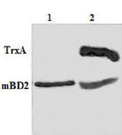
for expression fusion protein. In SDS-PAGE analysis, compared to
the negative control of Rosseta-gami(2)/pET32a, a foreign protein was expressed in Rosetta-gami(2)/pET32a-mBD2. Under the
optimization of fermentation parameters, the fusion protein TrxA-mBD2 reached 95% in the soluble fraction (Figure 2a). The
rEK-His cleaved fusion protein was further purified by rEK-His TrapTM HP
crude, and the mature mBD2 was released by the precise cleavage of TrxA-tag. The mBD2 was analyzed by Tricine-SDS-PAGE
(Figure 2b) and Western blot. Finally, the expression of soluble mature mBD2 was achieved 6 mg/L with a volumetric
productivity.
Figure 1. Schematic representation of the expression vector, pET32a-mBD2. The cDNA for mature mBD2 were amplified
using PCR from the pcDNA3.1(+)-mBD2 plasmid. PCR product
was cleaved by Xho I and Kpn I, and the mBD2 fragment was inserted into similarly digested pET32a(+) vector to construct the
Figure 2. Expression of fusion protein TrxA-mBD2 in E. coli Rosseta-gami (2)/pET32a-mBD2 cells and Tricine-SDS-PAGE analysis of enterokinase digested. The cDNA for mature mBD2 was inserted into pET32a(+) to construct the expression vector pET32a-mBD2, which was transformed into E. coli Rosseta-gami (2) for expression fusion protein. (a) Fusion protein was expressed in soluble and insoluble forms. Under the optimization of fermentation parameters: induce with 0.6 mM IPTG at 34 in the 2xYT medium and harvest at 6 h post-induction, fusion protein TrxA-mBD2 was high expressed in the soluble fraction (>95%). Lane l: soluble protein of gami (2)/pET32a-mBD2; Lane 2: insoluble protein of Rosseta-gami (2)/pET32a-mBD2; (b) The fusion protein was specifically digested with rEK-His, the mixture buffer was further purified by His TrapTM HP crude. The released mature rmBD2 protein was obtained by the effluent of loading sample of digestion mixture and further desalted and condensed by Amicon® Ultra-15 3K Centrifugal Filter Devices. Lane 1: purified mature mBD2; Lane 2: mixture of TrxA-mBD2 after rEK-His digestion.
Antibacterial assay
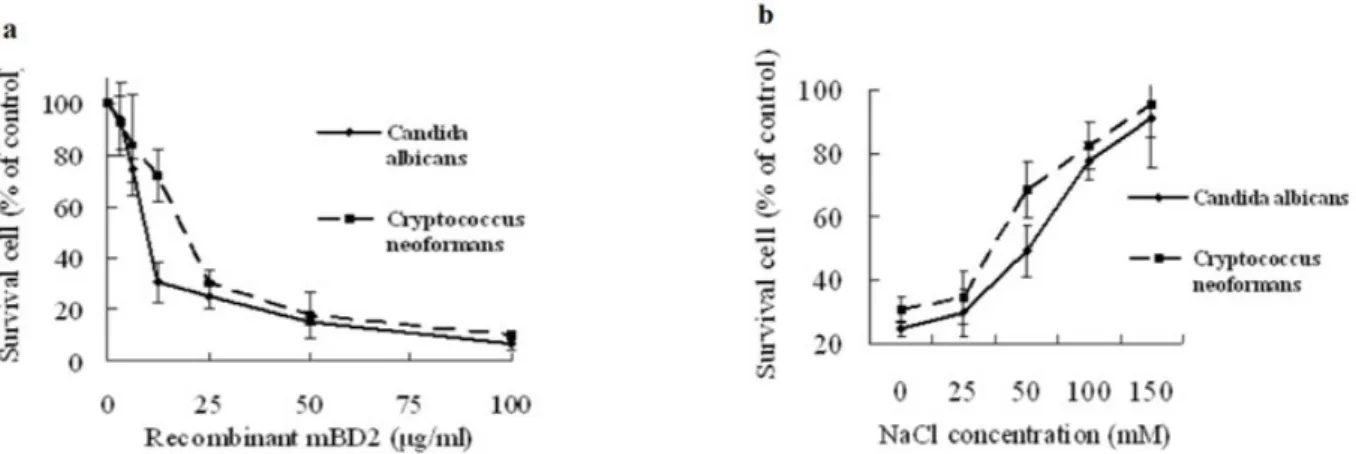
The result of broth dilution assay showed that MIC value of rmBD2 against Staphylococcus aureus was 50µg/ml, and the
concentration killing curves was shown in Figure 3a. The growth of Staphylococcus aureus was strongly suppressed with the increasing concentration of rmBD2. Thus, the rmBD2 showed antibacterial activity against Staphylococcus aureus. This result was in accordance with antibacterial assay of mBD2 in vivo (7). However, the antibacterial activity of rmBD2 against E. coli and Pseudomonas aeruginosa was not observed at the concentrations of 0.88-200 µg/ml (data not shown). In addition, the result of the effect of NaCl concentration on the antibacterial activity of mBD2 showed that mBD2 is a salt-sensitive peptide (Figure 3b). So far, most defensins and other antimicrobial peptides were described (15,16) as the salt-sensitive peptides, sodium chloride inhibited the antimicrobial activity. Tomita et al. (17) reported other salts with similar effects and a higher strength, not the specific ions are the cause of the inhibition. Currently, disruption of the initial interaction between the negatively charged membrane of microbe and the positively charged peptide is thought to be the cause of this dependency of ionic strength on activity (18).
Antifungal assays
results were shown in Figure 4a. The growth of C.albicans and C.
neoformans were significantly suppressed with the increasing concentration of rmBD2. Thus, our results suggest that mBD2
would play a role in the protection of fungal infection. However, the antimicrobial activity of fusion protein TrxA-mBD2 was not
observed at the concentrations 0.88-200 µg/ml (data not shown).
In addition, the NaCl concentration also had an effect on the antifungal activity of mBD2. And the antifungal effect was
suppressed by increasing NaCl concentration, when the
concentration was 150 mM, rmBD2 was almost totally inactive (Figure 4b). This salt-sensitive property was in accordance with
another peptides isolated from mouse (9, 16).
Figure 3. Antibacterial activity of rmBD2 using the broth dilution assay. Bacterial cells were inoculated into fresh broth and grown to mid-log
phage. The cells were washed three times and the final concentration was adjusted to 104 cfu/ml. 50 µl cells and serial dilutions of rmBD2 in 10
mM PB were added to each well of a microtiter plate, followed by incubation for 3 h at 37ºC. 100 µl of 2×M-H medium was added to each well,
and after 24 h of incubation at 37ºC, the cells growth was determined by visual inspection and by measurement of the OD600. Bacterial growth is
expressed as percentage of maximal OD observed in the absence of peptide. 0-150 mM NaCl were used in the culture with 50 µg/ml rmBD2 to
analyze the effect of NaCl concentration on the antibacterial activity. (a) Bacterial-killing curve of rmBD2 against Staphylococcus aureus. (b)
Effect of NaCl concentration on rmBD2 against Staphylococcus aureus. Data was obtained from three independent experiments performed each
in triplicate and reported as mean ± standard deviation.
Figure 4. Antifungal activity of rmBD2 using the colony counting assay. Fungal cells were grown overnight in medium YPG. The cells were
washed three times in buffer 10 mM PB and the final concentration was adjusted to 1×10⁶ cells/ml. The cell suspensions were mixed with serial
dilutions of rmBD2 in 10 mM PB and incubated for 3 h at 37ºC. Cell suspensions were diluted 200-fold in 10 mM PB, followed by spread onto
YPG agar plates and incubation at 25ºC. Control culture was incubated with 10 mM PB alone. After 48h, fungal colonies were enumerated. 0-l50
mM NaCl were used in the culture with 25 µg/ml rmBD2 to analyze the effect of NaCl concentration on the antifungal activity of against C.
Antiviral activity assay
In addition to the antibacterial and antifungal activity, antiviral activity of mBD2 was assessed against IAV. MDCK cells were incubated with serial double diluted rmBD2-treated PR-8, and the cytotoxicity was evaluated at 48 h postinfection with MTT kit. Under presented conditions, rmBD2 was effective to protect MDCK from infected with PR-8 and decreased virus-induced cell death in a dose-dependent manner (Fig.5) (12). The protective rate was 93.86% at the rmBD2 concentration of 100 µg/ml. However, the fusion protein TrxA-mBD2 had no activity of anti-influenza (data not shown). It suggested that rmBD2 has the clear antiviral activity.
In conclusion, firstly, we successfully established a high-level expression system of functional mBD2 peptide, this protocol could be applied for design and expression of other functional antimicrobial peptides. Secondly, this work firstly demonstrated that mBD2 exhibits broad-spectrum antimicrobial activity. It showed not only strongly antibacterial and antifungal activities but also antiviral activity. It may play an important role in the innate immune response against these pathogens in the airway of mice. Its pharmaceutical potential and medical importance are worth further exploring.
Figure 5. Antiviral activity of rmBD2 using the MTT assay. The viruses were preincubated with different concentrations (100 - 3.125 µg/ml) of rmBD2 for 1 h at 37ºC, followed by added the virus-peptide mixture to confluent monolayer
MDCK cells for 1 h at 37ºC. Infection mixture was discarded and cells were washed with PBS and overlaid with fresh DMEM. The cytotoxicity was measured with the MTT kit after infected 48 h. The percentage of antiviral activities of the rmBD2 was calculated as: protective rate = [(mean optical density of test mean optical density of virus controls)/(optical density of cell controls mean optical density of virus controls)] × 100%. At the concentration 100 µg/ml, rmBD2 protected 93.86% cells from infection by influenza virus PR-8 (12).
ACKNOWLEDGEMENTS
We are deeply grateful to Dr. De Yang for the gift of pcDNA3.1(+)-mBD2. This work was financially supported by National Natural Science Foundation of China (No. 30671964, People’s Republic of China).
REFERENCE
1. Bals, R.; Wang, X.; Wu, Z.; Freeman, T.M.; Bafna, V.; Zasloff, M.; Wilson, J.M. (1998). Human beta-defensin 2 is a salt-sensitive peptide antibiotic expressed in human lung. J. Clin. Invest. 102, 874-880. 2. Bals, R.; Goldman, M.J.; Wilson J.M. (1998). Mouse beta-defensin 1 is a
salt-sensitive antimicrobial peptide present in epithelia of the lung and urogenital tract. Infect Immunol. 66 (3), 1225–1232.
3. Berrouane, Y.F.; Herwaldt, L.A.; Pfaller, M.A. (1999). Trends in antifungal use and epidemiology of nosocomial yeast infections in a university hospital. J Clin Microbiol. 37 (3), 531-537.
4. Bessete, P.H.; Aslund, F.; Beckwith, J.; Georgiou, G. (1999). Efficient folding of proteins with multiple disulfide bonds in the Escherichia coli cytoplasm. Proc Nat Acad Sci. 96 (24), 13703–13708.
5. Burd, R.S.; Furrer, J.L.; Sullivan, J.; Smith, A.L. (2002). Murine Beta defensin 3 is an inducible peptide with limited tissue expression and broad–spectrum antimicrobial activity. SHOCK. 18 (5), 461–464. 6. De Smet, K.; Contreras, R. (2005). Human antimicrobial peptides:
defensins, cathelicidins, and histatins. Biotechnol Lett. 27 (18), 1337-1347.
7. Edwin J.A.; Veldhuizen, M.R.; Erwin, A.C.; van Dijk, A.; Henk, P.H. (2008). Procine -defensin 2 displays broad antimicrobial activity against pathogenic intestinal bacteria. 45, 386-394.
their mechanisms of action. Biochim Biophys Acta. 1462 (1-2), 11-28. 9. Ganz, T. (2003). Defensins: antimicrobial peptides of innate immunity.
Nat Rev Immunol. 3 (9), 710-720.
10. Gong, T.X.; Jiang, Y.; Wang, Y.L.; Yang, D.; Li, W.Y.; Zhang, Q.; Feng, W.; Wang, B.N.; Jiang, Z.H.; Li, M.Y.. (2010). Recombinant mouse beta-defensin 2 inhibits infection by influenza A virus by blocking its entry. Arch Virol. 155: 491-98.
11. Hobson, R.P. (2003). The global epidemiology of invasive Candida infections—is the tide turning? J Hosp Infect. 55 (3), 159-168.
12. Hussain, T.; Nasreen, N.; Lai Y.M.; Bellew, B.F.; Antony, V.B.; Kamal A. Mohammed, K.A. (2008). Innate immune responses in murine pleural mesothelial cells: Toll-like receptor-2 dependent induction of beta-defensin-2 by staphylococcal. Am J Physiol-lung Cell Physiol. 295 (3), 461-470.
13. Lehrer, R.I. (2004). Primate defensins. Nat Rev Microbiol. 2 (9), 727-738.
14. Morrison, G.M.; Davidson, D.J.; Dorin, J.R. (1999). A novel mouse beta defensin, Defb2, which is upregulated in the airways by lipopolysaccharide. FEBS Lett. 442 (1), 112-116.
15. Piers, K.L.; Brown, M.H.; Hancock, R.E.W. (1993). Recombinant DNA procedures for producing small antimicrobial cationic peptides in bacteria. Gene. 134, 7-13.
16. Schagger, H.; Jagow, G.V. (1987). Tricine-sodium dodecyl sulfatepolyacrylamide gel electrophoresis for the separation of proteins in the range from 1 to 100kDa, Anal Biochem. 166 (2), 368.-379. 17. Shafer W.M. (1997). Methods in Molecular Biology: Antibacterial
Peptide Protocols. Vol. 78. Totowa, NJ. Humana Press Inc. p, 35-48. 18. Tomia, T.; Hitomi, S.; Nagase, T.; Matsui, H.; Kimura, S.; Ouchi, Y.;
(2000). Effect of ions on antibacterial activity of human beta defensin 2.
Microbiol. Immunol. 44, 749-754.
19. Vylkova, S.; Li, X.S.; Berner J.C.; Edgerton, M. (2006). Distinct antifungal mechanisms: ß-Defensins require Candida albicans Ssa1 Protein, while Trk1p mediates activity of cysteine-freecationic peptides.
Antimicrob Agents Chemother. 50 (1), 324–331.
20. Yang, D.; Liu, Z.H.; Tewary, P.; Chen, Q.; de la Rosa, G.; Oppenheim, J.J. (2007). Defensin participation in innate and adaptive immunity.”
Currt Pharm Design. 13 (30), 3131-3139.