Inhibition of 11
β
-Hydroxysteroid
Dehydrogenase Type II Suppresses Lung
Carcinogenesis by Blocking Tumor COX-2
Expression as Well as the ERK and mTOR
Signaling Pathways
Jian Chang1,3☯, Min Xue1☯, Shilin Yang1, Bing Yao1, Bixiang Zhang3, Xiaoping Chen3, Ambra Pozzi1, Ming-Zhi Zhang1,2,4*
1Department of Medicine, Vanderbilt University School of Medicine, Nashville, TN, United States of America,2Department of Cancer Biology, Vanderbilt University School of Medicine, Nashville, TN, United States of America,3Hepatic Surgery Center, Tongji Hospital, Tongji Medical College, Huazhong University of Science & Technology, Wuhan, China,4Jiangsu Center for the Collaboration and Innovation of Cancer Biotherapy, Cancer Institute, Xuzhou Medical College, Xuzhou, China
☯These authors contributed equally to this work.
Abstract
Lung cancer is by far the leading cause of cancer death. Early diagnosis and prevention re-main the best approach to reduce the overall morbidity and mortality. Experimental and
clin-ical evidence have shown that cyclooxygenase-2 (COX-2) derived prostaglandin E2
(PGE2) contributes to lung tumorigenesis. COX-2 inhibitors suppress the development and progression of lung cancer. However, increased cardiovascular risks of COX-2 inhibitors limit their use in chemoprevention of lung cancers. Glucocorticoids are endogenous and
po-tent COX-2 inhibitors, and their local actions are down-regulated by 11β–hydroxysteroid
de-hydrogenase type II (11ßHSD2)-mediated metabolism. We found that 11βHSD2
expression was increased in human lung cancers and experimental lung tumors. Inhibition of 11βHSD2 activity enhanced glucocorticoid-mediated COX-2 inhibition in human lung car-cinoma cells. Furthermore, 11βHSD2 inhibition suppressed lung tumor growth and invasion in association with increased tissue active glucocorticoid levels, decreased COX-2 expres-sion, inhibition of ERK and mTOR signaling pathways, increased tumor endoplasmic reticu-lum stress as well as increased lifespan. Therefore, 11βHSD2 inhibition represents a novel approach for lung cancer chemoprevention and therapy by increasing tumor glucocorticoid activity, which in turn selectively blocks local COX-2 activity and/or inhibits the ERK and mTOR signaling pathways.
OPEN ACCESS
Citation:Chang J, Xue M, Yang S, Yao B, Zhang B, Chen X, et al. (2015) Inhibition of 11β-Hydroxysteroid Dehydrogenase Type II Suppresses Lung Carcinogenesis by Blocking Tumor COX-2 Expression as Well as the ERK and mTOR Signaling Pathways. PLoS ONE 10(5): e0127030. doi:10.1371/ journal.pone.0127030
Academic Editor:Pier Giorgio Petronini, University of Parma, ITALY
Received:September 25, 2014
Accepted:April 10, 2015
Published:May 26, 2015
Copyright:© 2015 Chang et al. This is an open access article distributed under the terms of the Creative Commons Attribution License, which permits unrestricted use, distribution, and reproduction in any medium, provided the original author and source are credited.
Data Availability Statement:All relevant data are within the paper files.
Funding:National Institutes of Health CA-122620, DK-95785, DK-51265 to MZ; CA-162433 to AP.
Introduction
Lung cancer is the second most common cancer in both men and women and is by far the lead-ing cause of cancer death among both men and women. The American Cancer Society estimates that about 230 000 new cases of lung cancer will be diagnosed along with approximate 160 000 deaths from lung cancer, accounting for about 27% of all cancer deaths in 2013 in the United States. Most patients present with advanced, non-curable disease. There are only 15% of patients still alive 5 years after diagnosis [1,2]. Therefore, early diagnosis and prevention remain the best approach to reduce the overall morbidity and mortality of lung cancer. There are two major types of lung cancer: small cell lung cancer (SCLC) and non-small cell lung cancer (NSCLC). NSCLC accounts for 85%-90% of lung cancers and contains three main subtypes: squamous cell (epidermoid) carcinoma, adenocarcinoma and large cell (undifferentiated) carcinoma.
Although the etiology of lung cancer is undoubtedly multifactorial, there is experimental and clinical evidence linking abnormalities in the cyclooxygenase/prostaglandin system to its pathogenesis. Cyclooxygenase (prostaglandin synthase G2/H2, COX) is the rate-limiting en-zyme in the metabolism of arachidonic acid to prostaglandin G2and subsequently to prosta-glandin H2(PGH2), which serves as the precursor for prostaglandin E synthetase to produce prostaglandins [3]. Two isoforms of cyclooxygenase exist in mammals,“constitutive”COX-1 and inflammatory-mediated and glucocorticoid-sensitive COX-2. COX-2 derived PGE2has been reported to promote tumor growth and metastasis through stimulation of cell prolifera-tion, cell migraprolifera-tion, cell invasion, angiogenesis and immunosuppression [4].
An increase in COX-2 expression has been associated with the development of different human NSCLC and possibly with acquisition of an invasive and metastatic phenotype, as well as with poor prognosis [5–7]. Notably, a single nucleotide polymorphism in the COX-2 promot-er region, a change of -1195 G to A (-1195 G/A SNP) that leads to increases in enzymatic activi-ty, is associated with poor survival and poor progression-free survival in unresectable locally advanced NSCLC [8]. In a randomized, double-blind, placebo-controlled trial, the selective COX-2 inhibitor celecoxib was found to be a potential chemoprevention agent in former-smok-ers [9]. COX-2 inhibitors have been reported as radiosensitizers for NSCLC patients [10]. How-ever, long-term use of selective COX-2 inhibitors has been found to be associated with an increased incidence of cardiovascular events, thought to be due to inhibition of endothelial cell-derived COX-2 activity, with selective inhibition of COX-2 cell-derived PGI2production but without inhibition of COX-1 mediated prothrombotic platelet thromboxane A2production [11–13].
COX-2 was initially described as an inflammatory-mediated and glucocorticoid-sensitive cyclooxygenase. Glucocorticoids (GCs) are the most potent, endogenous, specific COX-2 in-hibitors, acting to suppress COX-2 expression through stimulating glucocorticoid receptors [14–16]. In addition to inhibiting COX-2 expression, GCs also reduce prostaglandin produc-tion through inhibiproduc-tion of cytosolic phospholipase A2activity, which prevents the release of ar-achidonic acid from membrane phospholipids, and through inhibition of microsomal
prostaglandin E synthetase (mPGES-1) expression, a major terminal synthetase in PGE2 bio-synthesis [17,18]. In addition to their application in the treatment of hematologic malignancies, GCs inhibit solid tumor growth, regress tumor mass, and prevent metastasis by blocking angio-genesis [19,20]. However, the undesirable side effects of immune suppression limit their appli-cation in cancer chemoprevention and chemotherapy.
11ßHSD2 activity could suppress lung tumorigenesis due to increased tumor cell intracellular active glucocorticoids and subsequent inhibition of COX-2 expression/activity.
Materials and Methods
Ethics Statement
All animal experiments were performed according to animal care guidelines and were ap-proved by the Vanderbilt Institutional Animal Care and Use Committe (IACUC) (M/11/130).
Animals
KrasLA2 mice were a gift of Dr. T. Jacks, MIT [24]. Since theKrasLAallele is non-functional in the germline configuration, only heterozygous mice were maintained and used in the experi-ments. Age- and sex-matched KrasLA2 mice were treated with water (control) or the 11βHSD2 inhibitor, glyccyrrhetinic acid (GA, 10 mg/kg/day, i.p.) from 6 to 20 weeks of age and sacrificed at 20 weeks of age. Under anesthesia with Nembutal (60 mg/kg i.p.), the lungs were weighed, filled with fixative overnight, transferred to 70% ethanol for 24 h, and examined using a dissect-ing microscope to count surface polyps in a blinded fashion [22]. The tumor diameter on the surface of the lung was measured with a digital caliper. Tumor volume was expressed in mm3 and calculated as [l x w2] × 0.5, wherelandwdenote length and width, respectively [22]. After tumors were counted, lung tissues were processed for paraffin embedding and immunohisto-chemical analysis. A subset of mice was sacrificed for collection of adenomas and lung tissue for immunoblotting and determination of tissue levels of corticosterone and 11-keto-corticoste-rone. All animals were genotyped twice (after weaning and before sacrifice).
Survival analysis
KrasLA2 mice were treated with water or 11βHSD2 inhibitor, GA at 6 weeks of age. The ani-mals were monitored daily. The animal was sacrificed at the first sign of shortness of breath, re-duced locomotion and rere-duced body weight (greater than 20% total body weight). Lungs from these mice were analyzed for the presence of visible tumors on the lung surface. Only mice with visible lung tumors were used for the survival analysis. The animals were euthanized by overdose carbon dioxide.
Human lung cancer tissues
Slides of lung cancer tissue array with normal lung tissue were purchased from US Biomax (Rockville, MD).
Cell culture
according to the manufacturer’s protocol (BrdU cell proliferation assay kit, Cat#6813, Cell Sig-naling) and our recent report [26].
Primary tumor growth
For mouse LLC tumor experiments, male C57/B6 mice (8–12 weeks old) were given 2 dorsal s. c. injections (5 x 10⁵cells in 100μl PBS) with administration of water (control, i.p) or GA (10 mg/kg/day, i.p.) begun 1 day prior to cell injection. Tumors were harvested and weighed after 18 days of growth. In another set of experiment, LLC cell suspension containing 5 x 10⁵cells in 100μl PBS were injected into tail vein with or without GA administration as described above. The time of death, or when mice became moribund (by the same criteria as the endpoints de-scribed for survival studies), was recorded for survival analysis.
Measurement of corticosterone and 11-keto-corticosterone
Lung tissues from KrasLA2 mice were collected and stored at-80°C. Tissue levels of corticoste-rone (active form in rodent) and 11-keto-corticostecorticoste-rone (inactive form) were measured using high-performance liquid chromatography coupled with electrospray tandem mass spectrometry [22]. Corticosterone was from ICN Biomedicals, and 11-keto-corticosterone was from Steraloids.
Antibodies
Affinity-purified rabbit anti-11βHSD2 (catalog no. BHSD22-A) was purchased from Alpha Di-agnostic International; rabbit anti-murine COX-2 (catalog no. 160106) was from Cayman Chemicals; rabbit anti-CHOP (catalog no. 2895), rabbit anti-cleaved caspase-3 (catalog no. 9661), rabbit anti-p-ERK (T202/Tyr204, catalog no. 4370), p-mTOR (Ser2448, catalog no 2976), p-PERK (Thr980, Catalog no 3179) and p-eIF2α(Ser51, catalog no 9721) were from Cell Signaling; rabbit anti-cyclin D1 (catalog no. SC-753) was from Santa Cruz Biotechnology; mouse anti-mannose receptor (MR, CD206, catalog no.MAB25341) was from R&D; rabbit anti-Ki67 (ab15580) was from Abcam.
Immunohistochemical staining and immunoblotting
Immunostaining was carried out as in previous reports [27]. For immunostaining of phosphor-ylated proteins, antigen retrieval was achieved by boiling in citric acid buffer (100 mM, pH 6.0) for 3 x 5 min. Lung tumors isolated from 20 weeks old KrasLA2 mice were used for immuno-blotting as described previously [28].
Quantitative image analysis
Immunostaining was quantified by using the BIOQUANT image analysis system (R & M Bio-metrics, Nashville, TN) [22]. Bright-field images from a Leitz Orthoplan microscope with DVC digital RGB video camera were digitized and saved as computer files. Contrast and color level adjustment (Adobe Photoshop) were performed for the entire image; i.e., no region- or object-specific editing or enhancements were performed.
Statistical analysis
Results
To investigate whether 11ßHSD2 might be involved in lung tumorigenesis, its expression in normal mouse lung tissue, experimental lung cancer, normal human lung tissue and different human lung cancers was investigated with immunohistochemistry. In athymic nude mouse lung tissue, 11ßHSD2 was expressed in the epithelial cells of small airways and alveoli (Fig 1A). 11ßHSD2 expression was increased in A549 adenocarcinoma compared to adjacent normal athymic nude mouse lung tissue (Fig 1B). 11ßHSD2 immunostaining was strong in human adenocarcinoma and squamous cell carcinoma, moderate in papillary carcinoma as well as small cell lung cancer, but very weak in normal human lung tissue (Fig 1C). Negative control was performed by replacing the primary antibody with purified rabbit IgG and no
Fig 1. 11ßHSD2 expression in mouse and human lung tumors. AandB. Representative photomicrographs indicated 11ßHSD2 expression in small airway and alveolar epithelial cells of athymic nude mice(A)and in A549 lung tumor from athymic nude mice(B).C. Representative photomicrographs showed that 11ßHSD2 immunostaining was strong in lung adenocarcinoma and squamous cell carcinoma, moderate in papillary carcinoma and small cell lung cancer (SCLC), but very weak in uninvolved lung tissue. Original magnification: x 160 in all.
immunoreactivity was found (data not shown). Therefore, 11ßHSD2 expression is increased in both experimental lung cancers and human lung cancers.
To investigate whether inhibition of 11ßHSD2 activity was beneficial in lung tumorigenesis through inhibition of COX-2 expression due to increased intracellular active glucocorticoids, we screened lung cancer cell lines for 11ßHSD2 and COX-2 expression. As indicated inFig 2A, 11ßHSD2 was expressed in all investigated lung cancer cell lines; while COX-2 was only de-tected in LLC, H1435, A549 and H1395 cells. LLC cell line, which is highly tumorigenic in mice and expresses both COX-2 and 11ßHSD2, was chosen to investigate the role of 11ßHSD2 inhibition in COX-2 expression. As indicated inFig 2B, corticosterone (CS, glucocorticoid in rodent) inhibited LLC cell COX-2 expression in a dose-dependent pattern. However, even 1μM CS could not completely inhibited COX-2 expression. One possible reason for this partial inhibition is that intracellular CS is quickly inactivated by 11ßHSD2. It is also possible that some COX-2 is insensitive to glucocorticoid inhibition. CS-induced LLC cell COX-2 inhibition was markedly enhanced in the presence of GA, an inhibitor of 11ßHSD2 activity (Fig 2C). In
Fig 2. 11βHSD2 inhibition augmented corticosterone (CS)-induced COX-2 inhibition in lung cancer cells. A. Immunoblotting indicated that 11βHSD2 was expressed in all investigated cell lines while COX-2 was detectable in some of them, including LLC, H1435 and A549 cells.B. Immunoblotting indicated that CS inhibited LLC cell COX-2 expression in a dose-dependent manner.C. CS (1μM)-induced LLC cell COX-2 inhibition was enhanced by 11βHSD2 inhibitor, GA.D. CS (1μM)-induced LLC cell COX-2 inhibition was enhanced by carbenoxolone (CBX, 10μM), another 11βHSD2 inhibitor.E. CS (1μ M)-induced H1435 cell COX-2 inhibition was enhanced by GA (10μM).F. CS (1μM)-induced A549 cell COX-2 inhibition was enhanced by GA (10μM).
addition, CS-induced LLC COX-2 inhibition was also enhanced by carbenoxolone (CBX, 10μM), another inhibitor of 11ßHSD2 activity (Fig 2D) [29]. Furthermore, CS-induced COX-2 inhibition was also enhanced by GA treatment in both H1435 and A549 cells, which also ex-press both 11ßHSD2 and COX-2 (Fig2Eand2F).
To determine the effect of 11ßHSD2 inhibition on CS-mediated inhibition of mTOR and ERK signaling pathways, both LLC cells and A549 cells were treated with 1μM CS with or without 10μM GA for 24 hours and p-mTOR and p-ERK levels were determined with immunoblotting. As indicated inFig 3A, CS treatment resulted in decreases in both p-mTOR and p-ERK levels in LLC cells, which were enhanced by 11ßHSD2 inhibition with GA. Similarly, CS-mediated inhibi-tion of p-mTOR and p-ERK was also augmented by GA treatment in A549 cells (Fig 3B).
To investigate the direct effects of CS on lung cancer cell proliferation, we treated LLC cells (mouse) and A549 cells (human) with different dose of CS and cell proliferation was measured by using cell proliferation assay kit. As shown inFig 3C, CS inhibited LLC cell proliferation in a dose-dependent pattern. In addition, CS-mediated inhibition was significantly enhanced by addition of GA. Similarly, CS-mediated inhibition of A549 cell proliferation was also augment-ed by GA treatment in A549 cells (Fig 3D).
Fig 3. CS-induced inhibition of mTOR and ERK signaling pathways and cell proliferation was enhanced by 11βHSD2 inhibition with GA in lung cancer cell lines. (A and B). CS treatment led to decreased expression levels of p-mTOR and p-ERK in LLC cells(A)and A549 cells(B), which were augmented by GA treatment.(C and D). CS treatment led to inhibition of LLC(C)and A549(D)cell proliferation in a dose-dependent manner, which was enhanced by GA treatment.***P<0.001, n = 3.
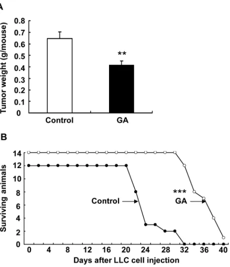
To determine whether 11ßHSD2 inhibition affected lung tumorigenesis, we examined LLC tumor growth in a Xenograft model. LLC cells were injected subcutaneously into C57BL/6 mice. GA treatment (10 mg/kg/day, i.p.) was initiated one day before LLC cell injection and tu-mors were harvested and weighted at the end of the experiment (18 days after injection). As shown inFig 4A, LLC tumor growth was markedly attenuated by GA treatment (tumor weight: 414 ± 38 vs. 646 ± 58 mg of vehicle, P<0.01, n = 8 in each group). Similarly, GA treatment
in-hibited tumor invasion to the lung in a LLC tail vein injection model. As indicated inFig 4B, GA treatment led to marked increase in survival probability (average survival days: 34.8 ± 3.3 vs. 24.5 ± 3.5 of control, P<0.001, n = 12 in vehicle group and n = 13 in GA group).
KrasLA2 mice carry an oncogenic mutation of theKrasgene (G12D) and develop spontane-ous primary tumors with features of NSCLC [24]. To further investigate the role of 11ßHSD2 inhibition in lung tumorigenesis, heterozygous KrasLA2 mice were treated with water (control) or GA (10 mg/kg/day, i.p.) from 6 weeks of age. At 20 weeks of age, a subset of mice was Fig 4. 11βHSD2 inhibition suppressed lung tumorigenesis. A. LLC tumor growth was significantly attenuated by 11βHSD2 inhibition with GA (**P<0.01, n = 8 in each group). LLC cell suspensions (100μl, 5 x 10⁵cells) were injected subcutaneously into the flank of C57/B6 mouse (2 sites). The mice were sacrificed
18 days later and tumor growth (tumor weight from two sites) was evaluated.B. Kaplan-Meier survival curve indicated that 11βHSD2 inhibition with GA increased survival probability in mice with tail vein injections of LLC cells (100μl of LLC cell suspensions containing 5 x 10⁵cells).
***P<0.001, n = 12 in control and n = 14 in GA group. In both models, GA was given at 10 mg/kg/day (i.p. injection) starting one day before LLC cell injections.
sacrificed, lung tissue kept for measurement of glucocorticoid levels and immunoblotting. An-other subset of mice was sacrificed and the lungs were filled with fixative to fix the tissue for analysis of tumor number, size and immunostaining. Lung tissue levels of glucocorticoids were measured by using high-performance liquid chromatography coupled with electrospray tan-dem mass spectrometry [22]. GA treatment led to marked increases in lung corticosterone lev-els (active glucocorticoid in rodent: 37.40 ± 12.50 vs. 5.06 ± 2.19 ng/g lung tissue of control, P
<0.05, n = 6 in each group), and led to marked decreases in 11-keto-corticosterone (inactive
glucocorticoid in rodent: 1.05 ± 0.08 vs. 2.63 ± 0.64 ng/g lung tissue of control, P<0.05) (Fig
5A). As expected, COX-2 levels were markedly reduced in lung tumors from GA treated Kra-sLA2 mice (26 ± 5% of control, P<0.001, n = 3 in each group) (Fig 5B). Immunostaining
con-firmed the reduction of tumor COX-2 expression in GA treatment mice. Glucocorticoids have been reported to induce mannose receptor (MR) expression in macrophages [30]. Therefore, we investigated MR expression in the tumor. As indicated inFig 5C, the number of tumor MR-Fig 5. 11βHSD2 inhibition with GA increased lung corticosterone levels in association with attenuation of tumor COX-2 expression in KrasLA2 mice. A. GA treatment markedly increased lung active corticosterone levels and decreased inactive 11-keto-corticosterone levels in KrasLA2 mice (20 weeks of age).*P<0.05 vs. control, n = 6.B. Lung tumor COX-2 expression was suppressed by 11ßHSD2 inhibition with GA.**P<0.01 vs. control, n = 3. Immunostaining showed markedly decreased tumor COX-2 with GA treatment. Original magnification: x 250.C. GA treatment increased mannose receptor (MR, CD206) expressing macrophages in tumor, an indication of increased tumor levels of active corticosterone.***P<0.001 vs. control, n = 4. Original
magnification: x 160.
expressing cells was markedly higher in GA treated tumors than in control tumors (16.77 ± 3.58 vs. 3.64 ± 0.96 cells/pf, P<0.001, n = 4 in each group).
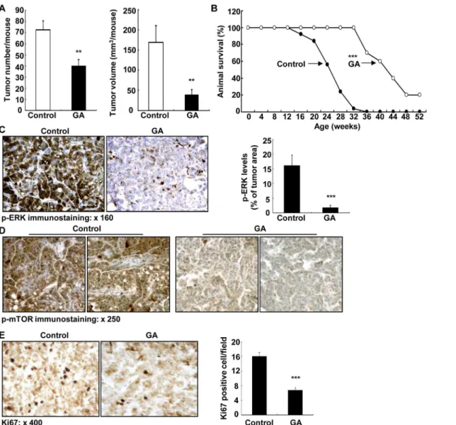
GA treatment caused significant inhibition of tumorigenesis in KrasLA2 mice, including de-crease in adenoma number on the lung surface (40.0 ± 5.8 vs. 72.7 ± 7.8 of control, P<0.01,
n = 9 in each group) and decrease in adenoma size (mm3/mouse: 40.7 ± 10.7 vs. 168.8 ± 41 of vehicle, P<0.01) (Fig 6A). GA treatment markedly increased lifespan of KrasLA2 mice
(weeks: 42.1 ± 2.6 vs. 24.1 ± 1.0 of control, P<0.01, n = 10 in GA group and n = 25 in control
group) (Fig 6B). Therefore, GA treatment inhibited lung tumorigenesis in KrasLA2 mice and increased KrasLA2 mouse lifespan.
Glucocorticoids have been reported to inhibit lung cancer cell growth through inhibition of ERK activity [31,32]. Therefore, we investigated tumor ERK activity with immunostaining of Fig 6. 11βHSD2 inhibition suppressed lung tumorigenesis in association with suppression of tumor ERK and mTOR activities in KrasLA2 mice. A. GA treatment led to decreases in both number and size of lesion in lung surface of KrasLA2 mice (20 weeks of age). n = 9,**P<0.01.B. Kaplan-Meier survival curve indicated that 11βHSD2 inhibition with GA increased survival probability of KrasLA2 mice.***P<0.001, n = 25 in vehicle group and n = 10 in GA group.C: GA treatment inhibited lung tumor levels of p-ERK.***P<0.001 vs. control, n = 4. Original magnification: x 160.D. GA treatment inhibited tumor p-mTOR expression. Original magnification: x 250.E. GA treatment inhibited tumor proliferation as indicated by decreased Ki67 positive cells in tumors.***P<0.001 vs. control, n = 4. Original magnification: x 400.
phosphorylated ERK. As indicated inFig 6C, the levels of tumor phosphorylated ERK were marked reduced by GA treatment (ratio of p-ERK area/tumor area: 1.79 ± 0.79 vs. 16.11 ± 3.61 of control, P<0.001, n = 4 in each group). GA treatment also inhibited tumor mammalian
tar-get of rapamycin (mTOR) signaling pathway (Fig 6D). Finally, tumor cell proliferation was in-hibited by GA treatment as indicated by decreased tumor cells with Ki67 positivity, a marker of cell proliferation (Fig 6E).
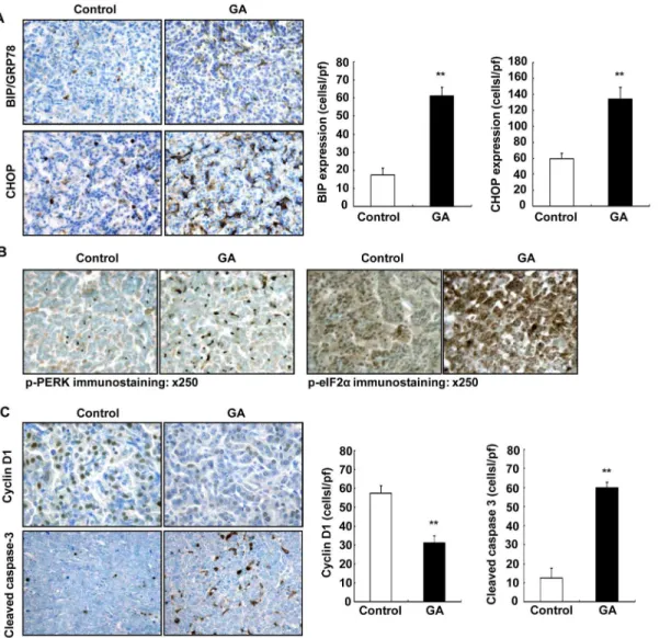
Recently, it was recognized that induction of endoplasmic reticulum stress (ER stress) and ac-tivation of the unfolded protein response (UPR) is an important pathway for antitumor agents to induce cancer cell death [33–35]. ER-stress has been reported to contribute to apoptosis induced by COX-2 inhibition in lung cancer cells as well as other cancer cells [36,37]. Furthermore, ER stress-mediated apoptosis requires eIF2αand PERK. Upon activation, PERK phosphorylates eIF2αto reduce global mRNA translation [38]. Therefore, we investigated the potential role of ER stress in 11ßHSD2 inhibition-induced suppression of lung tumorigenesis. C/EBP homolo-gous protein (CHOP) plays an important role in ER stress induced apoptosis [36,37]. 11ßHSD2 inhibition with GA led to marked increases in BIP/GPR78 (a marker of ER stress) and CHOP ex-pression in tumors from KrasLA2 mice (P<0.01, n = 4) (Fig 7A). In addition, 11ßHSD2
inhi-bition with GA led to increased phosphorylation of both PERK and eIF2α(Fig 7B).
To determine whether suppression of lung tumorigenesis by 11ßHSD2 inhibition was relat-ed to decreasrelat-ed cell proliferation and/or increasrelat-ed apoptosis, the expression levels of cyclin D1 (marker of proliferation) and cleaved caspase-3 (a specific marker of apoptosis) were investi-gated in KrasLA2 mouse tumors. GA treatment caused significant decreases in both cyclin D1 immunostaining density and the number of cyclin D1 positive cells in KrasLA2 lung tumors (cells/hpf: 31.1 ± 3.8 vs. 57.4 ± 4.0 of control, P<0.01, n = 4). In contrast, the number of
tumor cells that were positive for cleaved caspase-3 was significantly increased in GA-treated KrasLA2 lung tumors (cells/hpf: 59.8 ± 2.9 vs. 12.5 ± 5.2 of control, P<0.01, n = 4) (Fig 7C).
Therefore, inhibition of 11ßHSD2 activity with GA led to decreased cell proliferation and in-creased apoptosis in lung tumors.
Discussion
The major findings in the present studies include: 1) 11ßHSD2 is expressed in epithelial cells of small airways and alveoli of mouse lung and its expression is increased in experimental lung tu-mors and in human SCLC and NSCLC, 2) glucocorticoid-induced COX-2 inhibition in lung cancer cells is enhanced by 11ßHSD2 inhibition, 3) inhibition of 11ßHSD2 activity with GA re-duces LLC tumor growth and invasion, and 4) 11ßHSD2 inhibition with GA suppresses lung tumorigenesis and increases lifespan in KrasLA2 mice in association with increases in lung ac-tive corticosterone levels, decreases in tumor COX-2 expression, inhibition of the tumor ERK and mTOR signaling pathways and increased ER stress.
GCs are known to inhibit cell proliferation and induce cell differentiation through activation of glucocorticoid receptors. 11ßHSD2 has been thought to be pro-proliferative due to its ability to inactivate glucocorticoids [47–50]. We have recently reported that 11ßHSD2 expression is increased in epithelial cells and stromal cells in human colonic andApc+/minmouse intestinal adenomas and is correlated with increased COX-2 expression and activity, and that inhibition of 11ßHSD2 activity genetically or pharmacologically suppresses tumor COX-2 pathway and prevents adenoma formation, tumor growth, and metastasis as a result of increased tumor in-tracellular active glucocorticoids [22,51]. Sustained activation of the ERK signaling is impor-tant for lung cancer cell survival and proliferation [52,53]. GCs have been reported to inhibit lung cancer cell growth through inhibition of the ERK signaling pathway. Indeed, GA treat-ment led to inhibition of the tumor ERK signaling pathway in KrasLA2 mice (Fig 5C). Fig 7. 11ßHSD2 inhibition increased tumor endoplasmic reticulum (ER) stress in association with decreased cell proliferation and increased apoptosis in KrasLA2 mice. A. 11ßHSD2 inhibition led to increased expression of tumor BIP/GPR78 and CHOP, markers of ER stress. (**P<0.01 vs.
control, n = 4). Original magnification: x 160.B. Immunohistochemical staining indicated that tumor phosphorylated PERK and phosphorylated eIF2αlevels were increased in mice with GA treatment. Original magnification: x 250.C. Immunostaining demonstrated that cyclin D1 (a marker of proliferation) was primarily localized to tumor cell nuclei, and its expression was markedly decreased in GA treated KrasLA2 mice (**P<0.01 vs. control, n = 4). GA treatment
increased tumor cell apoptosis as indicated by increase in cleaved-caspase-3 positive cells, a specific marker of apoptosis in KrasLA2 mouse lung tumors (**P<0.01 vs. control, n = 4). Original magnification: x 250.
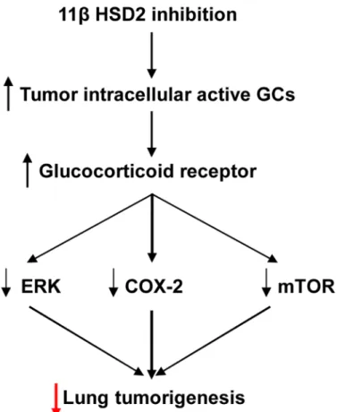
Oncogenic K-Ras regulates proliferation and cell functions in lung epithelial cells through induction of cyclooxygenase-2 and activation of metalloproteinase-9 [54]. In the current study, inhibition of 11ßHSD2 activity led to inhibition of lung tumorigenesis in KrasLA2 mice associ-ated with COX-2 inhibition and increased ER stress as well as inhibition of the ERK and mTOR signaling pathways due to increased tumor cell active glucocorticoids (Fig 8). Therefore inhibition of 11ßHSD2 activity with glycyrrhizic acid and its analogs may represent a novel ap-proach for lung cancer chemoprevention, particularly in long-term heavy cigarette smokers, with the following advantages [22]: 1) GA, a natural compound contained in licorice, is a non-toxic, inexpensive and powerful 11ßHSD2 inhibitor; 2) Physiologic 11ßHSD2 expression is largely restricted to colon and kidney and lung. Therefore, inhibition of 11ßHSD2 activity is not expected to incur the cardiovascular risk posed by 2 inhibitors that suppress COX-2-derived PGI2production in vascular endothelial cells; 3) Intracellular active GCs are only in-creased in tissues with elevated 11ßHSD2 expression. 11ßHSD2 inhibition will not produce immunosuppression or other systemic side effects of conventional glucocorticoid therapy; 4) In addition to inhibiting the COX-2 pathway, increased tumor active GCs also inhibit lung tu-morigenesis through inhibiting the ERK and mTOR signaling pathways as well as induction of G1 cell cycle arrest [51]. Although 11ßHSD2 inhibition may result in salt-sensitive hyperten-sion due to activation of mineralocorticoid receptors by GCs, development of locally acting 11ßHSD2 inhibitors that are not systemically absorbed would be a potential therapeutic means Fig 8. Proposed mechanism underlying 11ßHSD2 activity and lung tumorigenesis.11ßHSD2 inhibition leads to increased levels of tumor intracellular active glucocorticoid and activation of glucocorticoid receptors. The subsequent inhibition of the COX-2, ERK and mTOR pathways leads to suppression of
lung tumorigenesis.
to prevent lung tumorigenesis [55]. In the future, it is worthy investigating whether MEK in-hibitor or rapamycin can enhance GA-mediated anti-proliferative effect.
Acknowledgments
We would like to thank Dr. Raymond C. Harris for his comments about this manuscript.
Author Contributions
Conceived and designed the experiments: MZ. Performed the experiments: JC MX SY BY AP. Analyzed the data: MZ BZ XC AP. Contributed reagents/materials/analysis tools: AP. Wrote the paper: MZ AP BZ XC.
References
1. Houghton AM (2013) Mechanistic links between COPD and lung cancer. Nat Rev Cancer. 13: 233–
245. doi:10.1038/nrc3477PMID:23467302
2. Kwon MC, Berns A (2013) Mouse models for lung cancer. Mol Oncol 7: 165–177. doi:10.1016/j.
molonc.2013.02.010PMID:23481268
3. Harris RC, Zhang MZ (2011) Cyclooxygenase metabolites in the kidney. Compr Physiol. 1: 1729–
1758. doi:10.1002/cphy.c100077PMID:23733687
4. Wang D, Dubois RN (2010) Eicosanoids and cancer. Nat Rev Cancer. 10: 181–193. doi:10.1038/
nrc2809PMID:20168319
5. Hida T, Yatabe Y, Achiwa H, Muramatsu H, Kozaki K, Nakamura S, et al. (1998) Increased expression of cyclooxygenase 2 occurs frequently in human lung cancers, specifically in adenocarcinomas. Can-cer Res. 58: 3761–3764. PMID:9731479
6. Ho MY, Liang SM, Hung SW, Liang CM. (2013) MIG-7 controls COX-2/PGE2-mediated lung cancer metastasis. Cancer Res. 73: 439–449. doi:10.1158/0008-5472.CAN-12-2220PMID:23149922
7. Khuri FR, Wu H, Lee JJ, Kemp BL, Lotan R, Lippman SM, et al. (2001) Cyclooxygenase-2 overexpres-sion is a marker of poor prognosis in stage I non-small cell lung cancer. Clin Cancer Res. 7: 861–867.
PMID:11309334
8. Bi N, Yang M, Zhang L, Chen X, Ji W, Ou G, et al. (2010) Cyclooxygenase-2 genetic variants are asso-ciated with survival in unresectable locally advanced non-small cell lung cancer. Clin Cancer Res. 16: 2383–2390. doi:10.1158/1078-0432.CCR-09-2793PMID:20332326
9. Mao JT, Roth MD, Fishbein MC, Aberle DR, Zhang ZF, Rao JY, et al. (2011) Lung cancer chemopre-vention with celecoxib in former smokers. Cancer Prev Res (Phila). 4: 984–993. doi:
10.1158/1940-6207.CAPR-11-0078PMID:21733822
10. Komaki R, Liao Z, Milas L (2004) Improvement strategies for molecular targeting: Cyclooxygenase-2 in-hibitors as radiosensitizers for non-small cell lung cancer. Semin Oncol. 31: 47–53. PMID:15726523
11. Bresalier RS, Sandler RS, Quan H, Bolognese JA, Oxenius B, Horgan K, et al. (2005) Cardiovascular events associated with rofecoxib in a colorectal adenoma chemoprevention trial. N Engl J Med. 352: 1092–1102. PMID:15713943
12. Solomon SD, McMurray JJ, Pfeffer MA, Wittes J, Fowler R, Finn P, et al. (2005) Cardiovascular risk as-sociated with celecoxib in a clinical trial for colorectal adenoma prevention. N Engl J Med. 352: 1071–
1080. PMID:15713944
13. Cheng Y, Austin SC, Rocca B, Koller BH, Coffman TM, Grosser T, et al. (2002) Role of prostacyclin in the cardiovascular response to thromboxane A2. Science. 296: 539–541. PMID:11964481
14. Clark AR, Lasa M. (2003) Crosstalk between glucocorticoids and mitogen-activated protein kinase sig-nalling pathways. Curr Opin Pharmacol. 3: 404–411. PMID:12901950
15. Newton R (2000) Molecular mechanisms of glucocorticoid action: what is important? Thorax. 55: 603–
613. PMID:10856322
16. Zhang MZ, Harris RC, McKanna JA (1999) Regulation of cyclooxygenase-2 (COX-2) in rat renal cortex by adrenal glucocorticoids and mineralocorticoids. Proc Natl Acad Sci U S A. 96: 15280–15285. PMID:
10611376
18. Stichtenoth DO, Thoren S, Bian H, Peters-Golden M, Jakobsson PJ, Crofford LJ (2001) Microsomal prostaglandin E synthase is regulated by proinflammatory cytokines and glucocorticoids in primary rheumatoid synovial cells. J Immunol. 167: 469–474. PMID:11418684
19. Denis MG, Chadeneau C, Blanchardie P, Lustenberger P (1992) Biological effects of glucocorticoid hormones on two rat colon adenocarcinoma cell lines. J Steroid Biochem Mol Biol. 41: 739–745.
PMID:1562548
20. Schiffelers RM, Metselaar JM, Fens MH, Janssen AP, Molema G, Storm G (2005) Liposome-encapsu-lated prednisolone phosphate inhibits growth of established tumors in mice. Neoplasia. 7: 118–127.
PMID:15802017
21. Funder JW, Pearce PT, Smith R, Smith AI (1988) Mineralocorticoid action: target tissue specificity is en-zyme, not receptor, mediated. Science. 242: 583–585. PMID:2845584
22. Zhang MZ, Xu J, Yao B, Yin H, Cai Q, Shrubsole MJ, et al. (2009) Inhibition of 11beta-hydroxysteroid dehydrogenase type II selectively blocks the tumor COX-2 pathway and suppresses colon carcinogen-esis in mice and humans. J Clin Invest. 119: 876–885. doi:10.1172/JCI37398PMID:19307727
23. Yao B, Harris RC, Zhang MZ. (2005) Interactions between 11beta-hydroxysteroid dehydrogenase and COX-2 in kidney. Am J Physiol Regul Integr Comp Physiol. 288: R1767–1773. PMID:15718388
24. Johnson L, Mercer K, Greenbaum D, Bronson RT, Crowley D, Tuveson FA, et al. (2001) Somatic acti-vation of the K-ras oncogene causes early onset lung cancer in mice. Nature. 410: 1111–1116. PMID:
11323676
25. Yang L, Yamagata N, Yadav R, Brandon S, Courtney RL, Morrow JD, et al. (2003) Cancer-associated immunodeficiency and dendritic cell abnormalities mediated by the prostaglandin EP2 receptor. J Clin Invest. 111: 727–735. PMID:12618527
26. Chang J, Jiang L, Wang Y, Yao B, Yang S, Zhang B, et al. (2015) 12/15 lipoxygenase regulation of colo-rectal tumorigenesis is determined by the relative tumor levels of its metabolite 12-HETE and 13-HODE in animal models. Oncotarget. 6: 2879–2888. PMID:25576922
27. Zhang MZ, Yao B, Cheng HF, Wang SW, Inagami T, Harris RCl (2006) Renal cortical cyclooxygenase 2 expression is differentially regulated by angiotensin II AT(1) and AT(2) receptors. Proc Natl Acad Sci U S A. 103: 16045–16050. PMID:17043228
28. Harris RC, Zhang MZ, Cheng HF (2004) Cyclooxygenase-2 and the renal renin-angiotensin system. Acta Physiol Scand. 181: 543–547. PMID:15283769
29. Sandeep TC, Yau JL, MacLullich AM, Noble J, Deary IJ, Walker BR, et al. (2004) 11Beta-hydroxyster-oid dehydrogenase inhibition improves cognitive function in healthy elderly men and type 2 diabetics. Proc Natl Acad Sci U S A. 101: 6734–6739. PMID:15071189
30. Schmieder A, Schledzewski K, Michel J, Schonhaar K, Morias Y, Bosschaets T, et al. (2012) The CD20 homolog Ms4a8a integrates pro- and anti-inflammatory signals in novel M2-like macrophages and is expressed in parasite infection. European Journal of Immunology. 42: 2971–2982. doi:10.1002/eji.
201142331PMID:22806454
31. Morita M, Suyama H, Igishi T, Shigeoka Y, Kodani M, Hashimoto K, et al. (2007) Dexamethasone inhib-its paclitaxel-induced cytotoxic activity through retinoblastoma protein dephosphorylation in non-small cell lung cancer cells. Int J Oncol. 30: 187–192. PMID:17143528
32. Greenberg AK, Hu J, Basu S, Hay J, Reibman J, Yie TA, et al. (2002) Glucocorticoids inhibit lung can-cer cell growth through both the extracellular signal-related kinase pathway and cell cycle regulators. Am J Respir Cell Mol Biol. 27: 320–328. PMID:12204894
33. Jin HR, Zhao J, Zhang Z, Liao Y, Wang CZ, Huang WH, et al. (2012) The antitumor natural compound falcarindiol promotes cancer cell death by inducing endoplasmic reticulum stress. Cell. Death Dis 3: e376. doi:10.1038/cddis.2012.122PMID:22914324
34. Um HJ, Bae JH, Park JW, Suh H, Jeong NY, Yoo YH, et al. (2010) Differential effects of resveratrol and novel resveratrol derivative, HS-1793, on endoplasmic reticulum stress-mediated apoptosis and Akt in-activation. Int J Oncol. 36: 1007–1013. PMID:20198347
35. Jakobsen CH, Storvold GL, Bremseth H, Follestad T, Sand K, Mack M, et al. (2008) DHA induces ER stress and growth arrest in human colon cancer cells: associations with cholesterol and calcium ho-meostasis. J Lipid Res. 49: 2089–2100. doi:10.1194/jlr.M700389-JLR200PMID:18566476
36. Kim BM, Maeng K, Lee KH, Hong SH (2011) Combined treatment with the Cox-2 inhibitor niflumic acid and PPARgamma ligand ciglitazone induces ER stress/caspase-8-mediated apoptosis in human lung cancer cells. Cancer Lett. 300: 134–144. doi:10.1016/j.canlet.2010.09.014PMID:21067863
38. Teske BF, Wek SA, Bunpo P, Cundiff JK, McClintick JN, Anthony TG, et al. (2011) The eIF2 kinase PERK and the integrated stress response facilitate activation of ATF6 during endoplasmic reticulum stress. Mol Biol Cell. 22: 4390–4405. doi:10.1091/mbc.E11-06-0510PMID:21917591
39. Qiu R, Chen J, Sima J, Shen X, Liu D, Shen J (2012) NS398 induces apoptosis in non-small cell lung cancer cells. J Cancer Res Clin Oncol. 138: 119–124. doi:10.1007/s00432-011-1080-3PMID:
22048655
40. Krysan K, Dalwadi H, Sharma S, Pold M, Dubinett S. (2004) Cyclooxygenase 2-dependent expression of survivin is critical for apoptosis resistance in non-small cell lung cancer. Cancer Res. 64: 6359–
6362. PMID:15374938
41. Kim JI, Lakshmikanthan V, Frilot N, Daaka Y (2010) Prostaglandin E2 promotes lung cancer cell migra-tion via EP4-betaArrestin1-c-Src signalsome. Mol Cancer Res. 8: 569–577. doi:10.1158/1541-7786.
MCR-09-0511PMID:20353998
42. Kamei D, Murakami M, Sasaki Y, Nakatani Y, Majima M, Ishikawa T, et al. (2009) Microsomal prosta-glandin E synthase-1 in both cancer cells and hosts contributes to tumour growth, invasion and metas-tasis. Biochem J. 425: 361–371. doi:10.1042/BJ20090045PMID:19845504
43. Setia S, Vaish V, Sanyal SN (2012) Chemopreventive effects of NSAIDs as inhibitors of cyclooxygen-ase-2 and inducers of apoptosis in experimental lung carcinogenesis. Mol Cell Biochem. 366: 89–99.
doi:10.1007/s11010-012-1286-yPMID:22411738
44. Rioux N, Castonguay A (1998) Prevention of NNK-induced lung tumorigenesis in A/J mice by acetylsal-icylic acid and NS-398. Cancer Res. 58: 5354–5360. PMID:9850065
45. Kim BM, Won J, Maeng KA, Han YS, Yun YS, Hong SH (2009) Nimesulide, a selective COX-2 inhibitor, acts synergistically with ionizing radiation against A549 human lung cancer cells through the activation of caspase-8 and caspase-3. Int J Oncol. 34: 1467–1473. PMID:19360361
46. Hanaka H, Pawelzik SC, Johnsen JI, Rakonjac M, Terawaki K, Rasmuson A, et al. (2009) Microsomal prostaglandin E synthase 1 determines tumor growth in vivo of prostate and lung cancer cells. Proc Natl Acad Sci U S A. 106: 18757–18762. doi:10.1073/pnas.0910218106PMID:19846775
47. Rabbitt EH, Gittoes NJ, Stewart PM, Hewison M (2003) 11beta-hydroxysteroid dehydrogenases, cell proliferation and malignancy. J Steroid Biochem Mol Biol. 85: 415–421. PMID:12943730
48. Hundertmark S, Buhler H, Rudolf M, Weitzel HK, Ragosch V (1997) Inhibition of 11 beta-hydroxysteroid dehydrogenase activity enhances the antiproliferative effect of glucocorticosteroids on MCF-7 and ZR-75-1 breast cancer cells. J Endocrinol. 155: 171–180. PMID:9390020
49. Lipka C, Mankertz J, Fromm M, Lubbert H, Buhler H, Kuhn W, et al. (2004) Impairment of the antiproli-ferative effect of glucocorticosteroids by 11beta-hydroxysteroid dehydrogenase type 2 overexpression in MCF-7 breast-cancer cells. Horm Metab Res. 36: 437–444. PMID:15305225
50. Koyama K, Myles K, Smith R, Krozowski Z. (2001) Expression of the 11beta-hydroxysteroid dehydro-genase type II enzyme in breast tumors and modulation of activity and cell growth in PMC42 cells. J Steroid Biochem Mol Biol. 76: 153–159. PMID:11384873
51. Jiang L, Yang S, Yin H, Fan X, Wang S, Yao B, et al. (2013) Epithelial 11beta-hydroxysteroid dehydro-genase type II deletion inhibits Apc+/min mouse tumorigenesis via COX-2 pathway inhibition and in-duction of G1 cell cycle arrest. Mol Cancer Res.
52. Britson JS, Barton F, Balko JM, Black EP (2009) Deregulation of DUSP activity in EGFR-mutant lung cancer cell lines contributes to sustained ERK1/2 signaling. Biochem Biophys Res Commun. 390: 849–854. doi:10.1016/j.bbrc.2009.10.061PMID:19836351
53. Brognard J, Dennis PA (2002) Variable apoptotic response of NSCLC cells to inhibition of the MEK/ ERK pathway by small molecules or dominant negative mutants. Cell Death Differ. 9: 893–904. PMID:
12181740
54. Wang XQ, Li H, Van Putten V, Winn RA, Heasley LE, Nemenoff RA (2009) Oncogenic K-Ras regulates proliferation and cell junctions in lung epithelial cells through induction of cyclooxygenase-2 and activa-tion of metalloproteinase-9. Mol Biol of the Cell. 20: 791–800. doi:10.1091/mbc.E08-07-0732PMID:
19037103
55. Stewart PM, Prescott SM (2009) Can licorice lick colon cancer? J Clin Invest. 119: 760–763. PMID: