online | memorias.ioc.fiocruz.br
Purification and biochemical characterisation of endoplasmic
reticulum
α
1,2-mannosidase from
Sporothrix schenckii
Héctor M Mora-Montes1, Claudia I Robledo-Ortiz2, Laura C González-Sánchez2,
Adolfo López-Esparza2, Everardo López-Romero2, Arturo Flores-Carreón2/+
1School of Medical Sciences, Institute of Medical Sciences, University of Aberdeen, Scotland, United Kingdom 2Departamento de Biología, División de Ciencias Naturales y Exactas, Universidad de Guanajuato,Apartado Postal 187, Guanajuato, Gto. CP 36000, México
Alpha 1,2-mannosidases from glycosyl hydrolase family 47 participate in N-glycan biosynthesis. In filamentous fungi and mammalian cells, α1,2-mannosidases are present in the endoplasmic reticulum (ER) and Golgi complex and are required to generate complex N-glycans. However, lower eukaryotes such Saccharomyces cerevisiae con-tain only one α1,2-mannosidase in the lumen of the ER and synthesise high-mannose N-glycans. Little is known about the N-glycan structure and the enzyme machinery involved in the synthesis of these oligosaccharides in the dimorphic fungus Sporothrix schenckii. Here, a membrane-bound α-mannosidase from S. schenckii was solubi-lised using a high-temperature procedure and purified by conventional methods of protein isolation. Analytical zymograms revealed a polypeptide of 75 kDa to be responsible for enzyme activity and this purified protein was recognised by anti-α1,2-mannosidase antibodies. The enzyme hydrolysed Man9GlcNAc2 into Man8GlcNAc2 isomer
B and was inhibited preferentially by 1-deoxymannojirimycin. This α1,2-mannosidase was localised in the ER, with the catalytic domain within the lumen of this compartment. These properties are consistent with an ER-localised α1,2-mannosidase of glycosyl hydrolase family 47. Our results also suggested that in contrast to other filamentous fungi, S. schenckii lacks Golgi α1,2-mannosidases and therefore, the processing of N-glycans by α1,2-mannosidases
is similar to that present in lower eukaryotes.
Key words: α1,2-mannosidase - N-glycosylation - Sporothrix schenckii - endoplasmic reticulum
Financial support:CONACYT (2002-CO1-39528/A-1, 83414), Convocatoria
2008 Dirección de Investigación y Posgrado, Universidad de Guanajuato + Corresponding author: [email protected]
Received 20 July 2009 Accepted 19 November 2009
N-glycosylation of proteins is a common post-trans-lational modification in eukaryotic cells. Following the translocation of nascent proteins into the endoplasmic reticulum (ER), the Glc3Man9GlcNAc2 oligosaccharide
is transferred to asparagine residues and sequentially processed by ER α-glucosidases, which remove the three glucose residues and by α-mannosidases that trim at least one mannose residue (Kornfeld & Kornfeld 1985, Herscovics 1999a, b).
Alpha-mannosidases are grouped within families 38 and 47 of the glycosyl hydrolase classification (Henris-sat 1991, Henris(Henris-sat & Davies 1997). Members of fam-ily 38 are less specific than enzymes from famfam-ily 47, removing α1,2, α1,3 and α1,6-linked mannose units (Daniel et al. 1994, Herscovics 1999a). Enzymes from family 47 are α1,2-mannosidases that participate in the trimming of N-glycans and in ER-associated degrada-tion (Herscovics 1999a, b, 2001, Helenius & Aebi 2004). Two subgroups of enzymes comprise family 47: the ER α1,2-mannosidases that trim only one mannose resi-due from Man9GlcNAc2 (M9) generating Man8GlcNAc isomer B (M8B) (Ziegler & Trimble 1991, Tremblay &
Herscovics 1999, Mora-Montes et al. 2004, Movsichoff et al. 2005); and Golgi α1,2-mannosidases from mam-malian cells and filamentous fungi, which act after the ER enzyme to remove three α1,2-linked mannose units, generating Man5GlcNAc2, an intermediate required for the biosynthesis of complex and hybrid N-glycans (Yoshida et al. 1993, Herscovics et al. 1994, Schneik-ert & Herscovics 1994, Lal et al. 1998, Ichishima et al. 1999, Eades & Hintz 2000, Tremblay & Herscovics 2000, Lobsanov et al. 2002, Akao et al. 2006). Lower eu-karyotes, such as Saccharomyces cerevisiae, lack Golgi
α1,2-mannosidases and synthesise only high-mannose
N-glycans (Herscovics 1999b).
Sporothrix schenckii, the etiological agent of sporo-trichosis, is a dimorphic fungus that grows as filamen-tous and yeast cells during saprophytic and parasitic phases, respectively. This organism is closely related to
Ophiostoma stenoceras, a non-pathogenic fungus that grows on sapwood from coniferous and hardwood trees (de Beer et al. 2003). Sporotrichosis is a subcutaneous mycosis caused by traumatic inoculation of colonised materials (soil, wood, decomposed vegetable matter) or inhalation of the conidia (Ramos-e-Silva et al. 2007). The disease has been reported as an emerging mycosis in HIV-infected humans (Durden & Elewski 1997). The cell wall of S. schenckii is being actively researched, as
this structure has an important role in adhesion to host tissues and is the main source of antigens that can be ex-ploited in the immuno-diagnosis of the disease (Lopes-Bezerra et al. 2006). The cell wall of S. schenckii
2006) and glycoproteins with high amounts of rhamnose and mannose named peptidorhamnomannans (Lloyd & Bitoon 1971). The O-linked oligosaccharides of this glycopeptide contain α-D-mannose, α-D-glucuronic acid and α-L-rhamnose units, with up to five monosac-charides per glycan (Lopes-Alves et al. 1992). Despite advances in identification and characterisation of O -gly-cans, little is known about the N-glycosylation pathway in this organism.
In order to gain insight into the processing steps of N -glycan biosynthesis, a membrane-bound α-mannosidase from S. schenckii was targeted for purification and
char-acterisation. Biochemical characterisation and intracel-lular localisation studies indicated that this enzyme is an ER α1,2-mannosidase and therefore a new member of glycosyl hydrolase family 47. These results also sug-gested that, in contrast to other filamentous fungi, S. schenckii lacks Golgi α1,2-mannosidases.
MAtERIALS And MEtHOdS
Organism and culture conditions - S. schenckii
EH206 was maintained at 28°C in YPD medium [1% (w/v) yeast extract,2% (w/v) mycological peptone, 2% (w/v) glucose]. The mycelial form was propagated as described (Ruiz-Baca et al. 2005). The yeast form was induced and propagated by growing conidia in 2 L Er-lenmeyer flasks containing 600 mL of YPD medium, pH 7.3 and shaking at 120 rpm. After eight days of incubation at 37°C, nearly 100% of the cells were in the yeast form.
Preparation of cell-free extracts - Yeast cells were harvested by low-speed centrifugation, while mycelia were collected by filtration in a Buchner filter. Cells were then resuspended in ice-cold buffer A (50 mM MES-Tris buffer, pH 7.0) with or without protease in-hibitors and lysed with glass beads (0.45 diameter) in an MSK cell homogeniser (Braun, Melsungen, Germany) for 3 min, while cooling with a CO2 stream.
Homoge-nates were centrifuged for 10 min at 1,000 g and 4°C
and the resulting supernatant was further centrifuged for 1 h at 105,000 g and 4°C. The high-speed supernatant
was collected and freeze-dried. The pellet, consisting of a mixed membrane fraction (MMF), was homogenised in 2.0-2.5 mL of buffer A. Both fractions were kept at -20°C until use.
Enzyme solubilisation -Preparations containing MMF and either Triton X-100, Igepal or Lubrol in a protein: de-tergent ratio of 0.25, 0.5, 0.75, 1.0 or 2.0 were incubated at RT for 60 min, centrifuged for 1 h at 105,000 g and 4°C
and supernatant and pellet were kept at -20°C until use. For high-temperature extraction, the MMF was resus-pended in buffer A, incubated at 50°C for 2 h, frozen at -20°C for 30 min and finally centrifuged for 1 h at 105,000
g and 4°C. The high-speed supernatant was freeze-dried
and the MMF was processed as described above.
Enzyme purification -The freeze-dried, high-speed supernatant obtained after high-temperature extrac-tion was resuspended in 2.0 mL of buffer A and sep-arated by gel filtration on a column (0.8 x 104 cm) of
Ultrogel AcA 34 equilibrated with the same buffer. Sixty fractions of 1 mL were eluted with buffer A and the most active (usually fractions 18-26) were pooled, freeze-dried and resuspended in 1 mL of buffer A. This fraction was further separated by ion exchange chromatography on a column (0.9 x 2.0 cm) of DEAE Bio-Gel A equilibrated with buffer A. The sample was eluted with a discontinuos gradient of 5 mL each of 0, 0.1, 0.2 and 4 M NaCl in buffer A and 1 mL fractions were collected. Active fractions eluted with salt-free buffer were pooled and freeze-dried. For both columns, protein concentration was monitored by absorbance at 280 nm and enzyme activity was measured with the fluorogenic substrate 4-methylumbelliferyl-α-D-mannopyranoside (MUαMan) as described below.
Alpha-mannosidase assay - Enzyme activity was
measured using MUαMan, p-nitrophenyl-α-D-
mannopyranoside (p-NP-Man), M9 or Man5GlcNAc2
-Asn oligosaccharides as described previously (Jelinek-Kelly et al. 1985, Mora-Montes et al. 2004). For assays using oligosaccharides as substrates, 30 µL reactions (containing 5 µg of partially purified enzyme and 2 µg of oligosaccharide resuspended in buffer A) were incu-bated at 37ºC for either 12 h or 24 h, then boiled in water for 10 min and an equal volume of deionised water was added. Proteins, oligosaccharides and monosaccharides were applied to a column (0.3 x 105 cm) of Bio-Gel P-6 and eluted with deionised water containing 0.02% sodi-um azide. Fractions of 120 µL were collected and those containing oligosaccharides or monosaccharides were freeze-dried and resuspended in 50 µL of deionised water before analysis by high-performance anion ex-change chromatography. Sugars were separated with a CarboPac PA-100 column (4.6 x 250 mm) equipped with a CarboPac PA-100 guard column and analysed with a PED-2 pulse electrochemical detector. For oligosaccha-ride separation, a linear gradient of 10-100 mM sodium acetate in 100 mM NaOH was used, while monosaccha-rides were eluted in 8 mM NaOH.
Electrophoresis - Sodium dodecyl sulphate-poly-acrylamide gel electrophoresis (SDS-PAGE) was per-formed on10% polyacrylamide gels following standard protocols (Laemmli 1970). Proteins were revealed with Coomassie Blue. In situ enzyme detection was carried
out by analytical zymograms as described (Mora-Montes et al. 2004).
Immunoblots -Samples containing 10 µg of protein
were separated by SDS-PAGE on 10% polyacrylamide gels and transferred to Hybond-C extra nitrocellulose membranes following conventional protocols (Towbin et al. 1979). Ponceau S red staining was used to assess the equal loading of samples and efficiency of transfer. Immunodetection with an anti-α1,2-mannosidase an-tibody was conducted essentially as described (Mora-Montes et al. 2008a).
Differential centrifugation of protoplast homoge-nates - S. schenckii yeast cells were resuspended at an
OD600 of 2-3 in buffer B (50 mM Tris-HCl buffer, pH 7.5,
mL-1 lyticase and 15 mM β-mercaptoethanol and
incubated at 37°C for 35 min. The generation of proto-plasts was assessed by osmotic lysis (Diamond & Rose 1970). Protoplasts were disrupted and separated on a continuous 10-65% sucrose (wt/wt) density gradient as described (Mora-Montes et al. 2008a). In experiments with monensin, protoplasts were treated with 10 mM monensin for 1 h at 37°C before disruption.
Protease protection assay -Aliquots (20 µg protein)
of preparations enriched with ER vesicles and separated by sucrose density gradient (see above) were incubated with 5 µg proteinase K for 15 min at RT. The reactions were stopped by addition of 15 mM phenylmethylsul-phonylfluoride (PMSF) and α-mannosidase activity was then determined using the substrate MUαMan as described above.
Chemicals - M9, Man5GlcNAc2-Asn, MUαMan, p-NP-Man, PMSF, trans
-epoxysuccinyl-L-leucyl-amido(4-guanidino)butane, 1,10-phenantroline, pepstatin A, 1-deoxymannojirimycin, swainsonine, Ultrogel AcA 34, lyticase, sucrose, Igepal, Lubrol and Triton X-100 were from Sigma (St. Louis, MO). Man8GlcNAc2 isomers A
and C were a kind gift from Prof. A Herscovics (McGill University, Montreal, Canada). DEAE Bio-Gel A and all electrophoresis reagents were from Bio-Rad (Hercu-les, CA). Proteinase K was from Invitrogen (Carlsbad, CA). All other chemicals were of the highest purity commercially available.
RESuLtS
Enzyme purification and identification of the active polypeptide - Mycelia and yeast cells of S. schenckii were mechanically disrupted and fractionated by high-speed centrifugation as described in the Materials and Meth-ods. In both morphologies, the α-mannosidase activity was mainly associated with the MMF (about 70% of total enzyme activity), while the rest was found in the soluble fraction. This distribution was not affected when cells were broken in presence of PMSF, trans
-epoxysuccinyl-L-leucyl-amido(4-guanidino)butane or 1,10-phenantro-line, but addition of pepstatin A to the lysis buffer re-duced the levels of soluble α-mannosidase activity, with 96% of the total activity associated to the MMF (data not shown). Consequently, the membrane-bound form of the enzyme was chosen to purify. Incubation of MMF with Triton X-100, Igepal or Lubrol at different protein-detergent ratios failed to solubilise the α-mannosidase activity (data not shown). Because the enzyme activity was highly stable when the MMF was incubated at tem-peratures up to 60°C, an initial high-temperature extrac-tion of the enzyme was used (see Material and Methods), resulting in the solubilisation of about 35% of the total enzyme activity present in the MMF. The solubilised α-mannosidase from mycelia was purified on an Ultro-gel AcA 34 column, from which the enzyme activity was eluted as a symmetrical peak immediately following the void volume of the column, well separated from most of the protein present in the preparation (Fig. 1A). Fractions associated with α-mannosidase activity were pooled and passed through a column of DEAE Bio-Gel A. Enzyme
activity did not bind to the resin and so was separated from proteins eluted with the salt-containing buffer (Fig. 1B). At the end of this protocol, α-mannosidase was puri-fied 58-fold with a recovery of 29% (Table I). Analyti-cal electrophoresis revealed the presence of some barely detected protein bands with molecular masses ranging from 120-90 kDa and an enriched protein band of 75 kDa (Fig. 2, Lane a) that showed α-mannosidase activity using the fluorogenic substrate MUαMan (Fig. 2, Lane b). In addition, this protein was recognised by an anti-mannosidase antibody raised against the ER α1,2-mannosidase from Candida albicans (Fig. 2, Lane c).
Following the same purification protocol, the solubilised enzyme from yeast cells was purified 66-fold with a re-covery of 35% and a 75 kDa protein associated with the α-mannosidase activity was detected by immunostaining with anti-α1,2-mannosidase antibodies (data not shown).
Biochemical characterisation of purified α-man-nosidase - The purified α-mannosidase from
mycelial-form cells displayed optimal enzyme activity at pH 7.0 in 50 mM MES-Tris buffer and at 37°C. Hydroly-sis of non-natural substrates, such as MUαMan and p-NP-Man followed hyperbolic kinetics. For MUαMan, Lineweaver-Burk plots revealed Km and Vmax values of
0.04 mM and 24.3 nmoles of 4-methylumbelliferone min-1 mg of protein-1, respectively. In contrast, on the p-NP-Man substrate, the same analysis revealed Km and
Vmax values of 0.23 mM and 1.8 nmoles of p-nitrophenol
min-1 mg of protein-1, respectively. The enzyme activ-ity was not sensitive to the presence of Ca2+, Mg2+ and
Mn2+ at concentrations up to 3 mM, but a 50% reduc-tion in α-mannosidase activity was observed after ad-dition of either 3 mM ZnCl2 or CoCl2 (data not shown).
Hydrolysis of MUαMan was not affected by EDTA, but 1-deoxymannojirimycin and swainsonine inhibited the enzyme reaction, with IC50 values of 0.12 mM and 0.56
mM, respectively (data not shown).
Next, the ability of purified α-mannosidase from my-celial-form cells to hydrolyse the natural substrate M9
was assessed. Upon hydrolysis of M9, the M8B N -oligo-TABLE I
Purification of membrane-bound α-mannosidase from filamentous cells of Sporothrix schenckii
Total protein
(mg) SpecificActivitya total Purification (n-fold) Recovery (%)
MMF 139.0 0.7 97.3 1 100
Solubilization 26.4 1.3 34.3 2 35
Ultrogel AcA 34 1.6 20.1 32.2 29 33
DEAE Bio-Gel A 0.7 40.9 28.6 58 29
a: expressed as nmoles of 4-methylumbelliferone min-1 mg of protein-1; MMF: mixed membrane fraction.
Fig. 2: electrophoretic analysis and immunodetection of purified α-mannosidase from filamentous cells. Samples of purified enzyme (10 µg) were heat denatured (A) or not (B) and separated by electro-phoresis in 10% polyacrylamide gels. Protein was stained with
Coo-massie Blue (A) or enzyme activity was revealed with MUαMan (B).
Proteins separated by electrophoresis were transferred to nitrocel-lulose membranes and immunodetected with anti-α1,2-mannosidase
antibody (c). Controls run in parallel with rabbit pre-immune serum
(D) or without primary antibody (E) did not give detectable signals.
saccharide started to accumulate as function of the incu-bation time (Fig. 3, left panels). In parallel, the amount of free mannose present in the reactions increased pro-portionally to the incubation time (Fig. 3, right panels). After longer incubations (24 h), three other products, M7B and possibly M6 and M5 were detected (Fig. 3, left panels). Similar results were obtained using the purified α-mannosidase from yeast cells or the non-solubilised α-mannosidase from both yeast and filamentous cells (data not shown). Purified and non-solubilised enzymes from both cell forms did not process the Man5GlcNAc2 -Asn oligosaccharide (data not shown).
Subcellular localisation of α-mannosidase activity -To determine the intracellular localisation of α-mannosidase activity, protoplasts of S. schenckii were generated,
dis-rupted and subjected to centrifugation in a continuous, 10-65% sucrose density gradient (see Materials and Methods). Enzyme activity was distributed in two peaks obtained at densities of 1.132 ± 0.007 g cm-3 (fractions
18-20, peak I) and 1.184 ± 0.005 g cm-3 (fractions 24-26,
peak II) (Fig. 4) and a polypeptide of 75 kDa was im-munodetected with anti-α1,2-mannosidase antibodies in both peaks (data not shown). When similar experiments were conducted with monensin-treated protoplasts, en-zyme activity associated to peak I was barely detected and a concomitant increase in the α-mannosidase activity in peak II was observed (data not shown).
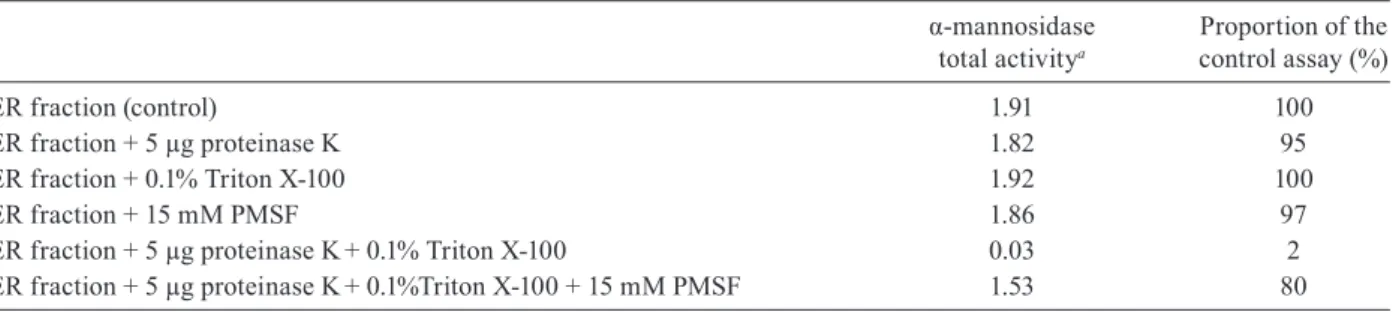
Orientation of the α1,2-mannosidase catalytic domain - In order to determine the orientation of the
α1,2-mannosidase catalytic domain, proteinase K pro-tection assays were carried out using the microsomal preparations from peak II separated by density gradi-ent. In the presence of Triton X-100 and proteinase K, α-mannosidase activity was absent whereas the addi-tion of PMSF to the reacaddi-tions prevented loss of enzyme activity (Table II). Therefore, α1,2-mannosidase is lo-calised in the ER, with the catalytic domain oriented towards the lumen of this compartment.
dISCuSSIOn
The present paper demonstrates that S. schenckii has only one major non-acidic α-mannosidase associated with the membrane fraction. As with α1,2-mannosidases from
Fig. 3: hydrolysis of M9 oligosaccharide by purified α-mannosidase from filamentous cells. A: 2 µg of each oligosaccharide or 200 ng mannose were separated as described in Materials and Methods; B:
aliquots containing 5 µg of purified enzyme were incubated with M9
oligosaccharide (2 µg) at 37°C and after the indicated times, reactions were stopped by heating in a water bath for 5 min and analyzed by high performance anion-exchange chromatography as described in the text. Elution profiles of oligosaccharides and mannose are indi-cated in left and right panels, respectively; M9: Man9GlcNAc2; M8B: Man8GlcNAc2 isomer B; M7B: Man7GlcNAc2 isomer B; M6 and M5:
probably Man6GlcNAc2 and Man5GlcNAc2, respectively; : GlcNAc
β1,4-; : Man β 1,4-; : Man α 1,6-; : Man α 1,3-; : Man α 1,2-.
Fig. 4: intracellular distribution of α-mannosidase activity in proto-plast of Sporothrix schenckii. Protoplast homogenates were loaded onto the top of a continuous sucrose density gradient and centrifuged
at 232,000 g at 4°C for 4 h, 1 mL fractions were collected and used
to quantify the α-mannosidase activity with the fluorogenic substrate MUαMan (open symbols) and the sucrose content (closed symbols).
activity purified here was susceptible to proteolysis by native aspartyl proteases. The thermostability of the en-zyme allowed a non-traditional solubilisation procedure that was similar to the approach used for the purification of ER α1,2-mannosidase from C. albicans (Mora-Montes
et al. 2008c) and other proteins from prokaryotic cells (Novikov et al. 1993, Heinz & Niederweis 2000).
The purified enzyme preferred MUαMan over p-NP-Man as a substrate, was recognised by anti-α1,2-mannosidase antibodies (Mora-Montes et al. 2008a) and was more sensitive to 1-deoxymannojirimycin over swain-sonine, suggesting that this α-mannosidase belongs to fam-ily 47 of the glycosyl hydrolases (Herscovics 1999a). The optimal pH for the enzyme activity confirmed that this en-zyme is not a vacuolar α-mannosidase, since these enen-zymes show an optimal activity at pH 4-5 (Herscovics 1999a).
S. cerevisiae α1,2-mannosidase is inhibited by Mn2+,
co2+, Zn2+ and Mg2+(Jelinek-Kelly & Herscovics 1988), whereas in C. albicans this activity is slightly stimulated
by Mn2+ and ca2+ (Vázquez-Reyna et al. 1999). Here, S. schenckii α-mannosidase was inhibited by Zn2+ and co2+. This may be explained by subtle differences in the structure of the α1,2-mannosidase from those organisms, which allow or preclude the accessibility of cations to the catalytic pock-et. The lack of stimulation by Co2+ indicated that this protein
is not similar to Co2+-dependent cytosolic α-mannosidases
described in mammalian cells (Herscovics 1999a).
Alpha-mannosidases purified in this study acted upon M9 as a typical, ER-localised α1,2-mannosidase,
producing M8B and mannose as the sole products of hy-drolysis after 12 h of incubation (Herscovics 1999a, b). As the incubation continued, M7B and most probably M6
and M5 were detected after 24 h. This result is in agree-ment with the hydrolysis reported for α1,2-mannosidases beloging to S. cerevisiae, C. albicans and human cells, in
which M8B can be processed to shorter oligosaccharides under both in vitro and in vivo conditions (Herscovics et al. 2002, Mora-Montes et al. 2004, 2008a, Avezov et al. 2008). The inability to trim Man5GlcNAc2-Asn was
expected, as this oligosaccharide lacks α1,2-linked man-nose residues. The M9 trimming profile is not consistent
arm at the end of the reaction (Herscovics 2001, Lob-sanov et al. 2002). Therefore, these results indicated that the purified enzyme from both filamentous and yeast cells of S. schenckii is an α1,2-mannosidase of glycosyl
hydrolase family 47 most likely located within the ER. The localisation of the enzyme was confirmed by den-sity gradient centrifugation. Alpha 1,2-mannosidase ac-tivity was separated in two peaks, with 32% of the acac-tivity associated with densities in the range reported for Golgi (1.122-1.142 g cm-3) and 68% of the activity associated
with densities reported for ER membranes (1.178-1.182 g cm-3) (Beaufay et al. 1974, Chrispeels et al. 1982, Harris & Waters 1996, Mora-Montes et al. 2008a), consistent with distribution of the enzyme between the Golgi complex and ER. Monensin blocks the vesicle transport within the Golgi complex (Rosa et al. 1992) and, as a consequence, proteins are accumulated in the ER. Hence, this compound only affects the intracellular localisation of proteins that are not Golgi residents. As α-mannosidase distribution was altered upon incubation of protoplasts with monensin, it is likely that the α-mannosidase activity associated with the Golgi complex is not resident in this compartment. ER α1,2-mannosidases lack ER retention signals and therefore its localisation depends on an interaction with Rer1, which allows the retrieval movement from the early Golgi compartments (Massaad & Herscovics 2001). A similar ER retention mechanism may be expected in S. schenckii and may explain the α1,2-mannosidase activity
detected in Golgi-derived vesicles. As with other α1,2-mannosidases involved in the biosynthesis of N-glycans, the catalytic domain of the S. schenckii enzyme was
lo-calised within the ER lumen.
The 75 kDa α1,2-mannosidase was the only protein immunodetected in the non-solubilised MMF and the trimming of M9 was similar to that carried out by the
purified enzymes. This suggests that both mycelial and yeast forms of S. schenckii do not contain Golgi
α1,2-mannosidases of glycosyl hydrolase family 47. These data contrast with reports from other filament fungi, in which both ER and Golgi α1,2-mannosidases are pres-ent and participate in the generation of hybrid and com-plex N-glycans (Herscovics 2001, Lobsanov et al. 2002). Thus, only high-mannose N-glycans may be present in the cell wall of S. schenckii, a finding that provides
in-sight into the structure of this cell wall.
ACknOwLEdGMEntS
To Conchita Toriello Nájera, for providing the Sporothrix
schenckii strain.
REFEREnCES
Akao T, Yamaguchi M, Yahara A, Yoshiuchi K, Fujita H, Yamada O, Akita O, Ohmachi T, Asada Y, Yoshida T 2006. Cloning and
ex-pression of 1,2-alpha-mannosidase gene (fmanIB) from filamentous
fungus Aspergillus oryzae: in vivo visualization of the
FmanIBp-GFP fusion protein. Biosci Biotechnol Biochem 70: 471-479.
Avezov E, Frenkel Z, Ehrlich M, Herscovics A, Lederkremer GZ 2008. Endoplasmic reticulum (ER) mannosidase I is compartmentalized and required for N-glycan trimming to Man5-6GlcNAc2 in
glyco-protein ER-associated degradation. Mol Biol Cell 19: 216-225.
Beaufay H, Amar-Costesec A, Thines-Sempoux D, Wibo M, Robbi M, Berthet J 1974. Analytical study of microsomes and isolated subcellular membranes from rat liver. 3. Subfractionation of the microsomal fraction by isopycnic and differential centrifugation in density gradients. J Cell Biol 61: 213-231.
Chrispeels MJ, Higgins TJ, Craig S, Spencer D 1982. Role of the endoplasmic reticulum in the synthesis of reserve proteins and the kinetics of their transport to protein bodies in developing pea cotyledons. J Cell Biol 93: 5-14.
Daniel PF, Winchester B, Warren CD 1994. Mammalian
alpha-man-nosidases - multiple forms but a common purpose? Glycobiology
4: 551-566.
de Beer ZW, Harrington TC, Vismer HF, Wingfield BD, Wingfield
MJ 2003. Phylogeny of the Ophiostoma stenoceras-Sporothrix
schenckii complex. Mycologia 95: 434-441.
Diamond RJ, Rose AH 1970. Osmotic properties of spheroplasts
from Saccharomyces cerevisiae grown at different temperatures.
J Bacteriol 102: 311-319.
Durden FM, Elewski B 1997. Fungal infections in HIV-infected pa-tients. Semin Cutan Med Surg 16: 200-212.
Eades CJ, Hintz WE 2000. Characterization of the class I
alpha-mannosidase gene family in the filamentous fungus Aspergillus
nidulans. Gene 255: 25-34.
Harris SL, Waters MG 1996. Localization of a yeast early Golgi man-nosyltransferase, Och1p, involves retrograde transport. J Cell Biol 132: 985-998.
Heinz C, Niederweis M 2000. Selective extraction and purification of a
mycobacterial outer membrane protein. Anal Biochem 285: 113-120.
Helenius A, Aebi M 2004. Roles of N-linked glycans in the
endoplas-mic reticulum. Annu Rev Biochem 73: 1019-1049.
TABLE II
Effect of proteinase K on the endoplasmic reticulum (ER) α1,2-mannosidase activity of Sporothrix schenckii
α-mannosidase total activitya
Proportion of the control assay (%)
ER fraction (control) 1.91 100
ER fraction + 5 µg proteinase K 1.82 95
ER fraction + 0.1% Triton X-100 1.92 100
ER fraction + 15 mM PMSF 1.86 97
ER fraction + 5 µg proteinase K+ 0.1% Triton X-100 0.03 2
ER fraction + 5 µg proteinase K+ 0.1%Triton X-100 + 15 mM PMSF 1.53 80
Henrissat B 1991. A classification of glycosyl hydrolases based on
amino acid sequence similarities. Biochem J 280: 309-316.
Henrissat B, Davies G 1997. Structural and sequence-based classifica-tion of glycoside hydrolases. Curr Opin Struct Biol 7: 637-644. Herscovics A 1999a. Importance of glycosidases in mammalian
gly-coprotein biosynthesis. Biochim Biophys Acta 1473: 96-107.
Herscovics A 1999b. Processing glycosidases of Saccharomyces
cerevisiae. Biochim Biophys Acta 1426: 275-285.
Herscovics A 2001. Structure and function of Class I alpha 1,2-man-nosidases involved in glycoprotein synthesis and endoplasmic reticulum quality control. Biochimie 83: 757-762.
Herscovics A, Romero PA, Tremblay LO 2002. The specificity of the yeast and human class I ER alpha 1,2-mannosidases involved in ER quality control is not as strict previously reported. Glycobiol -ogy 12: 14G-15G.
Herscovics A, Schneikert J, Athanassiadis A, Moremen KW 1994. Isola-tion of a mouse Golgi mannosidase cDNA, a member of a gene
fam-ily conserved from yeast to mammals. J Biol Chem 269: 9864-9871.
Ichishima E, Taya N, Ikeguchi M, Chiba Y, Nakamura M, Kawabata C, Inoue T, Takahashi K, Minetoki T, Ozeki K, Kumagai C, Gomi K, Yoshida T, Nakajima T 1999. Molecular and enzymic properties of recombinant 1, 2-alpha-mannosidase from Aspergillus saitoi over -expressed in Aspergillus oryzae cells. Biochem J 339: 589-597.
Jelinek-Kelly S, Akiyama T, Saunier B, Tkacz JS, Herscovics A 1985. Characterization of a specific alpha-mannosidase involved in
oligosaccharide processing in Saccharomyces cerevisiae. J Biol
Chem 260: 2253-2257.
Jelinek-Kelly S, Herscovics A 1988. Glycoprotein biosynthesis in
Sac-charomyces cerevisiae. Purification of the alpha-mannosidase
which removes one specific mannose residue from Man9GlcNAc.
J Biol Chem 263: 14757-14763.
Kornfeld R, Kornfeld S 1985. Assembly of asparagine-linked oligo-saccharides. Annu Rev Biochem 54: 631-664.
Laemmli UK 1970. Cleavage of structural proteins during the
assem-bly of the head of bacteriophage T4. Nature 227: 680-685.
Lal A, Pang P, Kalelkar S, Romero PA, Herscovics A, Moremen KW 1998. Substrate specificities of recombinant murine Golgi alpha1, 2-mannosidases IA and IB and comparison with endoplasmic
re-ticulum and Golgi processing alpha1,2-mannosidases. Glycobiol
-ogy 8: 981-995.
Lloyd KO, Bitoon MA 1971. Isolation and purification of a
peptido-rhamnomannan from the yeast form of Sporothrix schenckii.
Structural and immunochemical studies. J Immunol 107: 663-671.
Lobsanov YD, Vallee F, Imberty A, Yoshida T, Yip P, Herscovics A, Howell PL 2002. Structure of Penicillium citrinum alpha
1,2-mannosidase reveals the basis for differences in specificity
of the endoplasmic reticulum and Golgi class I enzymes. J Biol
Chem 277: 5620-5630.
Lopes-Alves LM, Mendonca-Previato L, Fournet B, Degand P, Previato
JO 1992. O-glycosidically linked oligosaccharides from
peptidor-hamnomannans of Sporothrix schenckii. Glycoconj J 9: 75-81.
Lopes-Bezerra LM, Schubach A, Costa RO 2006. Sporothrix schenckii
and sporotrichosis. An Acad Bras Cienc 78: 293-308.
Massaad MJ, Herscovics A 2001. Interaction of the endoplasmic re-ticulum alpha 1,2-mannosidase Mns1p with Rer1p using the split-ubiquitin system. J Cell Sci 114: 4629-4635.
Mora-Montes HM, Bader O, López-Romero E, Zinker S, Ponce-Noy-ola P, Hube B, Gow NA, Flores-Carreón A 2008a. Kex2 protease converts the endoplasmic reticulum alpha 1,2-mannosidase of
Candida albicans into a soluble cytosolic form. Microbiology
154: 3782-3794.
Mora-Montes HM, López-Romero E, Zinker S, Ponce-Noyola P,
Flores-Carreón A 2004. Hydrolysis of Man9GlcNAc2 and Man8
GlcNAc2 oligosaccharides by a purified alpha-mannosidase from
Candida albicans. Glycobiology 14: 593-598.
Mora-Montes HM, López-Romero E, Zinker S, Ponce-Noyola P, Flores-Carreón A 2006. Purification of soluble
alpha1,2-man-nosidase from Candida albicans CAI-4. FEMS Microbiol Lett
256: 50-56.
Mora-Montes HM, López-Romero E, Zinker S, Ponce-Noyola P, Flores-Carreón A 2008b. Conversion of alpha 1,2-mannosidase
E-I from Candida albicans to alpha1,2-mannosidase E-II by
lim-ited proteolysis. Antonie Van Leeuwenhoek 93: 61-69.
Mora-Montes HM, López-Romero E, Zinker S, Ponce-Noyola P, Flores-Carreón A 2008c. Heterologous expression and biochemical
char-acterization of an alpha 1,2-mannosidase encoded by the Candida
albicans MNS1 gene. Mem Inst Oswaldo Cruz 103: 724-730.
Movsichoff F, Castro OA, Parodi AJ 2005. Characterization of
Schizo-saccharomyces pombe ER alpha-mannosidase: a re-evaluation of
the role of the enzyme on ER-associated degradation. Mol Biol
Cell 16: 4714-4724.
Novikov OD, Fedoreeva LI, Khomenko VA, Portniagina O, Ermak IM, Likhatskaia GN, Moroz SV, Solov’eva TF, Ius O 1993. Ef-fect of the method of extracting the pore-forming protein from
Yersinia pseudotuberculosis on its macromolecular organization. Bioorg Khim 19: 536-547.
Ramos-e-Silva M, Vasconcelos C, Carneiro S, Cestari T 2007. Sporo-trichosis. Clin Dermatol 25: 181-187.
Rosa P, Mantovani S, Rosboch R, Huttner WB 1992. Monensin and brefeldin A differentially affect the phosphorylation and sulfation
of secretory proteins. J Biol Chem 267: 12227-12232.
Ruiz-Baca E, Villagómez-Castro JC, Leal-Morales CA, Sabanero-López M, Flores-Carreón A, Sabanero-López-Romero E 2005.
Biosynthe-sis of glycoproteins in the human pathogenic fungus Sporothrix
schenckii: synthesis of dolichol phosphate mannose and
manno-proteins by membrane-bound and solubilized mannosyl
trans-ferases. Antonie Van Leeuwenhoek 88: 221-230.
Schneikert J, Herscovics A 1994. Characterization of a novel mouse
re-combinant processing alpha-mannosidase. Glycobiology 4: 445-450.
Towbin H, Staehelin T, Gordon J 1979. Electrophoretic transfer of pro-teins from polyacrylamide gels to nitrocellulose sheets: procedure
and some applications. Proc Natl Acad Sci USA 76: 4350-4354.
Tremblay LO, Herscovics A 1999. Cloning and expression of a
spe-cific human alpha 1,2-mannosidase that trims Man9GlcNAc2 to
Man8GlcNAc2 isomer B during N-glycan biosynthesis. Glycobi -ology 9:1073-1078.
Tremblay LO, Herscovics A 2000. Characterization of a cDNA encod-ing a novel human Golgi alpha 1, 2-mannosidase (IC) involved in
N-glycan biosynthesis. J Biol Chem 275: 31655-31660.
Vázquez-Reyna AB, Ponce-Noyola P, Calvo-Méndez C, López-Romero E, Flores-Carreón A 1999. Purification and biochemical
characterization of two soluble alpha-mannosidases from
Can-dida albicans. Glycobiology 9: 533-537.
Yoshida T, Inoue T, Ichishima E 1993. 1,2-alpha-D-mannosidase
from Penicillium citrinum: molecular and enzymic properties of
two isoenzymes. Biochem J 290: 349-354.
Ziegler FD, Trimble RB 1991. Glycoprotein biosynthesis in yeast: purification and characterization of the endoplasmic reticulum