377 377 377 377 377 Mem Inst Oswaldo Cruz, Rio de Janeiro, Vol. 100(4): 377-383, July 2005
Subcellular localization of an intracellular serine protease of 68
kD a in
Leishmania (Leishmania) amazonensis
promastigotes
José Andrés M orgado-D íaz/+, Raquel Elisa da Silva-Lopez* , Carlos Roberto Alves* , M aurilio José Soares* * , Suzana Corte-Real* * , Salvatore Giovanni D e Simone* * * *
Grupo de Biologia Estrutural,Divisão de Biologia Celular, Centro de Pesquisas, Instituto Nacional de Câncer, Rua André Cavalcanti 37, 5° andar, 20231-050 Rio de Janeiro, RJ, Brasil *Laboratório de Bioquímica de Proteínas e Peptídios, Departamento
de Bioquímica e Biologia Molecular **Departamento de Ultra-estrutura e Biologia Celular, Instituto
Oswaldo Cruz-Fiocruz, Rio de Janeiro, RJ, Brasil ***Departamento de Biologia Celular e Molecular, Instituto de Biologia, Universidade Federal Fluminense, Niterói, RJ, Brasil
Here we report the subcellular localization of an intracellular serine protease of 68 kDa in axenic promastigotes of Leishmania (Leishmania) amazonensis, using subcellular fractionation, enzymatic assays, immunoblotting, and immunocytochemistry. All fractions were evaluated by transmission electron microscopy and the serine protease activity was measured during the cell fractionation procedure using α-N-r-tosyl-L-arginine methyl ester (L-TAME) as substrate, phenylmethylsulphone fluoride (PMSF) and L-1-tosylamino-2-phenylethylchloromethylketone (TPCK) as specific inhibitors. The enzymatic activity was detected mainly in a membranous vesicular fraction (6.5-fold enrichment relative to the whole homogenate), but also in a crude plasma membrane fraction (2.0-fold). Analysis by SDS-PAGE gelatin under reducing conditions demonstrated that the major proteolytic activity was found in a 68 kDa protein in all fractions studied. A protein with identical molecular weight was also recognized in immunoblots by a polyclonal antibody against serine protease (anti-SP), with higher immunoreactivity in the vesicular fraction. Electron microscopic immunolocalization using the same polyclonal antibody showed the enzyme present at the cell surface, as well as in cytoplasmic membranous compartments of the parasite. Our findings indicate that the internal location of this serine protease in L. amazonensis is mainly restricted to the membranes of intracellular compartments resembling endocytic/exocytic elements.
Key words: Leishmania - serine protease - subcellular localization - trypanosomatid
Study of parasitic proteases has received consider-able attention, since their physiological role elucidation may help to develop strategies for exploiting these en-zymes as novel chemotherapic targets (Cazzulo 2002, Sajid & McKerrow 2002). In eukaryotic organisms, serine pro-tease are the most studied propro-teases, while cysteine and metallo-type enzymes have been the most investigated in trypanosomatid protozoa of the Leishmania genus (Coombs & Mottram 1997, Mottram et al. 1998, Alves et al. 2000, Jaffe & Dwyer 2003). Serine peptidases are among the most extensively studied enzymes. They are found in all organisms studied and participate in blood clotting and complement activation cascade reactions, phage matu-ration and fertilization, as well as any number of other fields of biological phenomena (Rawling & Barret 1994). Protozoan serine proteases play crucial roles in host-para-site interaction (Rosenthal 1999). The most notorious serine proteases are found in species of Plasmodium. These malarial enzymes digest proteins of the cytoplas-matic membrane of the red blood cells thereby affording invasion and infection by the parasite performing several functions such as mediation of the merozoite entry into
Financial support: CNPq, Fiocruz - Papes III, Faperj
+Corresponding author. E-mail: jmorgado@inca.gov.br
Received 15 February 2005 Accepted 8 June 2005
host erythrocytes (Braun-Breton et al. 1992, Braun-Breton & Pereira da Silva 1993) and formation of the parasitophorous vacuole (Roggwille et al. 1996), afford-ing invasion and infection of erythrocytes by the para-sites.
Trypanosomatid protozoa serine peptidases have been identified and characterized, and play crucial roles in host infection (Burleigh & Woolsey 2002, Silva-Lopez & Giovanni De Simone 2004). Trypanosoma cruzi
proper-378 378 378 378
378 Serine protease in L. amazonensis • José Andrés M orgado-D íaz et al.
ties of 68 kDa Leishmania serine protease are known, its function in the parasite physiology and in the host-para-site interaction is still unclear. Studies on subcellular lo-calization will help to understand the function of this en-zyme. Recently, our group isolated a 56 kDa serine pro-tease from the culture supernatant of promastigotes of the same parasite, produced an antiserum against this enzyme and its intracellular location was analyzed by im-munocytochemistry (Silva-Lopez et al. 2004).
So, in order to improve the knowledge on the parasite physiology and create a possibility to develop a rational design of compounds that may be useful in the treatment of the leishmaniasis, here we report the subcellular local-ization of L. amazonensis promastigotes intracellular 68 kDa serine protease, on the basis of some biochemical properties.
MATERIALS AND METHODS
Parasite - Promastigote forms of L. (L.) amazonensis
(IOC 575; IFLA/BR/67/PH8) were maintained in brain heart infusion medium (Difco, Detroit, US) supplemented with 10% (v/v) heat-inactivated fetal-calf serum. The cultures were kept at room temperature (25°C) in Roller bottles using a Cel-Gro Rotator (Lab-Line Model, Thomas Scien-tific, New Jersey, US).
Parasite lysis and subcellular fractionation - Cells were harvested by centrifugation (Beckman centrifuge Model J2-21, Palo Alto, California, US) at 3800 g for 10 min and washed three times in P buffer (10 mM Tris-HCl, pH 7.0, and containing 0.25 M sucrose). Thereafter, the cellu-lar pellet was ressuspended for 30 min in hypotonic solu-tion (P buffer without sucrose) and the cells were dis-rupted in a Dounce type homogenizer (~ 80 strokes). The homogenization process was stopped while some unbro-ken cells were still present. A concentrated sucrose solu-tion (0.5 M), prepared in 10 mM Tris-HCl, pH 7.0, was immediately added to the lysate to make a final concentra-tion of 0.25 M, in order to minimize the osmotic damage caused by the hypotonic solution. Cell swelling and rup-ture was monitored by phase contrast microscopy. Lysed cells were submitted to a first centrifugation for 10 min at 4300 g, and the resulting pellet containing unbroken para-sites, nuclei, mitochondria, and some kinetoplast-mito-chondrial complexes was discarded. The post-nuclear supernatant was harvested and then centrifuged for 15 min at 12,000 g. The pellet was collected and the second supernatant was spun again for 45 min at 35,000 g. Aliquots of the last supernatant and the pellets (called P-12,000 g and P-35,000 g, respectively) were harvested to subse-quent analysis. All centrifugation steps and other opera-tions were performed at 4oC or on ice.
Assay of protease activity and effect of inhibitors -Enzyme activity was measured as previously described (Silva-Lopez & Giovanni De Simone 2004), with slight modifications. Briefly, the reaction mixtures, which com-prised 300 µl of 100 mM Tris-HCl buffer, pH 8.0, contain-ing 100 µl of sample preparations, were incubated with 100 µl of 0.1 mM (final concentration) α -N-r-tosyl-L-argi-nine methyl ester (L-TAME). Following incubation at room temperature (25oC) for 30 min, the solutions were
centri-fuged to remove insoluble material and the absorbance increase at 247 nm was measured in the supernatant (Hitachi U-2000 spectrophotometer, Schaumburg, Illinois, US). One unit of enzymatic activity was defined as the amount of enzyme required to cause an increase of 0.1 in the absorbance under standard conditions.
In order to investigate the serine protease presence in parasite subcellular fractions, inhibition assays were per-formed. Protease inhibitors were incubated with each frac-tion (50 mg protein/µl) for 30 min in Tris-HCl 0.1M pH 8.0 at room temperature. The following protease inhibitors were used: 10 mM phenymethanesulphonyl fluoride (PMSF), 1 mM L-1-tosylamino-2-phenylethylchloro-methylketone (TPCK), 1 mM trans-epoxysuccinyl-leucylamide (4-guanidino) butane (E-64), 2 mM ethylene-diaminetetraacetic acid (EDTA), 1mM o-phenanthroline (OP) and 25 µM Pepstatin-A (PA). The reaction started upon addition of the substrate (0.25 mM L-TAME) in Tris-buffer at 28°C for 30 min. The tests were performed in triplicate in standardized conditions (Silva-Lopez & Giovanni De Simone 2004). Appropriate controls were car-ried out in parallel using the same enzyme solutions with-out inhibitors. Inhibition was expressed as the percent-age of the appropriate control activity. All inhibitors and the substrate were purchased from Sigma Chemical Co. (St. Louis, Missouri, US).
Gelatin substrate gel electrophoresis - Protease ac-tivity in the different parasite fractions was also deter-mined using gelatin substrate gel electrophoresis. Samples were applied to 12% SDS-PAGE with 0.1% gelatin and electrophoresis conducted at 4oC in a Mini Protean II Apparatus (BioRad, Richmond) without prior reduction or heating, according to previously described conditions (Silva-Lopez & De Simone 2004). After electrophoresis, gels were washed under agitation for 60 min at room tem-perature with 2.5% Triton X-100, and incubated overnight at 37oC in 0.1 M Tris-HCl buffer, pH 8.0. The gel was then stained with 0.1% amide black and distained using metha-nol/acetic acid/distilled water (3:1:6).
Antiserum production - In order to produce the anti-body against the 68 kDa serine protease, the enzyme was purified from a whole extract of L. amazonensis pro-mastigotes by freezing-thawing. The purification was a combination of affinity chromatography on aprotinin-aga-rose and gel filtration high performance liquid chroma-tography, as previously described (Silva-Lopez & Giovanni De Simone 2004). The anti-L.amazonensis serine protease antiserum (anti-SP) was raised in rabbit by in-jecting 20 µg of the homogeneous heat inactivated (2 min at 100°C) 68 kDa enzyme emulsified in complete (first booster) and incomplete (subsequent boosters) Freud’s adjuvant. Rabbits were bled after the fourth injection and the antibody reactivity was checked by immunoblotting.
379 379 379 379 379 Mem Inst Oswaldo Cruz, Rio de Janeiro, Vol. 100(4), July 2005
For immune staining, the nitrocellulose sheets were incu-bated for 60 min with the rabbit anti-SP serum (1:400 dilu-tion). The sheets were then soaked for 60 min in peroxi-dase-conjugated goat anti-rabbit IgG and the blots devel-oped with 3,3'-diaminobenzidine/H2O2.
Electron microscopy and immunocytochemistry -Routine transmission electron microscopy analysis was carried out to control the subcellular fractionation pro-cess. Samples were fixed by the addition of an equal vol-ume of 5% glutaraldehyde in 0.2 M cacodylate buffer, pH 7.4, and containing 0.25 M sucrose. Post-fixation was car-ried out with 1% osmium tetroxide in cacodylate buffer containing 0.8% potassium ferricyanide and 5 mM CaCl2, followed by dehydration in acetone and embedding in PolyBed 812. Ultrathin sections were stained with uranyl acetate and lead citrate, and observed in a Zeiss EM10C transmission electron microscope.
The polyclonal antiserum (anti-SP) was used to deter-mine the subcellular localization of the serine protease in
L.amazonensis promastigotes, by using previously de-scribed methods for immunogold labeling (Bendayan et al. 1987). Briefly, the parasites were fixed in freshly pre-pared 4% paraformaldehyde/1% glutaraldehyde in 0.1 M sodium cacodylate buffer, pH 7.3. Samples were dehy-drated in methanol and embedded at progressively low-ered temperature in Lowicryl K4M resin. Thin sections were collected on 400 mesh uncoated nickel grids, incu-bated for 30 min at room temperature in phosphate buff-ered saline (PBS) containing 1.5% bovine serum albumin (BSA) and 0.01% Tween 20 (PBS-BSA-Tw), pH 8.0, and then for 60 min in the presence of the anti-SP antibody diluted 1:50 in PBS-BSA-Tw, pH 8.0. Grids were rinsed in PBS-BSA-Tw and finally incubated for 60 min with a 1:200 dilution of goat anti-rabbit antibody labeled with 10-nm gold particles (Bendayan et al. 1987). The grids were sub-sequently washed with PBS and distilled water, stained with uranyl acetate and lead citrate and observed in a Zeiss EM10C transmission electron microscope. Under these conditions, controls incubated only with the sec-ondary gold-labeled antibody showed little, if any, back-ground reaction.
Protein determination - The protein content in the different fractions was estimated with the BioRad (Rich-mond, UK) protein assay kit using bovine serum albumin (BSA) as standard.
RESULTS
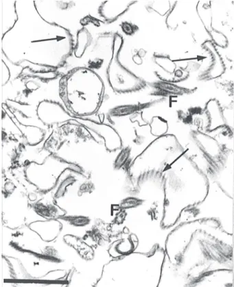
Isolation of subcellular fractions - Using the homog-enization procedure described here, 90% of cell disrup-tion was obtained as ascertained by phase contrast mi-croscopy. The fractionation methodology was also con-trolled by transmission electron microscopy. Nuclei and mitochondria in the resulting homogenate presented no apparent morphological membrane lesion (data not shown). The general aspect of the P-12,000 g and P-35,000 g fractions obtained by differential centrifugation is shown in Figs 1 and 2, respectively. The major elements found in the P-12,000 g fraction were plasma membrane profiles, as recognized by the attached subpellicular mi-crotubules, although some flagella could also be seen.
Fig. 1: electron micrograph of the 12,000 g pellet obtained by subcellular fractionation. Most isolated membranes are character-ized to be from surface, associated with microtubules (arrows). F: flagella. Bar = 1 µm.
380 380 380 380
380 Serine protease in L. amazonensis • José Andrés M orgado-D íaz et al.
Ultrathin sections of the P-35,000 g fraction showed that its main component was a population of spherical mem-brane bound vesicles of different sizes, which possibly represent elements of the endocytic/exocytic compart-ments.
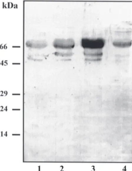
Immunoreactivity analysis - Specificity of the polyclonal serum to serine protease was evaluated by western blot analysis employing the four fractions ob-tained in the subcellular fractionation. The antibody to serine protease recognized a major band of 68 kDa in all fractions, with the highest immunoreactivity in the ve-sicular fraction (Fig. 3). Interestingly, the antiserum re-vealed two additional bands of 50 and 45 kDa in the whole homogenate, the membrane fraction and the vesicular frac-tion.
homogenate, in the P-12,000 gand P-35,000 g fractions, and in the final supernatant. Both the P-12,000 g fraction and the final supernatant showed some enrichment in ac-tivity (2- and 1.5-fold, respectively) in relation to the whole homogenate, but the highest activity was found in the P-35,000 g fraction. This later fraction corresponded to the membranous vesicles and displayed an enrichment of 6.5-fold in the enzymatic activity, with 42% recovery (Table I). Interestingly, the enzyme displayed no latency when the enzymatic activity was checked in samples of freshly iso-lated fractions incubated in the presence of Triton X-100 (data not shown).
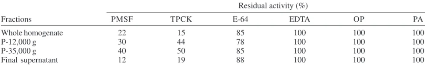
In order to identify the L. amazonensis protease activ-ity in the subcellular fractions, inhibition experiments were carried out using L-TAME as substrate. The activity was not inhibited by o-phenanthroline, EDTA or Pepstatin A, was weakly sensitive to E-64 (an inhibitor of cysteine pro-teinase), but the activity in the four fractions was PMSF-and TPCK-sensitive, both serine protease inhibitors. The enzymatic activity was reduced 60-88% in the presence of PMSF and 50-80% in the presence of TPCK (Table II), supporting the hypothesis that the activity was due to a serine-type peptidase. Similar results were observed for the purified serine enzyme (Silva-Lopez & Giovanni De Simone 2004).
Proteolytic activity of the fractions was also screened using SDS-PAGE gelatin at pH 8.0. As shown in Fig. 4, a single majority protein was associated with the four frac-tions analyzed, although noticeable differences in activ-ity could be seen among them. For instance, the enzy-matic activity was higher in the membranous vesicular fraction (lane 3) than in the other fractions. In all cases the protein migrated with the same electrophoretic mobility (68 kDa). Additionally, a minor protein about ≥ 100 kDa was also detected in the fractions.
Fig. 3: western blotting analysis of Leishmania amazonensis frac-tions using anti-SP antibody. Lanes - 1: whole homogenate; 2: P-12,000 g fraction; 3: P-35,000 g fraction; 4: final supernatant. Fifteen µg of protein were loaded in each lane. Values of the mo-lecular weight markers are indicated on the left side of gel.
In order to localize the serine protease in the para-sites, electron microscopy immunolocalization by label-ing the cells with the SP was performed. The anti-body reacted against the parasite surface and internal structures in most (about 95%) analyzed cells (Fig. 5A). Cytoplasmic gold particles were localized predominantly in vesicular structures close to the flagellar pocket re-gion, morphologically similar to that of the endocytic/ exocytic pathways (Fig. 5B). The gold markers were seen bound to the inner membrane leaflet of the cytoplasmic vesicles (Fig. 5C).
Enzymatic activity detection in subcellular fractions
- Previous experiments showed that extracts of L. amazonensis were able to hydrolyze L-TAME at pH 8.0. Thus, we have used this chromogenic substrate to evalu-ate protease activity in the different fractions obtained by the fractionation methodology. Table I shows the subcel-lular distribution profile of protease activity in the whole
381 381 381 381 381 Mem Inst Oswaldo Cruz, Rio de Janeiro, Vol. 100(4), July 2005
TABLE I
Distribution and recovery of the serine type protease activity in subcellular fractions of Leishmaniaamazonensis promastigotes prepared by differential centrifugation. Values are means of three independent experiments
Total proteina Total activity Specific activity Recovery Enrichment
Fractions (mg) (U) (U/mg protein) (%) (-fold-)
Whole homogenate 90.00 7.38 0.082 100 1.0
P-12,000 g 9.40 1.55 0.16 21 2.0
P-35,000 g 5.80 3.10 0.53 42 6.5
Final supernatant 15.00 1.90 0.12 25 1.5
a: from 2 × 10¹⁰ cells
TABLE II
Effects of inhibitors on the L-TAME activity of subcellular fractions of Leishmaniaamazonensis promastigotes. The fractions (50 µg protein/ml) were incubated previously for 30 min with the respective inhibitors and the residual activity was measured at
25oC at pH 8.0 as described in Materials and Methods. The percentage of inhibition was calculated taking the control values (without inhibitor) as 100%. The assays represent the means of at three independent determinations. Standard deviations
were always lower than 5%
Residual activity (%)
Fractions PMSF TPCK E-64 EDTA OP PA
Whole homogenate 22 15 85 100 100 100
P-12,000 g 30 44 78 100 100 100
P-35,000 g 40 50 85 100 100 100
Final supernatant 12 19 88 100 100 100
PMSF: phenymethanesulphonyl fluoride; TPCK: L-1-tosylamino-2-phenylethylchloromethylketone; E-64: trans-epoxysuccinyl-leucylamide (4-guanidino) butane; EDTA: ethylene-diaminetetraacetic acid; OP: O-phenanthroline; PA: Pepstatin-A
382 382 382 382
382 Serine protease in L. amazonensis • José Andrés M orgado-D íaz et al.
DISCUSSION
Parasite proteases have been demonstrated to play important roles in the pathogenesis of a variety of dis-eases (McKerrow et al. 1993), but their identification and subcellular localization are important prerequisites for understanding their function in the infection process. Immunolocalization studies associated with cell fraction-ation techniques are essential to achieve these goals, but in trypanosomatid protozoa, particularly in Leishmania
species, serious difficulties have been associated with cell rupture, due mainly to their own morphological char-acteristics (Coombs et al. 1982, Mottram & Coombs 1985). The cell rupture method used here has proved to be ideal in obtaining a homogenate in which the subcellular struc-tures were well preserved, as confirmed by transmission electron microscopy.
Subcellular distribution of serine protease in L. amazonensis promastigotes using the chromogenic sub-strate (L-TAME) indicated that the enzymatic activity was mainly associated to the membranous vesicular fraction. Protease activity was also recovered in the crude plasma membrane fraction, but in minor amounts, indicating that the enzyme may, in some way, be associated with mem-branes of intracellular compartments, instead of being enclosed in a soluble form in the lumen of these compart-ments. The finding that the enzymatic activity displayed no latency when vesicle fraction was incubated with Tri-ton X-100 (data not shown) together with the immunocy-tochemical data further suggests that the enzyme is mem-brane-bound. The small amount of activity in the final supernatant probably was derived from proteases released from vesicle membranes, as suggested by the identical molecular weight of the enzyme, although the possibility that it represents a different cytosolic form of the enzyme cannot be excluded. Previous studies in T. cruzi have dem-onstrated that an alkaline serine peptidase involved in the generation of a novel Ca2+-signaling factor for mam-malian cells was found in soluble form after disruption of parasites by freezing and sonication, but some activity was also associated with a crude membrane fraction (Burleigh & Andrews 1995, Burleigh & Woolsey 2002). Furthermore, since the activity could be removed by re-peated washing, it was concluded that the enzyme was not tightly associated to the membranes (Tardieux et al. 1994). Other efforts to obtain the same enzyme have also been carried out using different cell breakage methodolo-gies. For example, in T. cruzi and Crithidia fasciculata
both detergent Nonidet P-40 (Ashall et al. 1992, Healy et al. 1992) and freezing-thawing (Grellier et al. 2001) meth-ods were used. Freezing and thawing, followed by Triton X-100 treatment, was also used to rupture T. brucei brucei
(Troeberg et al. 1996). It is important to note that in these latter studies, as well as to L. amazonensis oligopeptidase (Ribeiro de Andrade et al. 1998), the enzyme was always recovered in a soluble form, but subcellular fractionation or immunocytochemistry investigations were not carried out.
Using gel enzymography the proteolytic activity of a
L.amazonensis serine protease corresponding to 68 kDa was observed as the main component in all four fractions.
Apparently, this protease activity corresponds to the mature protease, while the proteolytic activity detected at higher molecular mass (≥ 100 kDa) is likely due to associa-tions of the protease or precursor molecules, or both, con-sidering the relatively mild conditions used for protein separation. On the other hand, western blotting analysis using anti-SP revealed two proteins with 50 and 45 kDa. These proteins represent auto-proteolysis products yielded from the highly active 68 kDa serine protease dur-ing the purification procedures, since they do not display hydrolytic activity and are present after the chromatogra-phy steps and in stored samples (Silva-Lopez & Giovanni De Simone 2004).
By immunoelectron microscopy cytoplasmic labeled structures of L. amazonensis demonstrated to have strik-ing morphological similarities with compartments of the endocytic/exocytic pathways, as previously reported in cryosections of Leishmania species (Yahiaoui et al. 1993). Detailed examination of electron micrographs showed that the label was associated with membranes of cytoplasmic vesicles and tubules. Such endocytic/exocytic-like ele-ments have also been described in other trypanosomatids (Soares et al. 1992, Porto-Carreiro et al. 2000, McConville et al. 2002). It is possible that in L. amazonensis pro-mastigotes most of these structures co-sediment with the vesicular fraction obtained in the subcellular fractionation, as suggested by the higher enzymatic activity and immu-noreactivity to serine protease found in this frac-tion. Further studies using suitable biochemical markers for endocytic/exocytic components in these parasites (re-viewed in Overath et al. 1997, McConville et al. 2002) are necessary in order to characterize the cell compartments (and/or compartment domains) present in our subcellular fractions. Other interesting observation is that the struc-tures described in the present study were located between the nucleus and the flagellar pocket, which also resembles the localization of endocytic/exocytic structures of other trypanosomatids (Webster & Fish 1989, McConville et al. 2002). On the contrary of the 68 kDa intracellular serine protease here reported, to be mainly located in membranes of intracellular compartments and plasma membrane, the 56 kDa extracellular serine protease, previously described, reacted poorly with the parasite surface and moderately with internal structures. However, it was predominantly located in the flagellar pocket and structures that are mor-phologically similar to the compartments that found in mammalian endocytic/exocytic pathways (Silva-Lopez et al. 2004), which justified the released into the extracellular environment. If the intracellular location of this enzyme correspond a correlated enzymatic activity or intracellular trafficking, remains to be demonstrated.
In conclusion, we reported the subcellular localiza-tion of the 68 kDa intracellular serine protease in L. amazonensis promastigote forms, providing useful data in future investigations on cellular trafficking route in or-der to elucidate the role of this enzyme in the parasite.
ACKNOWLEDGMENTS
383 383 383 383 383 Mem Inst Oswaldo Cruz, Rio de Janeiro, Vol. 100(4), July 2005
REFERENCES
Alves CR, Corte-Real S, Rosa MF, De Simone SG 2000. Detec-tion of cysteine-proteinases in Leishmania amazonensis
promastigotes using a cross-reactive antiserum. FEMS Microbiol Lett 1186: 263-267.
Ashall F, Harris D, Roberts H, Healy N, Shan E 1992. Sub-strate specificity and inhibitor sensitivity of a try-panosomatid alkaline peptidase. Biochim Biophys Acta1035: 293-299.
Bendayan M, Nanci A, Kan FWK 1987. Effect of tissue pro-cessing on colloidal gold cytochemistry. J Histochem Cytochem35: 983-496.
Braun-Breton C, Pereira da Silva LH 1993. Malaria proteases and red blood cell invasion. Parasitol Today9: 92-96. Braun-Breton C, Blisnick T, Jouin H, Barale JC, Rabilloud T,
Langsley G, Pereira da Silva LH 1992. Plasmodium chabaudii: p68 serine proteinase activity required for mero-zoite entry into mouse erythrocyte. Proc Nat Acad Sci USA 89: 9647-9651.
Burleigh BA, Andrews NW 1995. A 120-kDa alkaline pepti-dase from Trypanosoma cruzi is involved in the generation of a novel Ca2+-signalling factor for mammalian cells. J Biol
Chem270: 5171-5180.
Burleigh BA, Woolsey AM 2002. Cell signaling and Trypano-soma cruzi invasion. Cell Microbiol 4: 701-711.
Cazzulo JJ 2002. Proteinases of Trypanosoma cruzi: potential targets for the chemotherapy of Chagas’ disease. Curr Top Med Chem2: 1261-1271.
Coombs GH, Mottram JC 1997. Parasite proteinases and amino acid metabolism: possibilities for chemotherapeutic exploi-tation. Parasitology114(Suppl.): S61-S80.
Coombs GH, Craft JA, Hart DT 1982. A comparative study of
Leishmaniamexicana amastigotes and promastigotes. En-zyme activities and subcellular locations. Mol Biochem Parasitol5: 199-211.
Grellier P, Vendeville S, Joyeau R, Bastos IM, Drobecq H, Frappier F, Teixeira AR, Schrevel J, Davioud-Charvet E, Sergheraert C, Santana JM 2001. Trypanosoma cruzi prolyl oligopeptidase Tc80 is involved in nonphagocytic mamma-lian cell invasion by trypomastigotes. J Biol Chem14: 47078-47086.
Healy NS, Greig S, Enahoro H, Roberts H, Drake L, Shaw E, Ashall F 1992. Detection of peptidases in Trypanosoma cruzi epimastigotes using chromogenic and fluorogenic sub-strates. Parasitology104: 315-322.
Jaffe CL, Dwyer DM 2003. Extracellular release of the surface metalloprotease, gp63, from Leishmania and insect trypanosomatids. Parasitol Res91: 229-237.
Laemmli UK 1970. Cleavage of structural proteins during the assembly of head of bacteriophage T4. Nature227: 680-685.
McConville MJ, Mullin KA, Ilgoutz SC, Teasdale RD 2002. Secretory pathway of trypanosomatid parasites. Microbiol
Mol Biol Rev66: 122-154.
McKerrow JH, Sun E, Rosenthal PJ, Bouvier J 1993. The pro-teases and pathogenicity of parasitic protozoa. Ann Rev Microbiol47: 821-853.
Mottram JC, Coombs GH 1985. Leishmaniamexicana:
subcel-lular distribution of enzymes in amastigotes and pro-mastigotes. Exp Parasitol59: 265-274.
Mottram JC, Brooks DR, Coombs GH 1998. Roles of cysteine proteinases of trypanosomes and Leishmania in host-para-site interactions. Curr Op Microbiol1: 455-460.
Overath P, Stierhof YD, Wiese M 1997. Endocytosis and se-cretion in trypanosomatid parasites. Tumultuous traffic in a pocket. Trends Cell Biol7: 27-33.
Porto-Carreiro I, Attias M, Miranda K, De Souza W, Cunha-e-Silva N 2000. Trypanosoma cruzi epimastigote endocytic pathway: cargo enters the cytostome and passes through an early endosomal network before storage in reservosomes.
Eur J Cell Biol79: 858-569.
Rawling ND, Barret AJ 1994. Families of serine peptidases.
Meth Enzymol244: 19-45.
Ribeiro de Andrade A, Santoro MM, Melo MN, Mares-Guia M 1998. Leishmania (Leishmania) amazonensis: purifica-tion and enzymatic characterizapurifica-tion of a soluble serine oligopeptidase from promastigotes. Exp Parasitol89: 153-160.
Roggwiller E, Betoule ME, Blisnick T, Braun-Breton C 1996. A role for erythrocyte band 3 degradation by the parasite gp76 serine protease in the formation of the parasitophorous vacuole during invasion of erythrocytes by Plasmodium falciparum. Mol Biochem Parasitol82: 13-24.
Rosenthal PJ 1999. Proteases of protozoan parasites. Adv Parasitol43: 105-159.
Sajid M, McKerrow JH 2002. Cysteine proteases of parasitic organisms. Mol Biochem Parasitol120: 1-21.
Silva-Lopez RE, Giovanni De Simone S 2004. Leishmania
(Leishmania) amazonensis: purification and characteriza-tion of a promastigote serine protease. Exp Parasitol107: 173-182.
Silva-Lopez RE, Morgado-Díaz JA,Alves CR, Côrte-Real S, Giovanni-De-Simone S 2004. Subcellular localization of an extracellular serine protease in Leishmania (Leishmania) amazonensis. Parasitol Res 93: 328-331.
Soares MJ, Souto-Padrón T, De Souza W 1992. Identification of a large pre-lysosomal compartment in the phatogenic protozoan Trypanosoma cruzi. J Cell Sci102: 157-167. Tardieux I, Nathanson MH, Andrews N 1994. Role in host
invasion of Trypanosoma cruzi-induced cytosolic-free Ca2+ transients. J Exp Med179: 1017-1022.
Towbin U, Staehelin T, Gordon J 1979. Electrophoretic trans-fer of protein from polyacrylamide gels to nitrocellulose sheets: procedure and some applications. Proc Nat Acad Sci USA76: 4350-4354.
Troeberg L, Pike RN, Morty RE, Berry RK, Coetzer TH, Lonsdale-Eccles JD 1996. Proteases from Trypanosoma brucei brucei. Purification, characterization and interactions with host regulatory molecules. Eur J Biochem 238: 728-736.
Webster P, Fish WR 1989. Endocytosis by African trypano-somes. II. Ocurrence in different life-cycle stages and intra-cellular sorting. Eur J Cell Biol49: 303-310.