147
CLINICS 2008;63(1):147-8
LETTER TO THE EDITOR
a Urology Department, Hospital Beneficência Portuguesa - São Paulo/SP,
Brazil.
b Urology Department, Hospital do Coração - São Paulo/SP, Brazil.
A WELL-DOCUMENTED CASE OF CHRONIC RENAL
FAILURE DUE TO MISPLACEMENT OF THE
TRANSPLANTED KIDNEY
Paulo Rodrigues a, Flavio Hering a, Antonio Gil b
INTRODUCTION
There may be many reasons for the malfunctioning of transplanted organs. Percutaneous biopsy can easily be un-dertaken to give greater specificity to possible immunologi-cal causes of rejection.1,2
Acute venous or arterial thrombosis frequently leads to graft loss after a short period of anuria which justifies a diagnosis of vascular occlusion. This causes graft
dysfunc-tion or loss in 3.5% to 12.5% of cases.3,4 Progressive
malperfusion with rising or stabilized serum creatinine at higher levels demands aggressive imaging investigation to exclude possible aneurismal formation, progressive steno-sis, or vascular fistula. This paper reports a rare case of kinking of the main artery due to organ rotation occurring after a simultaneous pancreas-kidney transplant.
CASE REPORT
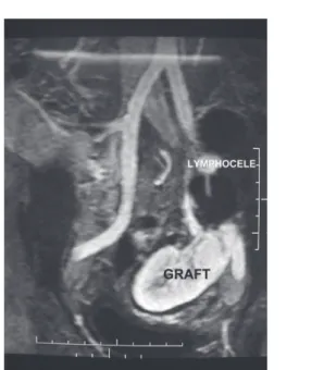
A 52-year-old non-dialytic man with long-standing dia-betes was submitted to simultaneous cadaveric pancreas-kidney transplant through peritoneal access. The right kid-ney and the pancreas from the same donor were sewn into the left flank by venous and arterial termino-lateral anas-tomosis in the iliac vessels. After an uneventful operation, the serum creatinine level dropped from 4.5 mg/dl and sta-bilized at 1.8 mg/dl on the seventh PO day in conjunction with a mild hypertension. Magnetic resonance angiogra-phy (MRA) showed a well perfused kidney (Figure 1). The patient had a renal biopsy revealing acute tubular necrosis (ATN). Cyclosporine, prednisone and mycophenolate mofetil were maintained. On clinical follow-up, creatinine level varied from 2.2 to 4.4 mg/dl and a new biopsy re-vealed persistent ATN. The cyclosporine level was kept at 150-250mg/dl. After three months, new MRA imaging re-vealed a well perfused, but dystopic kidney (Figure 2)
dis-Figure 1 - Magnetic resonance showing an upper pole lymphocele displacing the renal graft.
148
CLINICS 2008;63(1):147-8 A well-documented case of chronic renal failure due to misplacement of the transplanted kidney
Rodrigues P et al.
placed by a 500 ml lymphocele in the upper pole.
On the 120th PO day, the patient was submitted to open
surgical exploration but the arterial de-torsion was quite dif-ficult due to an intense fibrotic reaction, resulting in kid-ney retrieval after massive arterial lesion.
DISCUSSION
Complications arising from kidney transplants may
re-sult from immunological, urological, or vascular causes.5
Leakage of urine through the incision and/or persistent anu-ria are signs of early urological problems. These scenarios are usually investigated by percutaneous biopsy and ultra-sound exploration, either of which may indicate problems with causes ranging from immunological to surgical issues. Asymptomatic lymphocele collections are a well-known complication related to rejection or to improper donor lym-phatic ligation during the harvesting dissection.
Lymphocele formation is a frequent event in kidney transplantation, occurring in 22% of cases, although it does not seem to be associated with the source of the kidney,
but rather with rejections or ATN episodes.6
Most lymphocele formations are asymptomatic and do not harm the graft, but large or irregularly positioned for-mations may hamper the functioning of the ureter or im-pede vascular flow.
In our case, it is noteworthy that the decrease in the cre-atinine level ceased after the patient resumed walking, which possibly contributed to kidney displacement and torsion. Since fluid collection is more frequent in simultaneous pancreas-kid-ney grafting, some authors advocate intra-peritoneal placement of both organs in order to avoid retroperitoneal collections fre-quently seen from minor pancreatic collections.
The potential complication of renal pedicle torsion re-lated to this approach is a concern, though it has never been
reported in the literature.7 The kidney becomes loose in the
peritoneal cavity and, as seen in our case, the intra-perito-neal approach did not prevent lymphocele formation.
Although widely discussed, torsion or compression of the pedicle and displacement of the graft have rarely been re-ported in the literature8,9 due to inadequate methods of
visu-alization or restricted usage of imaging tools because of their invasiveness and lack of contrast. Arterial stenosis or kinking
at the vascular anastomosis may occur in 3.5% to 12.5%3,4
of grafts, but invasive investigation could not be justified un-less hypertension or progressive graft loss supervenes.
The advent of MRA has allowed for the study of the vascular pedicle with minimal clinical manipulation and no nephro-toxicity. Conventional arteriography, despite high resolution, is limited by its planar nature. MRA is a desir-able approach due to the possibility of image reconstruc-tion into a 3D image.
In our case, there was evident torsion of the pedicle in the post-operative period leading to stabilization of the de-creasing creatinine levels at an elevated level with fluctua-tions thereafter. The torsion was evident in the open field, but the fibrosis impeded a simple manual de-torsion. Knotty resection and re-anastomosis led to thrombosis and graft loss, revealing the difficulty in approaching the pedicle in surgical revisions for vascular complications. As reported by others, this results in a higher rate of transplant nephrec-tomies.3
It is impossible to determine the real cause of the de-scribed displacement, but it may be related to the intra-peri-toneal position of the kidney frequently used for simulta-neous pancreas-kidney transplants. This approach, as well
as peritoneal fenestration, has been advised10 in order to
minimize the formation of lymphoceles and to facilitate peritoneal absorption of intra-abdominal collections, but it did not provide absolute prevention in our case.
REFERENCES
1. Doyle AJ, Gregory MC, Terreros DA: Percutaneous renal biopsy: comparison of a 1.2 mm spring-driven system with a traditional 2 mm hard-driven system. Am J Kidney Dis. 1994;23:498-451.
2. Burstein DM, Korbet SM, Schartz MM: The use of the automatic core biopsy system in percutaneous renal biopsies: a comparative study. Am J Kidney Dis. 1993;22:545-548.
3. Roye SFS, van der Vliet JA, Hoitsma AJ, Reinaerts HHM, Buskens FGM: Early vascular complications of renal transplanation. Clin transplantation. 1993;7:496-500.
4. Rijksen JFW, Koolen MI, Warassewski JE: Vascular complications in 400 consecutive renal alotransplants. J Cardiovasc Surg. 1982;23:91-95. 5. Shoskes DA, Hanbury D, Cranston D, Morris PJ: Urological
complications in 1000 consecutive renal transplant recipients. J Urol. 1995;153:18-21.
6. Hauli RB, Stoff JS, Lovewell T: Post-transplant lymphoceles: a critical look into risk factors, pathophysiology and management. J Urol. 1993;150, 22-26.
7. Knight RJ, Matsumoto C: Renal pedicle torsion as a cause of acute allograft after kidney-pancreas transplantation. Abstract P1-09, Acta Chirurgica Astriaca, 8th Congress IPITA, Innsbruck. June 12-15, 2001, 33:174. 8. Williams SG,McVicar JP,Low RK: Endopyelotomy of ureteropelvic
junction obstruction caused by torsion of a renal allograf. J Urol. 1999, 161:1560-1564.
9. Marvin RG, Halff GA, Elshihabi I: Renal allograft torsion associated with Prune-Belly syndrome. Ped Nephrol. 1995;9:8-10.