Márlio Vinícius de Oliveira1
Skeletal and dental Class II malocclusion, with anterior open bite
and accentuated overjet*
Open bite is defined as a deficiency in normal vertical contact between antagonist teeth and may manifest in a limited region, or more rarely throughout the entire dental arch. If the lack of contact between teeth is located in the incisor and/or canine region when occlusion is in centric relation, it is called anterior open bite (AOB). Some studies have demonstrated that AOB is strongly associated with non-nutritional sucking habit. This article relates the treatment of a female African-Brazilian patient, with 20 years and 7 months of age, who presented Angle’s Class II, division 1 malocclusion, AOB, accentuated overjet, lingual interposition during swallowing and difficulty with pronouncing some phonemes. Orthodontic treatment began by mounting an Edgewise Standard fixed appliance system, with a fixed palatal crib appliance and extraction of maxillary first premolars. This case was presented to the Brazilian Board of Orthodontics and Facial Orthopedics (BBO), as part of the requisites to become a BBO Diplomate.
Keywords: Angle’s Class II malocclusion. Open bite. Tooth extraction.
How to cite this article: Oliveira MV. Skeletal and dental Class II malocclusion, with anterior open bite and accentuated overjet. Dental Press J Orthod. 2012 Mar-Apr;17(2):162-9.
Submitted: January 9, 2012 - Revised and accepted: March 11, 2012
» The author reports no commercial, proprietary, or financial interest in the products or companies described in this article.
» Patients displayed in this article previously approved the use of their facial and in-traoral photographs.
Contact address: Márlio Vinícius de Oliveira
R. Mato Grosso, 450 – Universal – CEP: 32686-050 – Betim/MG – Brazil E-mail: [email protected]
* Clinical Case Report, Category 7, approved by the Brazilian Board of Orthodontics and Facial Orthopedics (BBO).
1 Specialist in Orthodontics, Federal University of Alfenas (Unifal). Diplomate from
INTRODUCTION
The patient, a 20 years and 7 months Afro-Bra-zilian girl, presented for the initial exam in a good state of general health. Her medical and dental history contained no significant records. Her oral hygiene was good and her third molar had been ex-tracted. During anamnesis the patient reported that she had used a pacifier until she was eight years old. Her chief complaint was the “difficulty with pro-nouncing some words”.
DIAGNOSIS
The patient presented a symmetrical face, con-vex profile (upper lip – S line = 7 mm, Lower lip – S
line = 7 mm), acute nasolabial angle, lip sealing with contraction of the perioral muscles and chin muscle, lingual interposition when swallowing and difficulty with pronouncing some phonemes. Her smile, in spite of AOB, was pleasant, with a good contour, as well as an adequate exposure of the maxillary incisors.
Regarding her dental aspect, she presented Angle’s Class II division 1 malocclusion, 5.5 mm overjet, a 3 mm anterior open bite, coincident upper and lower midlines, triangular-shaped maxillary arch and para-bolic mandibular arch, both presenting symmetry in the anteroposterior and transverse directions. The patient presented no arch perimeter and Bolton dis-crepancy (Figs 1 and 2).
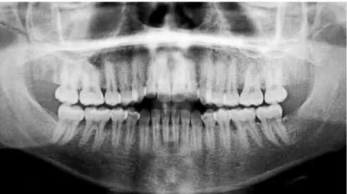
The periapical and panoramic radiographs dis-closed a normal bone, absence of third molars and a good root parallelism. With regard to the skeletal pattern, she presented a 5° ANB angle (SNA=79° and SNB=74°), showing evidence of disharmony between the bony bases, according to Steiner’s analysis. From the vertical aspect, there was an accentuated inclination of the mandibular plane (SN-G. Gn=40°) (Fig 4).
TREATMENT GOALS
The treatment goals were to establish canine Class I occlusion, correct the AOB, overjet and attain ideal functional occlusion. For this purpose extraction of the maxillary first premolars was necessary. In ad-dition, after closure of the OAB, and speech therapy, facial esthetics were expected to improve as well as the pronunciation of some sounds articulated by the teeth, lips and tongue.
TREATMENT PLAN
The plan was to correct canine Class II relation-ship and extract the maxillary first premolars. Regard-ing anterior open bite, the option was to use the fixed
palatal crib during the alignment, leveling and canine retraction phase. When incisor retraction began, the crib was removed and a lower spur was bonded on the lingual face of the incisors. At this time, when AOB and overjet had diminished, the patient was referred for speech therapy treatment.
The fixed appliance used was of the Edgewise Stan-dard type, slot 0.022 x 0.028-in, stainless steel arches from 0.014 to 0.020-in were used for alignment and leveling, and maxillary canine retraction began in 0.018-in arch. For incisor retraction a 0.019 x 0.025-in rectangular stainless steel arch with “bull” loops was used distal to the lateral incisors.
For finishing, a rectangular 0.019 x 0.025-in maxil-lary and mandibular arches were used, with first and third order bends, individualized according to the needs of the case. After the active treatment stage, up-per and lower retention appliances would be placed.
TREATMENT PROGRESS
Orthodontic bands with brackets welded on the buccal region and lingual tubes were adapted to the first maxillary molars. Transfer impressions were tak-en in order to fabricate the crib appliance.
A B
Figure 3 -Initial panoramic and periapical radiographs.
Figure 4 - Initial cephalometric profile radiographs and cephalometric tracings.
After cementation of the orthodontic bands, the crib was fitted into the lingual tubes and tied with metal ligatures (Fig 5). The remainder of the fixed ap-pliance was installed on the maxillary and mandibu-lar dental arches, Edgewise standard system with slot 0.022 x 0.028-in.
After the appliances were installed, extraction of the maxillary first premolars was requested, and in se-quence, round stainless steel arches (0.014 and 0.020-in) for alignment and leveling were inserted.
In the maxillary arch, the posterior teeth were tied together, on both sides, using 0.018-in wire. Elas-tic chains were placed from the second premolars to the canines to distalize them. After canine distaliza-tion, the stainless steel 0.019 x 0.025-in arch made of a rectangular section with bull loops located in the distal region of the lateral incisors for retraction of
the canines. At this stage the fixed palatal crib was re-moved and spurs were bonded on the lingual surface of the mandibular incisors to control tongue position (Fig 6). The patient was referred to speech therapy.
Figure 5 - Fixed palatal crib. Figure 6 - Bonded mandibular spurs.
Figure 7 -Final facial and intraoral photographs.
ACHIEVED RESULTS
When evaluating the patient’s records after the end of active treatment, it was observed that the main goals had been attained. Facial esthetics was enhanced by the reduction in lip protrusion, functional balance
between the lips resulting from closure of the AOB and speech therapy. The smile remained pleasant in spite of the small increase in exposure of the maxillary incisors (Fig 7).
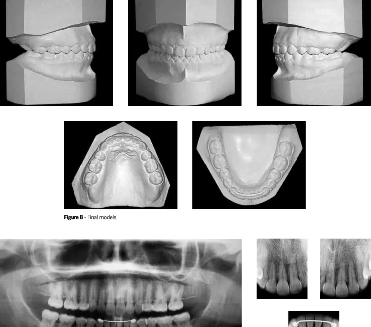
Figure 8 - Final models.
Figure 9 - Final panoramic and periapical radiographs.
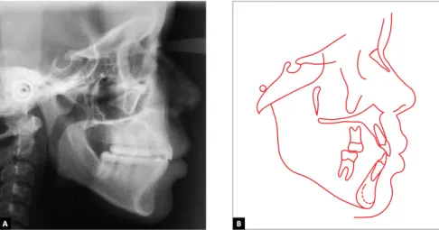
remained unaltered even with retraction of the in-cisors. The mandibular position remained verti-cally unaltered, however, SNB angle increased by 2 degrees, demonstrating a more anterior position at the end of treatment (Fig 10 and Table 1). This anti-clockwise rotation of the mandible, verified in the total superimposition of the initial and final trac-ings (Fig 11), may have occurred as a result of the im-provement in the cusp-fossa relationship at the end
of treatment, and/or as a result of the loss of anchor-age of the maxillary molars.
A
A
B
B
Measures Normal A B A/B Difference
Skeletal pattern
SNA (Steiner) 82° 79° 79° 0
SNB (Steiner) 80° 74° 76° 2
ANB (Steiner) 2° 5° 3° 2
Convexity angle (Downs) 0° 10° 6° 4
Y axis angle (Downs) 59° 61° 66° 5
Facial angle (Downs) 87° 87° 83° 4
SN–GoGn (Steiner) 32° 40° 40° 0
FMA (Tweed) 25° 29 35° 6
Dental pattern
IMPA (Tweed) 90° 94° 88° 6
1–NA (degrees) (Steiner) 22° 31° 22° 9
1–NA (mm) (Steiner) 4 mm 9 mm 6 mm 3
1–NB (degrees) (Steiner) 25° 31° 25° 6
1–NB (mm) (Steiner) 4 mm 8 mm 7 mm 1
1
1 – Interincisal angle (Downs) 130° 109° 130° 21
1–APo (mm) (Ricketts) 1 mm 5 mm 5 mm 0
Profile Upper lip – S line (Steiner) 0 mm 7 mm 5 mm 2
Lower lip – S line (Steiner) 0 mm 7 mm 6 mm 1
Table 1 -Summary of cephalometric measurements.
Figure 10 -Final cephalometric profile radiographs and cephalometric tracings.
FINAL CONSIDERATIONS
Open bite is defined as a deficiency in normal ver-tical contact between the antagonist teeth, and may be manifested in a limited region, or more rarely, throughout the entire dental arch.1,2 If lack of contact
of the teeth is located in the region of the incisors and/ or canines, when occlusion is in centric relation, then this is denominated anterior open bite.1,3
Some studies have shown that anterior open bite is strongly associated with non-nutritional sucking habit.4,5,6 In the case of this patient, who
reported having used a pacifier up to the age of 8 years, it is believed that the open bite was main-tained by lingual interposition after pacifier re-moval. Regarding the functional alteration caused by the anterior open bite, it was planned to use the fixed palatal crib at the beginning of treatment, and afterwards, when the open bite had been reduced, mandibular spurs would be bonded to the lingual surface of the incisors and the patient would be re-ferred for speech therapy.
In this patient, the option was to extract the max-illary first premolars to correct the overjet and Class II canine relationship. Treatment of Class II without extractions requires extensive distal movement of the maxillary posterior teeth, which frequently becomes complicated, particularly in adult patients7. Recent
studies have demonstrated that for the same age and degree of severity, the protocol for Class II malocclu-sion treatment with extraction of the two maxillary premolars is more efficient than the treatment proto-col without extractions.8,9
Regarding stability, it is consensus that anterior open bite is ranked among the most challenging treat-ments and its end results have been shown to be less stable. However, it is believed that with the establish-ment of a correct overjet, over bite and muscular bal-ance between the lips and tongue there will be no re-lapse of the malocclusion.
As may be seen in the record obtained after the end of active treatment, all the goals were attained from both a functional and facial esthetic point of view.
1. Moyers RE. Ortodontia. 4th ed. Rio de Janeiro: Guanabara Koogan; 1991.
2. Van der Linden FP. Genetic and environmental factors in dentofacial morphology. Am J Orthod. 1966;52(8):576-83.
3. Graber TM, Vanarsdal RL. Ortodontia: princípios e técnicas atuais. Rio de Janeiro: Guanabara Koogan; 2002.
4. Forte FDS, Bosco VL. Prevalência de mordida aberta anterior e sua relação com hábitos de sucção não nutritiva. Pesq Bras Odontop Clin Integr. 2001;1(1):3-8. 5. Katz CRT. Relação entre hábitos de sucção, mordida aberta anterior, mordida cruzada posterior e morfologia facial em pré-escolares de Recife/PE: um estudo longitudinal [dissertação]. Camaragibe (PE): Universidade de Pernambuco; 2003. 6. Serra-Negra JMC, Pordeus IA, Rocha Junior JF. Estudo da associação entre
aleitamento, hábitos bucais e maloclusões. Rev Odontol Univ São Paulo. 1997;11(2):79-86.
REFERENCES
7. Bishara SE, Cummins DM, Jakobsen JR. The morphologic basis for the extraction decision in class II, division 1 malocclusions: a comparative study. Am J Orthod Dentofacial Orthop. 1995;107(2):129-35.
8. Janson G, Barros SE, de Freitas MR, Henriques JF, Pinzan A. Class II treatment efficiency in maxillary pre-molar extraction and nonextraction protocols. Am J Orthod Dentofacial Orthop. 2007;132(4):490-8.