Department of Orthopedics, University of São Paulo, Medical School - São Paulo/SP, Brazil.
Email: [email protected]
Received for publication on October 14, 2005. Accepted for publication on April 19, 2006
ORIGINAL RESEARCH
ALTERATION OF THE LOAD-RESPONSE MECHANISM
OF THE KNEE JOINT DURING HEMIPARETIC GAIT
FOLLOWING STROKE ANALYZED BY
3-DIMENSIONAL KINEMATIC
Paulo Roberto Garcia Lucareli, Julia Maria D’Andrea Greve
Lucarelli PRG, Greve JMA. Alteration of the load-response mechanism of the knee joint during hemiparetic gait following stroke analyzed by 3-dimensional kinematic. Clinics. 2006;61(4):295-300.
PURPOSE: The aims of this study were to evaluate the variables found in the alteration of the load-response mechanism on 3-dimensional kinematic analysis of the knee joint during hemiparetic gait following stroke.
METHODS: We evaluated 66 adult patients (33 men and 33 women), aged 45.4 ± 8.5 years (mean ± SD), with a diagnosis of ischemic cerebrovascular accident either right or left hemiparesis and brachial prevalence. All the participants underwent 3-dimensional gait evaluation with a Vicon 370, and the values of the angular kinematics of the knee joint were selected for analysis.
RESULTS: There were no statistically significant differences (by the Kruskal-Wallis test) between the subjects regarding the following variables: angular knee position at initial contact and time of peak knee flexion in the stance. The clinically relevant characteristics found were: an increase in knee joint flexion during the initial contact and a movement amplitude below that anticipated in this phase of the walking cycle. These should be taken into account when choosing the best treatment, because they are the ones which exhibit the most important alteration in the load-response mechanism in all patients.
CONCLUSION: There is still no consensus among the different specialists regarding the variations in kinematics during the hemiparetic gait. One of the most frequently discussed joints is the knee—the way the main changes take place during the gait cycle and whether the gait velocity changes the patterns of joint mobility.
KEYWORDS: Hemiplegia, Knee, Biomechanics, Gait, Neurologic Gait Disturbance.
INTRODUCTION
The leading gait cycle changes that take place as se-quelae of neurological disorders and the walking patterns in cerebral palsy are the most studied The large number of gait studies in cerebral palsy led the scientific community to a better understanding of how the pattern of biomechan-ics of normal gait is changed in this population. As a re-sult, some changes were found and some walking patterns were defined in an attempt to find the best treatment strat-egy for each kind of change1-8. One of the most clarifying
studies on walking pattern in cerebral palsy is the one which defines walking according to knee movement throughout the walk cycle. In other populations with neu-rological sequelae, gait is also one of the greatest–if not the greatest–problems during rehabilitation. Craik and Oatis9 note that approximately 70% of the patients
surviv-ing a brain stroke regain their walksurviv-ing capacity. This abil-ity needs to be understood for better treatment and reha-bilitation of these patients.
The challenge is that so far there is not a consensus among the several experts on the kinematic joint variables during hemiparetic gait. The most discussed joint is the knee regarding how the most important changes behave during the walk cycle10-14 and whether the walking speed
changes the mobility patterns of this joint.15,16 The
aims to reduce the impact on the lower limb and smoothen the mass center displacement, thus reducing energy ex-penditure.
The aim of this study was to describe and evaluate the angular kinematic variables of the knee joint during load response in hemiparetic gait from brain stroke.
MATERIALS AND METHODS
Sixty-six adult patients (33 women and 33 men) were involved in this study. Their ages were 45.4 ± 8.5 years (mean ± SD; range, 31-60). Their weight averaged 67.6 ± 16 kg (range, 44-110), and their height averaged 161.3 ± 9.7 cm (range, 136-189).
The inclusion criteria were diagnosis of ischemic brain stroke sequela with right or left hemiparesis, being at least 12 months post injury; being a community walker; not need-ing walkneed-ing aids; beneed-ing able to walk on bare feet, and not having undergone previous orthopedic surgery.
All study subjects had a clinical indication for tridimentional gait evaluation and were referred to the labo-ratory by medical request. Having met the above inclusion criteria, the patients were asked to consent to the use of the final report of their test in this study and sign an in-formed consent form.
The data were collected at the Gait Laboratory of the Associação de Assistência à Criança Deficiente – AACD, in the city of São Paulo.
After anamnesis and evaluation regarding inclusion cri-teria, the subjects underwent the tridimentional computer-ized gait analysis protocol. Six infra-red VICON/PEAK®
370 movement analysis cameras and the kinematic mark-ers (Helen Hayes marker system) for the pelvis, hip, knee, and ankle joints were used.17,18
For each cycle in the exam track, a single walking cy-cle was selected. Using the Vicon Clinical Manager soft-ware, the tridimentional kinematic data of the pelvis, hip, knee, and ankle joints were expressed in graphs of the an-gle positioning of the joint versus time, thus generating 12 graphs for each subject. From the total of 12 cycles, only 1 was chosen for analysis. This selection was based on the protocols of the Gait Analysis Laboratory of AACD, tak-ing as a parameter the mean value of angular kinematics and the mean walking speed. The same cycle was used for angular kinematics analysis of the knee in the sagittal plane. Three variables were defined based on the knee joint in sagital plane graph: knee angular position at initial con-tact, knee first peak flexion in the stance phase (degrees), time of peak knee flexion in the stance (% of the stance).
The comparison groups were formed from the walking speed parameter, taking into consideration the median
(nonparametric distribution) of all speeds measured, mak-ing up 2 large groups: one with a speed slower or equal to the median and the other with a speed higher than the me-dian. Each of these groups was divided into 2 subgroups, right and left, according to the hemiparesis-affected side.
Four groups were therefore formed: Group 1 – speed slower than the median, and left hemiparesis (LSLH), with 23 subjects; Group 2 – speed slower than the median, and right hemiparesis (LSRH), with 10 subjects; Group 3 – speed higher than the median, and left hemiparesis (HSLH) with 7 subjects; and Group 4 – speed higher than the me-dian and right hemiparesis (HSRH), 26 subjects.
The possible differences among the groups, according to predefined variables were evaluated by the Kruscal-Wallis nonparametric test for independent samples, com-pleted, as necessary, by the multiple comparisons test. The means were calculated and presented for information pur-poses. Standard deviation was not calculated, because of the use of a nonparametric test. The evaluated variables do not have a normal distribution; thus, there would have been no meaning to its calculation.
The rejection level for the null hypothesis was estab-lished as equal to or less than 0.05 (5%).
RESULTS
The results of angular kinematics for each studied sub-ject were distributed into 4 groups according to the evalu-ation parameters of walking speed median and affected side.
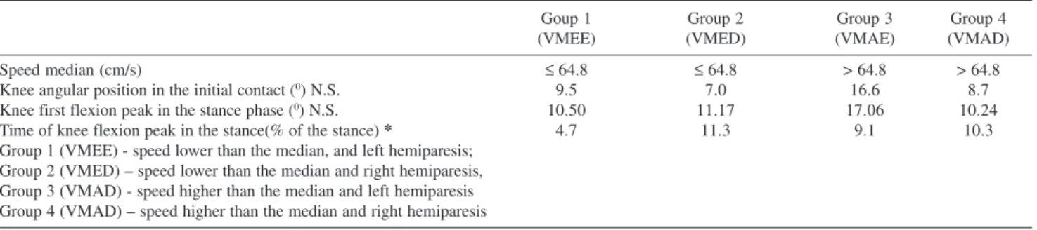
No significant difference was noted among the groups for the angular position variables at knee initial contact and peak knee flexion at loading response phase (Table 1).
The time-point of peak knee flexion in stance was sig-nificantly different in the 4 groups studied. After applica-tion of the complementary test of the absolute values of the differences between means, it was possible to note a smillarity between group 1 (LSLH) and group 4 (HSRH): the slower gait velocity changed the time-point in which the peak flexion occurred (Table 2).
DISCUSSION
Evaluation of joint and segment kinematics is a valu-able resource for clinical practice, since it can precisely measure the angular variation between the segments, clari-fying and quanticlari-fying what the human eye, even with great clinical experience, is unable to do.
When a patient is affected by a brain stroke, alterations in motor control make understanding the walking mechan-ics more complex. In most cases, such alterations affect the entire lower limb, and, all too frequently they are superfi-cially evaluated. It is not uncommon that clinicians make a wrong, or a partially wrong diagnosis, turning their at-tention to secondary changes, which are frequently more evident; consequently, the primary cause of the disability goes undetected. In these cases, the patient is submitted to interventions that do not actually treat the problem, caus-ing the gait patterns to return. Eventually, new patterns may appear.
The most effective alternative is to evaluate each joint individually and later understand the influence they have on each other; for instance, excessive knee flexion may affect the hip joint or even lead to postural changes of the body.
This work describes the angular kinematic changes of the knee joint during hemiparetic gait, in search for simi-larities among the evaluated variables, and trying to define a pattern for the knee joint.
Initially, the result of knee angular movement during the walk cycle was subdivided into 11 variables contain-ing values of angular positioncontain-ing durcontain-ing the cycle, speed values of angular movement, and values representing the cycle period when these events occurred.
The first characteristic found in the evaluated groups was the knee positioning at the start of the cycle, that is, at initial contact. The values found in our samples corre-sponded to those in the literature for normal gait19
inde-pendently of walking speed changes. Burdett et al10
de-scribed an increase in knee joint flexion during the initial contact, when subjects walked at “normal” speed. Olney,16
however, notes that speed variation affects the knee flexion: with increased flexion at faster walking speeds. Knutsson,11,12 Lehmann,13 Cozean,14 and Intiso,15 report that
the values found for the knee position at initial contact are different from the previous ones, with less knee flexion at initial contact. Thus, the literature data9-16 show
discrepan-cies, and this subject has not yet been completely clarified. However, it is known that changes in knee positioning may interfere with step length, compromising the walking cy-cle at its beginning, and may be reflected later in the walk-ing cycle.
The first peak knee flexion takes place in the bearing phase and is called load response. We found values agree-ing with the findagree-ings of Burdett et al,10 Olney,16 and
Kerrigan.20 These authors also described for patients with
hemiplegia a movement amplitude below that anticipated in this phase of the walking cycle when compared subjects with no neurological afflictions. Olney21 however,
disa-greed, stating that there is difference in the angular posi-tion of the knee in load response when the walking speed is increased or reduced.
The load response contributes to a smoothening of the sudden changes in the intersections of the track arcs of the Table 2 - Absolute value of the difference between the mean values of patients with ischemic brain stroke and hemiparesis of the four groups and least significant difference
Group 1, Group 2 Group 1, Group 3 Group 1, Group 4
VADMP DMS VADMP DMS VADMP DMS
Time to knee flexion peak in load response (% of load response) 14.52 19.18 12.14 21.86 12.90 § 14.50 Time to knee flexion peak in load response (% of load response) 2.38 24.96 1.62 18.85 0.76 21.57 VADM - absolute value of the means difference; DMS - least significant difference; § Groups values approximation
Table 1 - Speed median value and mean value of the kinematic variables of knee joint of patients with ischemic brain stroke sequela
Goup 1 Group 2 Group 3 Group 4
(VMEE) (VMED) (VMAE) (VMAD)
Speed median (cm/s) ≤64.8 ≤ 64.8 > 64.8 > 64.8
Knee angular position in the initial contact (0) N.S. 9.5 7.0 16.6 8.7
Knee first flexion peak in the stance phase (0) N.S. 10.50 11.17 17.06 10.24
Time of knee flexion peak in the stance(% of the stance) * 4.7 11.3 9.1 10.3
mass center. More specifically, the weight load response flat-tens the arc that the mass center displaces throughout the movement. The change produced by this mechanism reduces the quality and quantity of ground shock absorption.
Changes in this walk phase can be due to several causes: (i) weakness of the quadriceps muscle resulting in excessive knee flexion; (ii) spasticity of the quadriceps muscle lead-ing to stretchlead-ing the quadriceps when startlead-ing flexion, in turn leading to a hypertonic response and consequent premature knee extension; (iii) excessive plantar flexion (plantar flexion contracture and spasticity of the soleous muscle); (iv) fail-ure to make make the initial contact with the rear foot, caus-ing inhibition or blockcaus-ing of the rollcaus-ing mechanism of the tibia over the foot during load bearing.21-24
The time-point of the peak knee flexion during load bearing, that is, an event which occurred while the knee was in flexion, was significantly different among the 4
stud-ied groups. Although the evaluated groups presented a sig-nificant difference in the duration of the flexion, their av-erage values all ranged from 4.7% to 10.3% of the walk-ing cycle. These values are below the normal gait pattern described in literature,19,22 where the reference values for
duration of flexion are 15% of the gait cycle. The loss of selective control, the absence of efficacious contraction of knee extensor muscles, or even the spasticity of plantar flexors, could be pulling the tibia backwards.
Therefore, an important alteration in the load response caused to the angular kinematics of the knee joint of the knee during hemiparetic gait following cerebral vascular accident modifies in an important way one of the prereq-uisites for the normal gait and increases the energy con-sumption during deambulation. Further studies are needed to detect the possible causes of this alteration to direct sur-gical treatment and therapeutic choices for these patients.
RESUMO
Lucarelli PRG, Greve JMA. Alteração do mecanismo de resposta à carga da articulação do joelho durante a marcha hemiparética oriunda de AVC, analisada por cinemática tri-dimensional. Clinics. 2006;61(4):295-300.
OBJETIVO: O objetivo deste estudo foi avaliar as variáveis resultantes da alteração do mecanismo de resposta à carga por análise tridimensional da cinemática angular da articulação do joelho durante a marcha hemiparética oriunda de acidente vascular cerebral.
MÉTODO: Estudo retrospectivo com 66 pacientes adultos de ambos os sexos (33 masculino e 33 feminino), com idade 45,4 ± 8,5 anos, com diagnóstico de acidente vascu-lar cerebral isquêmico com hemiparesia em lado direito ou esquerdo. Todos os pacientes foram submetidos ao protocolo de realização do exame tridimensional computadorizado de marcha por meio de VICON/PEAK® 370. Os valores da cinemática angular do joelho foram selecionados para análise.
estatisticamente significantes (teste de Kruskal Wallis) en-tre os sujeitos para as variáveis: posição angular e pico de flexão do joelho (p<0,05). As caracterísitcas clínicas relevantes encontradas foram: aumento da flexão do joelho no contato inicial e amplitude de movimento abaixo do esperado nesta fase do ciclo de marcha, estas são as que devem ser levadas em conta na seleção do melhor tratamento, pois são as que apresentam alterações importantes no mecanismo de resposta à carga na totalidade dos pacientes.
DISCUSSÃO: O mecanismo de resposta à carga é
funda-REFERENCES
mental para o andar porque tem como objetivo reduzir o impacto no membro inferior e suavizar o deslocamento do centro de massa no espaço reduzindo assim o consumo energético. Esta capacidade necessita ser compreendida para ser então melhor tratada e reabilitada. A dificuldade é que ainda não há consenso entre os diversos estudiosos do tema sobre as variáveis cinemáticas das articulações du-rante a marcha hemiparética.
UNITERMOS: Hemiplegia, Joelho, Biomecânica, Marcha, Distúrbios Neurológicos da Marcha.
1. Arnold AS, Liu MQ, Schwartz MH, Ounpuu S, Delp SL.The role of estimating muscle-tendon lengths and velocities of the hamstrings in the evaluation and treatment of crouch gait. Gait Posture. 2005 Jun 16 [Epub ahead of print].
2. Arnold AS, Liu MQ, Schwartz MH, Ounpuu S, Delp SL. Do the hamsrings operat at increased muscle tendon lengths and velocities after surgical lengthening? J. Biomech. 2006;39:1498-506.
3. Lyon R, Liu X, Schwab J, Harris G. Kinematic and kinetic evaluation of the ankle joint before and after tendo achilles lengthening in patients with spastic diplegia. J Neurosurg. 2005;102(4 Suppl):385-9. 4. Wong AM, Pei YC, Lui TN, Chen CL, Wang CM, Chung CY.
Comparison between botulinum toxin type A injection and selective posterior rhizotomy in improving gait performance in children with cerebral palsy. Dev Med Child Neurol. 2005;47:329-36.
5. Rose J, McGill KC. Neuromuscular activation and motor-unit firing characteristics in cerebral palsy. Dev Med Child Neurol. 2005;47:329-36.
6. Gage JR. Gait Analysis in cerebral palsy. London: Mac Keith Press; 1991.
7. Gage JR. Treatment of gait problems in cerebral palsy, London: Mac Keith Press; 2004.
8. Sutherland DH, Davids JR. Common gait abnormalities of the knee in cerebral palsy. Clin Orthop Relat Res. 1993;288:139-47.
9. Craik RL, Oatis CA. Gait analysis: theory and applications. St. Louis: Mosby-Year Book; 1995.
10. Burdett RG, Borello-France D, Blatchly C, Poptter C. Gait comparison of subjects with hemiplegia walking unbraced, with ankle-foot orthosis, and with Air-Stirrup brace. Physical Therapy. 1988;68:1197-203. 11. Knutsson E, Richards C. Different types of disturbed motor control in
gait of hemiparetic patients. Brain. 1979;102:405-30.
13. Lehmann JF, Condon SM, Price R, Delateur BJ. Gait abnormalities in hemiplegia: their correction by ankle-foot orthoses. Arch Phys Med Rehabil. 1987;68:763-71.
14. Cozean CD, Pease WS, Hubbell SL. Biofeedback and functional electric stimulation in stroke rehabilitation. Arch Phys Med Rehabil. 1988;69:401-5.
15. Intiso D, Santilli V, Grasso MG, Rossi R, Caruso I. Rehabilitation of walking with electromyographic biofeedback in foot-drop after stroke. Stroke. 1994;25:1189-92.
16. Olney SJ, Griffin MP, Monga TN, Mcbride ID. Work and power in gait of stroke patients. Arch Phys Med Rehabil. 1991;72:309-14. 17. Davis RB, Ounpuu S, Tyburski D, Gage, JR. A gait analysis data collection
and reduction technique. Hum Mov Sci. 1991;10:575-87.
18. Kabada MP, Ramakrishnan HK, Wootten ME. Measurement of lower extremity kinematics during level walking. J Orthop Res. 1990;8:383-92.
19. Gamble JG, Rose J. Marcha humana. Baltimore: Premier; 1998. 20. Kerrigan DC, Karvosky ME, Riley PO. Spastic paretic stiff-legged gait:
joint kinetics. Am J Phys Med Rehabil. 2001;80:244-9.
21. Olney Sj, Richards C. Hemiparetic gait following stroke. Part I: Characteristics. Gait and Posture. 1996;4:136-48.
22. Perry J. Gait analysis, normal and pathological function. Thorofare, NJ: Charles B. Slack; 1992.
23. Shiavi R, Bugle Hj, Limbird T. Electromyographic gait assessment, Part 2: Preliminary assessment of hemiparetic synergy patterns. J Rehabil Res Dev. 1987;24:24-30.