Debonding forces of different pads in a
lingual bracket system
Valter O. Arima1, Mario Vedovello Filho1, Heloísa C. Valdrighi1, Adriana S. Lucato1, Milton Santamaria Jr.1, Silvia A. S. Vedovello1
Objective: To evaluate the shear bond strength of lingual orthodontic brackets with resin or metal pads, the location of bond failure and the adhesive remnant index (ARI). Methods: A total of 40 extracted upper premolars were randomly divided into two groups of 20 each: bonding with brackets having (1) pads with extended resin directly on the lingual surface of teeth, and (2) pads with metal custom base on the lingual surface of teeth. The debonding force was measured with an Instron universal testing machine. A Student’s t-test was used to assess the difference between groups (α = 0.05). Results: The results showed a significant difference between the groups (p < 0.001). The shear bond strength of metal pads was significantly higher than resin pads. Conclu-sions: Within the limitations of this in vitro study, it was concluded that the bond strength of lingual brackets with metal pads was higher than that of brackets with composite resin pads, due to the metal part being a single unit and welded. The failure location in the region between the bracket and the resin pad affected a higher percentage of the resin pads than the metal pads.
Keywords:Orthodontic brackets. Dental debonding. Shear strength.
1 UNIARARAS, Fundação Hermínio Ometto (Araras/SP, Brasil).
Submitted: April 10, 2016 - Revised and accepted: March 09, 2017
» The authors report no commercial, proprietary or financial interest in the products or companies described in this article.
DOI: https://doi.org/10.1590/2177-6709.22.4.034-040.oar
How to cite: Arima VO, Vedovello Filho M, Valdrighi HC, Lucato AS, San-tamaria Jr. M, Vedovello SAS. Debonding forces of different pads in a lin-gual bracket system. Dental Press J Orthod. 2017 July-Aug;22(4):34-40. DOI: https://doi.org/10.1590/2177-6709.22.4.034-040.oar
Contact address: Silvia A. S. Vedovello
Av. Dr. Maximiliano Baruto, 500 - Jardim Universitário Araras/SP, Brasil – E-mail: [email protected]
Objetivo: avaliar a resistência de união ao cisalhamento de braquetes ortodônticos linguais com pads em resina e me-tálicos, assim como os locais de ocorrência das falhas, além do Índice de Adesivo Remanescente (ARI). Métodos: 40 pré-molares superiores extraídos foram aleatoriamente divididos em dois grupos de 20 espécimes cada, para a colagem de braquetes com: 1) pads em resina, estendida diretamente na face lingual dos dentes; e 2) pads com base metálica cus-tomizada, na face lingual dos dentes. A força de descolagem dos braquetes foi mensurada por meio de uma máquina de ensaio universal (Instron). O teste t de Student foi utilizado para avaliar diferenças intergrupos (α = 0,05). Resultados:
os resultados demonstram haver diferença significativa intergrupos (p < 0,001). A resistência de união ao cisalhamento dos pads metálicos foi significativamente maior em comparação aos pads em resina. Conclusões: dentro das limitações desse estudo in vitro, foi possível concluir que a resistência de união de braquetes linguais com pads metálicos foi superior à resistência de braquetes com pads em resina composta, uma vez que os pads metálicos customizados e os braquetes são fundidos, formando uma única unidade. A porcentagem de falhas de colagem na região entre o braquete e o pad foi maior para os pads em resina, em comparação aos pads metálicos.
INTRODUCTION
The lingual technique offers significant aesthetic advantages for patients submitted to orthodontic treatment, and therefore demands continuous
devel-opment of brackets1,2 and related materials, aiming
to obtain results similar to those from conventional
labial orthodontic treatment.3
For better adaptation to the lingual surface of teeth, compensations have been created at the bracket bases. Also, denominated pads, these com-pensations are fabricated during the set-up of mal-occlusion and used to correct torque, angulation
and in/out.4 However, pads made of composite resin
have shown the disadvantage of a high debonding rate, attributed to the fact that the contact area with
the lingual surface of the tooth is smaller.5
There are several options available for lingual systems, and due to the wide variation in the mor-phology of tooth surface, making an individual cus-tomized base for each bracket has shown to be the most adequate option, with the purpose of avoiding
bends in the arches during the course of treatment.6
At present, there are systems available in which the lingual bracket is fabricated by means of computer-aided design (CAD) and computer-computer-aided manu-facturing (CAM). This technology minimizes the bracket profile and reduces discomfort to the patient, because customized metal pads and brackets are case
in a single unit.7 In spite of the technology provided
by 3D digitization in lingual appliances,8,9 their cost
is still high in comparison to the conventional lin-gual technique. However, the metal pad fabricated in a personalized manner has been reported to show
greater resistance to fracture.7 It is worth pointing out
that the literature is not conclusive with respect to the resistance of brackets with different bases or pads used
in the lingual orthodontic technique.6,10
The aim of this study was to evaluate the influ-ence of different pads (resin and metal) on the shear bond strength to human enamel,as well as the Ad-hesive Remnant Index (ARI) and site of failures. The hypothesis tested was that there is difference on the shear bond strength among different pads.
MATERIAL AND METHODS
The present study was conducted ater being ap-proved by a Research Ethics Committee (988.213/2015).
Sample size was determined in a pilot study (n = 5) using a power analysis of 0.80 and 5% signiicance level. At least, 12 specimens would have been required to analyze debonding force. In this study, 40 speci-mens were used to analyze debonding force and the power analysis was found to be 0.90.
The 40 upper premolars were selected from among teeth extracted for orthodontic treatment, through a careful examination, using as exclusion criteria the presence of restorations, cracks or surface defects and previous orthodontic treatment. Ater that, they were cleaned with periodontal curette (Gracey 7/8, Golgran
Millenium®, São Caetano do Sul, São Paulo, Brazil),
washed with physiological serum, kept in test tubes
with thymol 0.1% chlorochemicals (Biotec®, Campo
Mourão, Paraná, Brazil), and stored in a refrigerator at
4oC until they were used.
The STb™ lingual brackets (Light Lingual
Sys-tem, Ormco®, Orange, California, USA) were
ran-domly divided into two groups with 20 each. Group 1 (n= 20) received personalized extended pads made of Transbond XT composite resin (3M Unitek, Mon-rovia, California, USA) and Group 2 (n = 20), metal pads. The area was standardized by measuring the greatest vertical and horizontal length of each lin-gual face of all premolars used, by means of the
cal-culation A = π R2. Then, the total area (28.66 mm²)
average was used as the default (7.06 mm²) (Fig 1).
A digital caliper (Starret®, São Paulo, São Paulo,
Brazil) was used to ensure fidelity.
The teeth were embedded in PVC tubes (Tigre®,
Santa Catarina, Brazil) measuring 25 mm in diameter by 20 mm high. To ix the teeth, chemically activated
acrylic resin Jet (Classic®, São Paulo, São Paulo,
Bra-zil) in the sandy phase was used. The lingual surface remained perpendicular to the base of the PVC tube.
The 20 customized pads of Group 1 were prepared
by applying a thin layer of Cel Lac (SS White®, Rio
de Janeiro, Rio de Janeiro, Brazil) insulator on the lingual surface of the teeth, with the use of a dispos-able paint brush, and drying the region with a light jet of compressed air for 15 seconds. Transbond XT composite resin was used at a 0.5-mm thickness
checked with an Iwanson spectrometer (Golgran®,
Figure 1 - Original bracket (A), bracket with resin pad (B), bracket with a metal pad (C).
and in/out were corrected. An extended pad was fabricated around the bracket, covering the lingual surface and crown of the tooth, delimited at approxi-mately 28.66 mm², however not covering the bracket slot region. After this, the resin was light-activated
with the FlashMax P3 (Rocky Mountain®, Denver,
Colorado, USA) appliance for 40 seconds.
Metal pads were fabricated for Group 2 following the same procedure for isolation of the tooth enamel. In this group, a layer of medium consistency wax was
sculpture (Babinete®, Curitiba, Paraná, Brazil) and
applied with the aid of a Hollenback 3S instrument
(Quinelato®, Rio Claro, São Paulo, Brazil),
cover-ing the lcover-ingual surface of the tooth crown, delimitcover-ing the same area as that in the previous group. The per-sonalized wax bases were sent for casting in a den-tal laboratory. The 20 cast parts were polished with
a metal polishing disc (Dedeco®, New York, New
York, USA) and welded to the bracket by means of a
laser welding appliance Model LM-D 60 (Sisma®,
It-aly). The weld was calibrated to 0.5mm with Iwanson spectrometer and the diameter of each welding spot was approximately 0.2 mm. All the materials used for joining the weld, orthodontic wire with diameter of
0.020 mm (Morelli®, Sorocaba, São Paulo, Brazil),
brackets and cast metal parts were made of nickel-chrome. The laser weld filled the space between the customized metal base and bracket, simulating the metal pad on which the torque, angulation and in/out
were corrected. It is important to note that torque, in/out and angulation could only be controlled if the orthodontic wire was in position. In this way, all pads were standardized to maintain the size and thickness in relation to the lingual surface.
Before cementing the pads of Groups 1 and 2, they were sandblasted with aluminum oxide par-ticles (50 micrometers) at a distance of 20mm and pressure of 2 bars for 5 seconds. The teeth were washed with water and dried, treated with acetone
(Equate®, São Paulo, São Paulo, Brazil), aiming at
removing residues and creating an area of mechani-cal retention.
The protocol for fixation of the brackets with metal pads was similar to that of the composite resin pad, with the difference in application of a bonding
agent (Metal Primer, Ivoclar, Vivadent®,
Liechten-stein, Germany) on the entire metal pad. The teeth were cleaned and polished with extra fine pumice
stone paste (SS White®, Paraná, Brazil) and water,
by using a Robson type brush (Microdont®, São
Paulo, São Paulo, Brazil) for 10 seconds; afterwards washed in running water for 15 seconds and dried with jets of oil- and water-free compressed air. The lingual surfaces were etched with phosphoric
acid 37% (Magic Acid Coltene®, Rio de Janeiro,
Rio de Janeiro, Brazil) for 30 seconds; washed with distilled water for 15 seconds and dried with a jet of compressed air.
During bracket bonding (pads made of resin and metal), a thin layer of Maxcem Elite resin cement
(Kerr®, Orange, California, USA) was applied on
the bases of the pads in the two groups. The brack-ets were pressed on manually and resin excess was removed with an exploratory probe. Light activa-tion was performed (20 seconds), being 5 seconds for each surface of the bracket, by using a halogen
FlashMax P3 (Rocky Mountain®, Denver,
Colo-rado, USA) light-activation appliance and light intensity of 4000-6000 mW/cm². The tip of the light-activating appliance was placed as close as pos-sible to the brackets and the specimens were stored
in deionized water in an oven (Olidef®, São Paulo,
Brazil) at 37oC, for 24 hours.
After the storage period had elapsed, the speci-mens were submitted to the shear bond strength test performed in an universal testing machine (Model 4411; Instron, Canton, MA, USA), at a speed of
1mm/minute11. The protocol used for all stages of
the study was based on previous literature.12
The shear bond strength values were obtained in (N) until failure occurred. The failure location was evaluated and classified as follows: (1) failure between the cement and tooth; (2) failure between the cement and customized pad; (3) failure between the base and pad; (4) failure between the pad and bracket; (5) fracture of the tooth.
The surface of the bracket and tooth were ob-served under an optical microscope (Olympus Corp, Tokyo, Japan) at 8x magnification. The Ad-hesive Remnant Index (ARI) was ranked in the se-quence, as follows: score 0 = absence of any resin on the enamel surface; score 1 = less than half the resin on the enamel surface; score 2 = more than half the resin on the enamel surface; score 3 = all the resin on the enamel surface, with the impression of the
bracket base.10
DATA ANALYSIS
The shear bond strength values were evaluated as regards the normality by the Shapiro-Wilk test and homoscedasticity of the variances by the Bartlett test. It was verified that distribution was not nor-mal and variances differed. Therefore, the original data were transformed into log and presented
nor-mal distribution (p = 0.3851) and equal variances
(p = 0.3291). A Student’s t-test was used to assess the
difference between groups (α = 0.05).
RESULTS
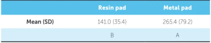
The results (Table 1) showed significant
dif-ference between the brackets (p = 0.001). Figure 2
shows the force required to remove the brackets with metal pads was significantly higher than the force to remove the brackets with resin pads.
Failure location analysis was performed in the two groups to verify the maximum limit of stress force used for shearing to occur. Failures were ob-served between the pad and bracket for both groups, with this percentage being 70% for the resin pads, and 45% for the metal pads. There was 30% frac-ture of the tooth in the customized metal pads, due to the high resistance of force required for shear-ing (Fig 3). According to Figure 4, there was a high-er phigh-ercentage of ARI with score 3 in both groups.
Resin pad Metal pad
Mean (SD) 141.0 (35.4) 265.4 (79.2)
B A
Table 1 -Shear bond strength (N) required to remove the lingual orthodontic
brackets with different pads.
DISCUSSION
This study compared the shear bond strength of lingual brackets with different customized pads (composite resin and metal). In the literature, studies that evaluate the shear bond strength of convention-al metconvention-al brackets are more frequently conducted in
comparison with those on the lingual technique,14,15
while those that evaluate the bond strength of
cus-tomized brackets are scarce.6
To compensate the great anatomic variability of the lingual surface of teeth, the purpose of the resin-based composite pad included at the base of the bracket is to correct the torque, angulation and in/out. However, on the customized metal pad, the
compensations are fabricated on the arches,7
differ-ently from the resin pads.
There is no previous study in the literature that has evaluated the shear bond strength of lingual brackets with customized pad in comparison with the metal pad in the lingual orthodontics technique. In the present study, metal pads were fabricated and included by means of welding between the metal pad and bracket, favoring lower laboratory costs.
The results of this study showed that the lin-gual bracket with metal pad had higher shear bond strength in comparison with those made of compos-ite resin, and the most fragile point of fracture for both groups was between the pad and bracket. Fur-thermore, it is pointed out that for the resin pads, this percentage was 70%, and for the metal pads, 45%. This high percentage may be due to various factors. Two probable hypotheses are the mechan-ical retention provided by the bracket base or the type of resin used in this study. Whereas the fail-ure in the metal pads could result from the welded union preformed during the process of fabricating the pad. The pulsed laser process is performed with superimposition of welding spots around the parts, so that there is contact between the customized pad and bracket. This enables better control of the
weld-ing energy,16 with the consequence of greater
fragil-ity of the parts. When polishing around the weld, this region becomes more regular, and may make it more vulnerable to failures.
During the tests, 30% of fractures at the custom-ized metal bases were in the tooth, a value that may be related to the high resistance obtained in this group.
Figure 2 - Graphic illustration of the shear bond strength (N) required to
remove the lingual orthodontic brackets with different pads.
Figure 4 - Frequency distribution (%) of Adhesive Remnant Index (ARI) for
both groups.
Adhesive Remnant Index (ARI)
Resin 100% Tooth fracture Score 3 Score 2 Score 1 Score 0 80% 60% 40% 20% 90% 70% 50% 30% 10% 0%
Figure 3 - Percentage (%) and failure sites in the experimental groups.
Failure mode
Resin pad 100%
5 - fracture of tooth
4 - failure between bracket an pad
3 - failure between cus-tomized base and pad 2 - failure between cement and customized base 1 - failure between ce-ment and enamel
80% 60% 40% 20% 90% 70% 50% 30% 10% 0% Metallic pad Metallic Debonding Force (N)
An increased frequency of fracture may be consid-ered a disadvantage. Clinically, this is unlikely to oc-cur, because the forces are distributed more homo-geneously in the oral cavity, and the force (265N)
used in this in vitro study is unlikely to be released.17
This is a limitation of in vitro studies. On the other
hand, during removal of the brackets, some care must be taken, observing the point of force application and avoiding tensile tensions on the enamel.
A previous study comparing the debonding force and residual adhesives of three different lingual sys-tems (conventional pad resin, Incognito-metal and
extended KommonBase-resin)6 showed that all the
customized lingual bracket systems withstood the orthodontic and masticatory forces. However, the brackets with extended resin pads require a shear force of 104.35(N); with metal pads, 69.29(N); and with conventional resin pad, 60.83(N). The shear bond strength of the customized metal pad was lower in comparison with that of the other brack-ets, probably due to the fact that the light does not reach the internal region of the metal. In the present
study, dual resin cement Maxcem Elite (Kerr®) was
used with physical and chemical activation, and the results were higher than those observed in a previous
study.6 Therefore, in the regions where the light is
unable to attain adequate polymerization, this may be complemented by chemical activation,
guaran-teeing better cement properties.18
According to a study reference,13 an ideal clinical
force is around 40 to 120 N. Bond forces used in this study were acceptable for the bond strength of brack-ets, although the metal pads showed higher values in comparison with the extended pads made of compos-ite resin. The ARI values indicated that the major-ity of the failures were score 3 (all the resin on the enamel surface) for both groups, indicating that the resin cement used showed a good bond to both the resin pad to tooth enamel and the metal base to tooth enamel. Score 2 occurred two times more frequently in the resin pads than in the metal pads, while score 1 occurred only in 5% of the resin pads.
The limitations of this research are due to the scarcity of studies that evaluate the shear bond strength in the lingual technique. However, the re-sults of this study may contribute to the choice of the lingual accessory most suitable for orthodontic
treatment, which will benefit both clinician and pa-tient, with less possibility of debonding and reduc-tion of orthodontic treatment time.
CONCLUSION
Within the limitations of this in vitro study, it was
concluded that the bond strength of lingual brack-ets with metal pads was higher than that of brackbrack-ets with composite resin pads, due to the metal part be-ing a sbe-ingle unit and welded. Therefore, the hypoth-esis tested was accepted.
Author contributions
1. Slater RD. Speech and discomfort during lingual orthodontic treatment. J Orthod. 2013 Sept;40 Suppl 1:S34-7.
2. Pereira GO , Gimenez CMM, Prieto L, Prieto MGL, Basting RT. Inluence of ligation method on friction resistance of lingual brackets with diferent second-order angulations: an in vitro study. Dental Press J Orthod. 2016 July-Aug;21(4):34-40.
3. Gorman JC. Treatment of adults with lingual orthodontic appliances. Dent Clin North Am. 1988 July;32(3):589-620.
4. Hiro T, Iglesia F, Andreu P. Indirect bonding technique in lingual orthodontics: the HIRO system. Prog Orthod. 2008;9(2):34-45.
5. Wiechmann D, Rummel V, Thalheim A, Simon JS, Wiechmann L. Customized brackets and archwires for lingual orthodontic treatment. Am J Orthod Dentofacial Orthop. 2003 Nov;124(5):593-9.
6. Sung JW, Kwon TY, Kyung HM. Debonding forces of three diferent customized bases of a lingual bracket system. Korean J Orthod. 2013 Oct;43(5):235-41. 7. Wiechmann D. A new bracket system for lingual orthodontic treatment.
Part 1: Theoretical background and development. J Orofac Orthop. 2002 May;63(3):234-45.
8. Fukawa R. Lingual orthodontics in the new era treatment according to criteria for occlusion and aesthetics. Int Orthod. 2009 Dec;7(4):370-402.
9. Kwon SY, Kim Y, Ahn HW, Kim KB, Chung KR, Kim Sunny SH. Computer-aided designing and manufacturing of lingual ixed orthodontic appliance using 2D/3D registration software and rapid prototyping. Int J Dent. 2014(2014):164164. 10. Paul W. Bonding techniques in lingual orthodontics. J Orthod. 2013 Sept;40
Suppl 1:S20-6.
REFERENCES
11. Matos NR, Costa AR, Valdrighi HC, Correr AB, Vedovello SA, Santamaria M Jr, et al. Efect of acid etching, silane and thermal cycling on the bond strength of metallic brackets to ceramic. Braz Dent J. 2016 Oct-Dec;27(6):734-738. 12. Correr AB, Costa AR, Lucato AS, Vedovello SA, Valdrighi HC, Vedovello Filho M,
et al. Efect of activation mode on shear bond strength of metallic brackets. Braz Dent J. 2013 Sept-Oct;24(5):513-6.
13. Artun J, Bergland S. Clinical trials with crystal growth conditioning as an alternative to acid-etch enamel pretreatment. Am J Orthod. 1984 Apr;85(4):333-40.
14. Lombardo L, Wierusz W, Toscano D, Lapenta R, Kaplan A, Siciliani G. Frictional resistance exerted by diferent lingual and labial brackets: an in vitro study. Prog Orthod. 2013 Oct 18;14:37.
15. Sifakakis I, Pandis N, Makou M, Katsaros C, Eliades T, Bourauel C. A comparative assessment of forces and moments generated by lingual and conventional brackets. Eur J Orthod. 2013 Feb;35(1):82-6.
16. Duley WW. Laser Welding. 1st ed. New Jersey: John Wiley & Sons; 1999. 17. Reynolds IR. Composite illing materials as adhesives in orthodontics. Br Dent J.
1975;138:83.