Tatiana Feres Assad-Loss1, Mônica Tostes2, José Nelson Mucha3
Influence of saliva contamination on the shear bond strength of
adhesives on enamel
Objective: To evaluate shear bond strength of 3 adhesive systems (Single Bond, Transbond™ MIP and Trans-bond™ XT) applied on bovine enamel under saliva contamination condition.
Method: One hundred and twenty enamel surfaces of bovine incisors were divided into 6 groups (n = 20) accord-ing to the adhesive system used (Transbond™ XT, Transbond™ MIP and Saccord-ingle Bond) with or without saliva con-tamination. For each adhesive system, there were two groups defined as no contamination group (NC): 37% H3PO4 conditioning for 30 seconds and two layers of adhesive systems; saliva contamination group (SC): After the first ad-hesive layer application, the examined areas were contaminated with saliva. Samples were mounted appropriately for testing and stored in deionized water at 37 °C for 7 days. Samples were then submitted to shear bond strength trials at a speed of 0.5 mm/min. The Adhesive Remnant Index (ARI) was evaluated under stereomicroscopy. Two-way analysis of variance and the Tukey test were used to compare mean values (α = 0.05).
Results: Groups XT (NC) = 26.29 ± 7.23; MIP (NC) = 24.47 ± 7.52 and SB (NC) = 32.36 ± 4.14 XT (SC) = 19.59 ± 6.76; MIP (SC) = 18.08 ± 6.39 and SB (SC) = 18.18 ± 7.03 MPa. ARI 0 and 1 were the most prevalent scores in all study groups examined.
Conclusion: Saliva contamination significantly decreased bond strength of the three adhesive systems examined (p <0.05). However, the comparison of groups with and without saliva contamination did not reveal any significant differences, and, therefore, the three systems may be considered equivalent.
Keywords: Enamel bonding. Saliva contamination. Shear bond strength.
How to cite this article: Assad-Loss TF, Tostes M, Mucha JN. Influence of saliva contamination on the shear bond strength of adhesives on enamel. Dental Press J Orthod. 2012 Mar-Apr; 17(2):30.e1-6.
Submitted: August 12, 2008 - Revised and accepted: November 24, 2008
» The authors report no commercial, proprietary, or financial interest in the products or companies described in this article.
Contact address: Tatiana Feres Assad-Loss
Praia de Icaraí 469 – Ap. 602 – CEP: 24230-008 – Niterói / RJ – Brazil E-mail: [email protected]
1 MSc in Dentistry, UFF. Specialist in Orthodontics, UFF. Specialist in Pediatric
Dentistry, UFF.
2 Associate Professor of Pediatric Dentistry, UFF.
A B C D
INTRODUCTION
Adhesive failures may occur during orthodontic bracket bonding due to saliva contamination. The development of hydrophilic primers has minimized this problem because they ensure acceptable bond strength even in a moist environment. The formula-tion of hydrophilic primers includes ethanol and ac-etone, substances that can displace moisture from the enamel surface. One of these primers, Transbond™ MIP (3M Unitek Dental Products, Monrovia, CA, USA) is recommended for bonding in a moist environ-ment either with or without light-curing adhesives.
Studies using Transbond™ MIP adhesive system found acceptable strength values for bonding in con-taminated environments.5,6 However, when used in
moist environments, its advantages over Transbond™ XT (3M Unitek Dental Products, Monrovia, CA), a con-ventional adhesive system, remain controversial.4,15,16
In a dry environment, both have similar bond strength,4,5,15,17 but Transbond™ MIP may present a
significantly higher bond strength than necessary for bonding (18.31 MPa),13 which may damage the enamel
during debonding. Adhesive materials should have shear bond strengths compatible to the clinical needs.12
According to Grandhi et al,5 the hydrophilic
Trans-bond™ MIP adhesive system is chemically identical to the Single Bond system (3M, St. Paul, MN, USA), but the effect of contamination when using Single Bond has not been fully defined.
This study evaluated the shear bond strength of three adhesive systems, Single Bond, Transbond™ XT and Transbond™ MIP, on bovine enamel with and without saliva contamination.
MATERIAL AND METHODS
This study was approved by the Ethics in Research Committee of the Center of Medical Sciences, Uni-versidade Federal Fluminense, Niterói, Brazil (CEP CMM/HUAP n. 36/03).
Shear bond strength trial
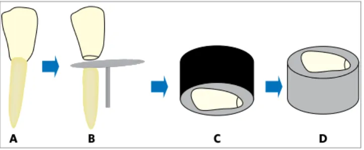
The root of 120 bovine incisors stored in distilled water at room temperature until the time of use were separated from the crowns using a double-faced dia-mond disk (KG Sorensen, Curitiba, Brazil) mounted on a micro motor handpiece under constant cooling (Figs 1A and 1B). The buccal surfaces were polished
under cooling (DPU-10, Struers, Copenhagen, Den-mark) using 400 and 600-grit silicon carbide sanding discs to produce a flat and smooth enamel surface.
The teeth were embedded in polystyrene resins (UC 2120) into PVC cylinders, with the flat enamel surfaces facing the base of the cylinders (Figs 1C and 1D). After the polystyrene resin polymerization, the enamel surfaces were polished with a 600-grit sand-ing paper to standardize the smear layer. The enamel surfaces underwent prophylaxis with pumice and wa-ter applied with a rubber cup at low speed for 15 sec-onds and rinsed with water-air sprays for 30 secsec-onds (Fig 1D). The specimens were randomly divided into six groups (n=20) according to the adhesive system used (Table 1 and Fig 2).
Groups XT (NC), MIP (NC) and SB (NC)
The enamel surfaces were etched by applying 37% phosphoric acid for 30 seconds, rinsed for 60 sec-onds and dried for 20 secsec-onds (Fig 2A); then, the ad-hesives were applied according to the instruction of the manufacturer (Fig 2B).
Groups XT (SC), MIP (SC) and SB (SC)
After receiving the same surface treatment used in the previous groups (Fig 2A) and the application of an adhesive layer (Fig 2B), the surfaces were contami-nated for 10 seconds with non-stimulated fresh saliva collected from the operator after brushing the teeth with a non-fluoride toothpaste after one-hour fasting lapse (Fig 2C). Excess saliva was removed with gauze, the specimen was air sprayed for 3 seconds, and a new adhesive layer was applied.
After adhesive application, 3x3 mm cylinders
Figure 1 -Schematic representation of storage of 120 bovine incisors
(A); roots separated from crowns using double-faced diamond disc
(B). Teeth embedded in polystyrene resin into PVC cylinders, with
the enamel surfaces facing the base of cylinders (C). After curing
polystyrene resin, enamel surfaces were polished for surface
of Transbond™ XT composite resin were prepared and placed in the center of the enamel surfaces (Fig 2E and 2F). The cement was inserted, in a single in-crement, into a metal matrix divided into two parts (Fig 2D) and light cured for 20 seconds.
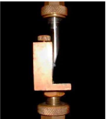
After storage in distilled water at 37 °C for 7 days, the specimens underwent shear bond strength trials (DL 10.000, EMIC, Curitiba, Brazil) at a cross-head speed of 0.5 mm/min and 50 N load (Fig 3). Data were collected for means and standard deviations analysis, as shown in Table 1.
Evaluation of adhesive remnant index score
After the shear bond strength trial, the enamel surfaces were examined under stereomicroscopy (Coleman) at 15x magnification to evaluate adhe-sive remnants according to the adheadhe-sive remnant index (ARI) score:1
1 - Less than half of the adhesive remained on the surface;
2 - More than half of the adhesive remained on the surface.
The type of failures at the interface was also evalu-ated using the following criteria:
A - Cohesive fracture - with damage to the enamel; B - Adhesive Fracture - without damage to the enamel.
STATISTICAL ANALYSIS
The shear bond strength data (in MPa) were ana-lyzed using the Shapiro-Wilk test to normal distribu-tion and the Levene test for homogeneity variance. As the normal distribution was confi rmed, data were analyzed using two-way analysis of variance and the Tukey test to compare means (α = 0.05).
RESULTS
Mean bond strength data (MPa) are shown in Table 1. Analysis of variance revealed a signifi cant difference in surface treatment (p = 3.15 x 10⁻¹¹). There was a
sta-tistically signifi cant reduction in bond strength in all adhesive systems under test when the enamel surface was contaminated with saliva. There was also a sta-tistically signifi cant difference in the comparison of adhesive systems (p < 0.05), as shown in Table 1 and Figure 4. The Tukey test revealed that the SB(NC) system had the best performance (32.36 ± 4.14) and a
Figure 3 -Specimen being submitted to shear bond strength trials (DL 10,000, EMIC, Curitiba, Brazil) at cross-head speed of 5 mm/ min and 50 N load.
Figure 2 -Schematic sequence of etching with 37% phosphoric acid for 30
seconds (A) Specimens divided into 6 groups, and application of 3 adhesive
systems according to manufacturer instructions (B). Contamination of
sur-faces in SC groups with saliva (C) Adaptation of metal matrix (D), insertion
of Transbond XT composite resin into matrix (E) and cylinders placement on
enamel surface for testing (F).
Table 1 - Data (MPa ± standard deviation) of the Transbond XT, Transbond MIP and Single Bond adhesive systems with no contamination (NC) or with saliva contamination (SC).
Data followed by the same letter were not statistically different (p < 0.05).
Adhesive system NC SC
XT a26.29
± 7.23 b.c19.59
± 6.76
MIP a.b24.47
± 7.52 c18.08
± 6.39
SB 32.36 ± 4.14 c18.18
significantly higher bond strength than the other ad-hesives with no contamination. Transbond™ XT and Transbond™ MIP systems had no statistically signifi-cant differences in the groups with no saliva contami-nation (NC). In the groups with saliva contamicontami-nation (SC), there were no differences between the adhesive systems under study.
Data followed by the same letter were not statisti-cally different (p < 0.05).
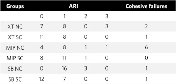
Table 2 shows the adhesive remnant index (ARI) score and the number of cohesive failures.
The most prevalent ARI scores were 0 and 1 for all groups. The highest number of cohesive failures was in the Transbond™ MIP group with no saliva contam-ination (MIP NC) (n=6).
DISCUSSION
This study compared the shear bond strength of Single Bond, Transbond™ MIP and Transbond™ XT adhesive systems used together with the Trans-bond™ XT composite resin in an environment with or without saliva contamination.
Mean shear bond strength data for the groups with no contamination (NC) were 26.3 MPa, 24.5 MPa and 32.4 MPa for Transbond™ XT, Transbond™ MIP and Single Bond, respectively. Regarding groups with saliva contamination, mean data were 19.6 MPa, 18.1 MPa and 18.2 MPa. The results showed no statis-tically significant differences for the comparison of the materials tested, which indicated that they are equivalent, except for the group of the Single Bond system with no contamination, which showed a very high shear bond strength mean, probably due to the adhesive light-curing procedure before the compos-ite resin specimen was inserted.
There was a statistically significant difference be-tween systems when groups with and without saliva contamination were compared. Although the bond strength data in the saliva contamination groups were significantly lower than in the groups with no contamination, the results were still acceptable for orthodontic bracket bonding.
Other studies also found statistically significant differences between the Transbond™ MIP and the Transbond™ XT in a dry environment.4,5,16
Enamel surface contamination may occur at two critical time points during bonding: After etching or after adhesive application. When that happens, bonding may be affected.4
Some studies found a greater decrease in bond strength when contamination occurred after acid-etching; in such cases, re-etching of the enamel sur-face was necessary.5,14,16
The contamination of the etched area by saliva or blood leads to the sealing of most of the porosity created by enamel acid-etching, which prevents the penetration of the adhesive material, resulting in in-sufficient resin tags in both number and length, and compromises bonding.7
Other studies also found statistically significant differences when materials were compared before and after saliva contamination, with no differences regarding the material employed,6,16 although some
studies have suggested differently about whether Transbond™ MIP may be the best choice for moist environments.11
According to Grandi et al,5 the hydrophilic primer
(Transbond™ MIP, 3M Unitek) is chemically identical to the dental adhesive that contains ethanol (Single
Table 2 -Shows the adhesive remnant index (ARI) score and the number of cohesive failures.
Groups ARI Cohesive failures
0 1 2 3
XT NC 7 8 0 3 2
XT SC 11 8 0 0 1
MIP NC 4 8 1 1 6
MIP SC 8 11 1 0 0
SB NC 0 16 3 0 1
SB SC 12 7 0 0 1
Figure 4 - Means and standard deviations of the 3 systems under study, with and without saliva contamination.
35
26.29 24.47 32.36
19.59
18.08 18.18 30
25
20
15
10
5
0
No contamination Saliva contamination
Transbond XT
Transbond MIP
Bond, 3M). In our study, bond strength results were similar for both materials when the enamel surface was contaminated with saliva, which suggests that Single Bond may be used as a replacement for Trans-bond™ MIP in a moist environment.
No similar comparison could be made in dry en-vironments because the material was used strictly as recommended by the manufacturer, and, there-fore, our study methods differed from those used by other authors. The Single Bond adhesive system used in a dry environment showed a higher bond strength result than the other materials, possibly due to its previous curing.
It is difficult to compare results of bond strength obtained in different studies because of the differenc-es in tooth storage methodology, specimen prepara-tion, cross-head speed and load, and storage of speci-mens before debonding.10
One of the factors that may affect the bond strength of adhesive systems is the storage time after specimen bonding. In this study, the shear bond strength trial was conducted 7 days after bonding, and the samples were stored in distilled water at 37 °C during that time.2,5 In other studies, shear bond strength trials
were conducted 24 hours after bonding.3,4,11,17
The substrate used in our study was bovine tooth enamel, as in other studies.4,5,13,16 Bovine teeth are
ana-tomically, histologically and chemically similar to hu-man teeth, although bond strength result shows to be lower than those of human teeth.8,9
In this study, the samples used for the bond strength trial were cylinders made of Transbond™ XT composite resin. The objective of this study was to evaluate the interface between enamel-adhesive-composite resin. Therefore, the use of a bracket would add another adhesive interface (composite resin-bracket base), which would add several vari-ables; such as bracket material, retention system, form and structure of the bracket base.
The ARI score is used to evaluate enamel surface after debonding. A greater amount of remaining ma-terial means that the probability of damage to the enamel surface is lower. Adhesive fractures between the bracket and the composite resin are preferable because there is no enamel damage and the dentist can remove remnants without any risk of affecting the enamel. Damage to the enamel may occur in the case of cohesive fractures, which may be correlated14
or not6 with bond strength values.
In the Transbond™ MIP group with no contami-nation, a higher number of specimens had enamel damage, although this was not the group with the highest mean bond strength value. Other studies found higher bond strength results for Transbond™ MIP in dry environments,3 and some authors
be-lieved that they were higher than recommended for orthodontic bracket bonding.13
Therefore, not only bond strength, but also bond-ing characteristics, such as mechanisms and depth of penetration of different adhesives in the tags formed during enamel etching, are factors that should be eval-uated and taken into consideration when making a de-cision about which materials to use.
Further studies should analyze bonding mecha-nism and the surface generated by these materials, both in dry environments and in cases of saliva con-tamination. Also, in vivo studies should test whether bond strength values in saliva contaminated environ-ments are acceptable for orthodontic bracket bonding.
CONCLUSIONS
1. Artun J, Bergland S. Clinical trials with crystal growth conditioning as an alternative to acid-etch enamel pretreatment. Am J Orthod. 1984;85:333-40.
2. Bishara SE, Laffoon JF, Vonwald L, Warren J. Effect of time on the shear bond strength of cyanoacrylate and composite orthodontic adhesives. Am J Orthod Dentofacial Orthop. 2002;121(3):297-300.
3. Cacciafesta V, Sfondrini MF, Angelis M, Scribante A, Klersy C. Effect of water and saliva contamination on shear bond strength of brackets bonded with convencional, hydrophilic and self-etching primers. Am J Orthod Dentofacial Orthop. 2002;123(6):633-40.
4. Cacciafesta V, Sfondrini MF, Angelis M, Scribante A, Klersy C. Effect of blood contamination on shear bond strength of convencional, hydrophilic primers. Am J Orthod Dentofacial Orthop. 2004;126(2):207-12.
5. Grandhi RK, Combe EC, Speidel TM. Shear bond strength of stainless steel orthodontic brackets with a moisture-intensive primer. Am J Orthod Dentofacial Orthop. 2001;119(3):251-5.
6. Hobson RS, Ledvinka J, Meechan JG. The effect of moisture and blood contaminationon bond strength of a new orthodontic bonding material. Am J Orthod Dentofacial Orthop. 2001;120(1):54-7.
7. Hormati AA, Fuller JL, Denehy GE. Effect of contamination and mechanical disturbance on the quality of acid-etched enamel. J Am Dent Assoc. 1980;100(1):34-8.
8. Nakamichi I, Iwaku M, Fusayama T. Bovine teeth as a possible substitutes in adhesion test. J Dent Res. 1983;62(10):1076-81.
REFERENCES
9. Oesterie LJ, Shellhart WC, Belanger GK. The use of bovine enamel in bonding studies. Am J Orthod Dentofacial Orthop. 1998;114(5):514-9.
10. Pessoa CN, Souza ROA, Valença AMG, Nóbrega CBC, Mendes ACR, Nascimento ABL. Resistência ao cisalhamento de diferentes sistemas restauradores à dentina. Pesq Bras Odontoped Clin Integr. 2004;4(1):59-63.
11. Rajagopal R, Padmanabhan S, Gnanamani J. A comparison of shear bond strength and debonding characteristics of convencional, moisture-intensitive and self-etching primers in Vitro. Angle Orthod. 2004;74(2):264-8.
12. Reynolds IR. A review of direct orthodontic bonding. Br J Orthod. 1975;2(2):171-8. 13. Rosa CB, Pinto RAC, Habib FAL. Colagem ortodôntica em esmalte com presença
ou ausência de contaminação salivar: é necessário o uso de adesivo auto-condicionante ou de adesivo hidrofílico? Rev Dental Press Ortod Ortop Facial. 2008;13(3):34-42.
14. Schaneveldt S, Foley TF. Bond strength comparison of moisture-insensitive primers. Am J Orthod Dentofacial Orthop. 2002;122(3):267-73.
15. Tortamano A, Naufi F, Nacaratto SRF, Vigorito JW. Avaliação do sistema adesivo ortodôntico MIP na presença de água e saliva. Ortodontia. 2001;34(3):66-9. 16. Webster MJ, Nanda RS, Duncanson MG, Khajotia SS, Sinha PK. The effect of saliva
on shear bond strengths of hydrophilic bonding systems. Am J Orthod Dentofacial Orthop. 2001;119(1):54-8.