The Dementia Rating Scale (DRS)
in the diagnosis of vascular dementia
Cláudia Sellitto Porto
1, Paulo Caramelli
2, Ricardo Nitrini
3Abstract – The Dementia Rating Scale (DRS) is considered a very useful instrument to assess patients with dementia. The tasks are grouped into fi ve subscales, each one evaluating different cognitive areas, namely: Atten-tion, Initiation/Perseveration (I/P), ConstrucAtten-tion, Conceptualization and Memory. Objective: To verify the ability of the DRS in discriminating vascular dementia (VaD) patients from healthy controls and VaD from Alzheimer disease (AD) patients. Methods: The DRS was applied to 68 patients with mild dementia (12 with VaD and 56 with AD) and 60 healthy controls. The clinical diagnosis was made by two neurologists based on the patients´ history, laboratory and neuroimaging results and neuropsychological tests. Results: In the comparison between VaD patients and controls, the subscales I/P, Memory, Conceptualization and Attention were those displaying best discrimination between the two groups. The cutoff <124 yielded 93.3% of sensitivity and 91.7% of specifi c-ity for the diagnosis of VaD. Only the I/P subscale differentiated VaD from AD patients. Conclusions: The DRS was found to be a useful instrument to discriminate VaD patients from controls. VaD patients showed worse performance in tasks of executive functions than AD patients. Executive dysfunction, evaluated through the I/P subscale of the DRS, might be useful in differentiating between VaD and AD patients.
Key words: dementia, neuropsychological tests, cognitive disorders, Alzheimer disease, vascular dementia.
A Escala de Avaliação de Demência (DRS) e o diagnóstico de demência vascular
Resumo – A Escala de Avaliação de Demência (Dementia Rating Scale – DRS) é considerada um instrumento de grande valor para avaliação de pacientes com demência. As tarefas estão agrupadas em cinco subescalas, as quais avaliam diferentes domínios cognitivos: Atenção, Iniciativa/Perseveração (I/P), Construção, Conceituação e Memória. Objetivo: Verifi car a capacidade da DRS em discriminar pacientes com demência vascular (DV) de controles saudáveis e pacientes com DV de pacientes com doença de Alzheimer (DA). Métodos: A DRS foi administrada a 68 pacientes com demência leve (12 com DV e 56 com DA) e a 60 indivíduos controles saudáveis do ponto de vista cognitivo. Os diagnósticos de DV e de DA foram realizados por dois neurologistas baseando-se na história clínica, em exames laboratoriais e de neuroimagem e em testes neuropsicológicos.Resultados: Na comparação de pacientes com DV e controles, as subescalas I/P, Memória, Conceituação e Atenção foram as que melhor diferenciaram os dois grupos. A nota de corte <124 demonstrou sensibilidade de 93,3% e 91,7% de es-pecifi cidade para o diagnóstico de DV. Em relação à comparação de pacientes com DV e DA, apenas a subescala I/P apresentou signifi cância estatística na diferenciação dos dois grupos.Conclusões: A DRS mostrou ser um instrumento útil na discriminação entre pacientes com DV e controles. Pacientes com DV demonstraram pior desempenho em tarefas de funções executivas que pacientes com DA. Disfunção executiva, avaliada através das tarefas da subescala I/P, pode ser útil na diferenciação entre pacientes com DV e DA.
Palavras-chave: demência, testes neuropsicológicos, transtornos cognitivos, doença de Alzheimer, demência vascular.
1PhD, Behavioral and Cognitive Neurology Unit, Department of Neurology of the University of São Paulo School of Medicine and Cognitive Disorders Reference Center (CEREDIC), Hospital das Clínicas of the University of São Paulo School of Medicine, São Paulo, SP, Brazil. 2MD, PhD, Behavioral and Cognitive Neurology Unit, Department of Internal Medicine, Faculty of Medicine, Federal University of Minas Gerais, Belo Horizonte, MG, Brazil. 3MD, PhD, Behavioral and Cognitive Neurology Unit, Department of Neurology of the University of São Paulo School of Medicine, and Cognitive Disorders Reference Center (CEREDIC), Hospital das Clínicas of the University of São Paulo School of Medicine, São Paulo, SP, Brazil.
Claudia Sellitto Porto – Rua Itapeva, 378 / cj. 92 - 01332-000 São Paulo SP - Brazil . E-mail: [email protected]
The Dementia Rating Scale (DRS)1,2 is a measure of
general cognitive status and has been used both in clinical practice and research. The scale includes 36 tasks which are grouped into fi ve subscales assessing different cognitive
domains, namely: Attention, Initiation/Perseveration (I/P), Construction, Conceptualization and Memory.
diagno-sis and discrimination of patients with Alzheimer disease (AD) from those with other forms of dementia. Previous investigators have reported that the DRS is able to
differen-tiate patients with AD from cognitively healthy controls,3,4
as well as AD from dementia associated with Parkinson´s
disease,5 Huntington´s disease from AD,6 and patients with
vascular dementia (VaD) from patients with AD.7,8
The main objective of this work is to verify the ability of the DRS to discriminate VaD patients from controls, and VaD from AD patients.
Methods
The study involved 68 patients (39 women and 29 men), aged 54 to 84 years (mean=72.35±7.78), with schooling ranging from 3 to 17 years (mean=9.40±4.78), attended by members of the Behavioral and Cognitive Neurology Unit of the Department of Neurology at the University of São Paulo School of Medicine, in Brazil. All patients were submitted to appropriate laboratory tests and to structural neuroimaging (computed tomography (CT) or magnetic resonance (MR) of the skull). Moreover, they were sub-mitted to a comprehensive neuropsychological evaluation, which included the following tests: the Mini-Mental State
Examination (MMSE),9,10 the Brief Cognitive Screening
Battery (BCSB),11 visual and verbal memory tests (subtest
Visual Reproduction of the Wechsler Memory Scale –
Re-vised (WMS-R),12 Rey Complex Figure – delayed recall,13
subtest Logical Memory (WMS-R),12 Rey Auditory Verbal
Learning Test (RAVLT),14 constructive abilities (subtest
Block Design –Wechsler Adult Intelligence Scale (WAIS),15
Rey Complex Figure copy,13 visual perception (Hooper
Vi-sual Organization Test16 and Raven´s Progressive Matrices,17
language (Boston Naming Test),18 and executive functions
(Trail Making Test versions A and B,19 Stroop Test,19
Wis-consin Card Sorting Test (WCST)19 and phonemic verbal
fl uency (F.A.S.).19 Information on performance in daily life
activities was obtained through the Pfeffer Functional
Ac-tivities Questionnaire,20 which was applied to an informant.
The clinical diagnosis of mild dementia was based on the criteria of the Diagnostic and Statistical Manual of
Mental Disorders, Third Edition, revised (DSM-III-R)21
and was made by two neurologists (PC and RN), who were blind to DRS and BCSB results, and based on the patients’ history, laboratory and neuroimaging results, MMSE scores and on results of the following neuropsychological tasks: constructive abilities (Block Design (WAIS), memory (Rey Auditory Verbal Learning Test (RAVLT) – sum of scores from trials 1 to 5 and the number of words recalled after 30 minutes), language (Boston Naming Test), executive functions (phonemic verbal fl uency and Trail Making Test (versions A and B)).
The AD group was composed of 56 individuals, aged 54 to 84 years (mean=72.98±7.43), with schooling rang-ing from 3 to 17 years (mean=9.62±4.68), comprisrang-ing 35 women and 21 men. The diagnosis of probable AD was based on the criteria of the National Institute of Neurologi-cal Disorders and Communicative Disorders and Stroke-Alzheimer Disease and Related Disorders Association
(NINCDS-ADRDA).22
Twelve patients were included in the VaD group, aged 54 to 80 years (mean=69.41±8.99), with schooling rang-ing from 4 to 16 years (mean=8.33±5.30), comprisrang-ing 4 women and 8 men (nine cases of subcortical VaD and three cases of multiple infarct dementia). Eleven patients were submitted to MRI of the skull and one to CT. The diagnosis of probable VaD was based on the criteria of the National Institute of Neurological Disorders and Stroke – Associa-tion InternaAssocia-tionale pour la Recherche et l´Enseignement
en Neurosciences (NINDS- AIREN).23
The control group (60 subjects; mean age=68.90±7.48; mean schooling=10.72±4.74; 42 women and 18 men) was composed of spouses or consorts of the patients, or vol-unteers from the community, with no memory disorders and who were self-suffi cient in terms of daily life activities. Subjects with neurological disease, history of alcoholism, depression, or any other psychiatric disorder, non-correct-ed visual or auditory disorders, motor disorders, or users of psychotropic drugs that could affect cognitive functions were excluded. Chronic diseases such as arterial hyperten-sion, diabetes mellitus and cardiac disorders, if adequately controlled, were not criteria for exclusion. All controls were submitted to the MMSE, the BCSB and to the Memory Complaint Questionnaire (MAC-Q) (24) or to the Infor-mant Questionnaire on Cognitive Decline in the Elderly
(IQCODE),25,26 administered to an informant.
The Portuguese version of the DRS17 was administered
to all patients and controls. The tasks are presented in a fi xed order, as recommended by the author, and only the Attention tests are not grouped in a sequence, as they also serve as distractors for the Memory subscale. Within each subscale, the most diffi cult tests were presented in fi rst and second, and if performed well, subsequent items of the sub-scale were automatically scored as having been performed correctly. The advantage of this procedure is that it shortens total testing time for individuals who are relatively intact.
In the two groups studied, the scale was applied indi-vidually in a single session. The time of application for the group of patients was, on average, 40 minutes, and for the control group, from 20 to 30 minutes.
The study was approved by the Research end Ethics Committee of Hospital das Clínicas of the University of São Paulo School of Medicine. All subjects who agreed to participate signed a written informed consent.
Statistical analysis
In order to evaluate associations between the categorical variables and the results, the Pearson Chi-Squared test was performed. When the variables were continuous, the com-parisons were made for two samples by the Mann-Whitney test, and for more than two, by the Kruskall-Wallis test.
Sensitivity and specifi city calculations were performed for each subscale and for the total scale. The cutoff score,
calculated through ROC (receiver operator characteristics)
curves, was defi ned as the value presenting the best rela-tionship between sensitivity and specifi city.
Alpha risk was considered to be less than or equal to 5% for type 1 error and beta risk greater than or equal to 20% for type II error.
All statistical analysis was carried out using the pro-gram Statistical Package for the Social Sciences (SPSS), version 10.0.
Results
There were no statistically signifi cant differences be-tween controls and both patient groups in relation to schooling (p=0.213) and gender (p=0.055), but there was a signifi cant difference in relation to age (p=0.011). A
statis-tically signifi cant difference was found between mean total DRS scores of controls and both patients´ groups (Table 1).
VaD patients and controls
In the analysis of the mean total scores on the total scale and each subscale, the I/P subscale (p<0.001) as well as Memory (p<0.001), Conceptualization (p<0.001) and At-tention (p=0.021) subscales differentiated VaD from con-trols. The scores in the Construction subscale (p=0.150) were not signifi cantly different between the two groups. The same phenomenon occurred in the analysis of the ar-eas under curves obtained through the ROC curves (Figure 1) (Table 2).
Table 1. Performance of patients with VaD and controls, and VaD and AD, DRS total and subscales.
VaD controls p AD p
N 12 60 56
Total
Mean (SD) 110.1 (11.0) 136.2 (6.3) <0.0001 113.8 (12.4) 0.314
Attention
Mean (SD) 35.2 (0.6) 35.7 (1.3) 0.021 34.9 (1.6) 0.818
I/P
Mean (SD) 24.6 (4.2) 35.1 (1.9) <0.001 29.1 (5.8) 0.010
Construction
Mean (SD) 5.8 (0.5) 5.8 (0.3) 0.150 5.6 (1.0) 0.549
Conceptualization
Mean (SD) 28.2 (4.8) 34.5 (4.0) <0.001 29.4 (6.0) 0.600
Memory
Mean (SD) 16.3 (4.0) 24.0 (1.5) <0.001 14.7 (3.9)
0.325
N, subjects; I/P, initiation/perseveration; SD, standard deviation; p<0.05.
VaD and AD patients
The performance of the AD group on the DRS was compared to the VaD patients’ scores (Table 1).
In the comparison between VaD and AD patients, only the I/P subscale was able to signifi cantly differentiate between the two groups (p=0.010) (AUC=0.739±0.064; p=0.010).
Discussion
In the present study, the DRS was able to accurately discriminate VaD patients from controls, while only the I/P subscale differentiated VaD from AD patients.
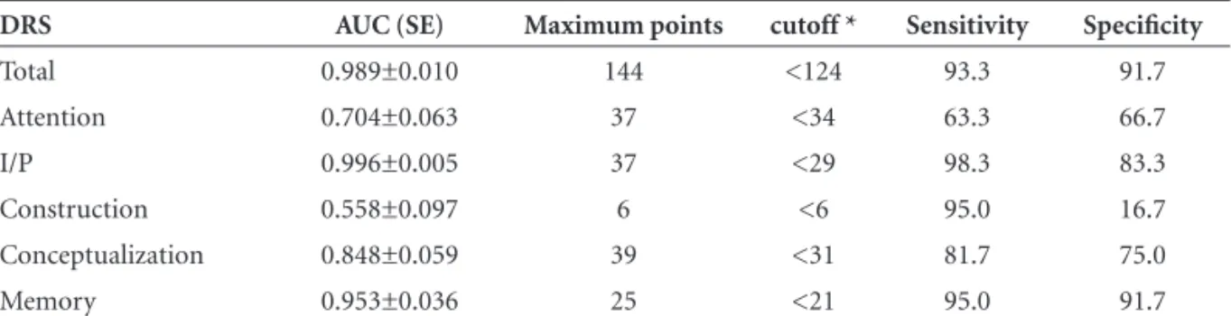
In the discrimination between VaD patients and control individuals, the cutoff score <124 in the DRS showed good sensitivity (93.3%) and specifi city (91.7%) values.
Both in the analysis of the areas under the curves (AUC) and comparison between the means scores of the two groups, I/P, Memory, Conceptualization and Attention subscales also allowed good discrimination between VaD patients and controls.
The Memory subscale differentiated VaD patients
from normal elderly. Lukatela et al.8 verifi ed, in their study
comparing DRS scores in VaD, AD and controls, that the group with AD and the group with VaD presented sig-nifi cant impairment in comparison to the control group.
Price et al.29 concluded that tests of executive control and
memory, along with neuroimaging evidence of involve-ment of around one-fourth of the cerebral white matter as measured by the Leukoaraiosis Scale, may be suffi cient for the diagnosis of subcortical VaD.
The results of the Inasaridze et al.34 study demonstrated
that attentional defi cits are characteristic of VaD. Impaired
at-tention was also observed in other studies,35,36 a feature in
agree-ment with our results on the Attention subscale of the DRS. The Construction subscale was not able to discriminate VaD patients from controls. This fi nding seems to be in
con-trast with the work of Lukatela et al.8 in which VaD patients
showed greater impairment on this subscale compared to AD individuals. According to these authors, the results
dem-onstrated that problems in simple graphomotor construc-tion and coordinaconstruc-tion are more pronounced in VaD than in AD. Perhaps due to the small number of patients in our se-ries, our results differ from those published in the literature. Patients with VaD and controls also showed different performances on the Conceptualization subscale in the
present study. Giovannetti et al.33 investigated different
mechanisms that may underlie defi cits in verbal concept formation among patients with AD and ischemic VaD. The test utilized by the authors was the Wechsler Adult Intel-ligence Scale – Revised (WAIS-R). The Similarities subtest, which contains similar tasks as the Conceptualization sub-scale, did not differentiate between the two groups. None-theless, AD patients produced a greater proportion of very vague superordinate concepts for the word pairs (for ex-ample: dog/lion: “they´re alive”) while the errors produced by VaD patients demonstrated an inability to provide a su-perordinate concept for the same word pairs (for example: dog/lion: “the lion roars and the dog barks”). The errors produced by VaD patients showed impairment in concept formation associated with defi cits in executive systems nec-essary to monitor responses and to sustain mental set. The AD patients´ errors were associated with measures of de-layed recognition memory and semantic intrusion errors, indicating that the defi cit of concept formation appears to be secondary to impaired verbal response selection.
The comparison between the performances of VaD and AD patients on the DRS showed that only the I/P subscale was able to differentiate between the two groups.
Similar results were reported by two independent
in-vestigations. Kertesz and Clydesdale7 compared AD and
VaD patients performances on the DRS. VaD patients were signifi cantly worse on motor performance subtests of the I/P subscale than AD patients. The authors concluded that these subtests might be useful in discriminating between
VaD and AD. In the above-mentioned Lukatela et al. study,8
VaD patients with multiple infarcts demonstrated signifi -cantly lower scores on the I/P subscale than AD patients. Table 2. Areas under the curves, cutoff, sensitivity and specifi city for the DRS between VaD patients and controls.
DRS AUC (SE) Maximum points cutoff * Sensitivity Specifi city
Total 0.989±0.010 144 <124 93.3 91.7
Attention 0.704±0.063 37 <34 63.3 66.7
I/P 0.996±0.005 37 <29 98.3 83.3
Construction 0.558±0.097 6 <6 95.0 16.7
Conceptualization 0.848±0.059 39 <31 81.7 75.0
Memory 0.953±0.036 25 <21 95.0 91.7
The I/P subscale of DRS is composed by verbal fl uency for semantic categories (supermarket items), double simul-taneous hand movements and design copy tasks. These two latter tasks evaluate bimanual coordination and motor per-severation, which are recognized to be associated to frontal
lobes defi cits. According to some authors,27-31 the executive
dysfunction might serve as diagnostic marker for VaD,
es-pecially for the subcortical subtype. Villardita32 verifi ed that
attention processes, planning and fi ne motor coordination tasks were more severely impaired in VaD than in AD pa-tients, concluding that these disturbances resemble some of those occurring in frontal lobe syndromes. VaD patients were signifi cantly disadvantaged in executive functions which in-clude planning and sequencing, speed of mental process-ing, performance on unstructured tasks, and also attention. In conclusion, the DRS in the present study proved a useful instrument to discriminate between VaD patients and controls. Our results suggest that executive dysfunc-tion, evaluated through the I/P subscale tasks, is helpful in differentiating VaD from AD patients. Further studies involving larger samples of patients are necessary in order to confi rm these initial fi ndings.
References
1. Mattis S. Mental Status Examination for Organic Mental Syn-drome in the Elderly Patient. In: Bellak L, Karasu TB, editors. Geriatric Psychiatry. A Handbook for Psychiatrists and Prima-ry Care Physicians. New York: Grune & Stratton; 1976:77-121. 2. Mattis S. Dementia Rating Scale. Professional manual. Florida:
Psychological Assessment Resources; 1988.
3. Monsch AU, Bondi MW, Salmon DP, et al. Clinical Validity of the Mattis Dementia Rating Scale in Detecting Dementia of the Alzheimer Type. Arch Neurol 1995;52:899-904. 4. Porto CS, Charchat-Fichman H, Caramelli P, Bahia VS,
Ni-trini R. Brazilian version of the Mattis Dementia Rating Scale. Diagnosis of mild dementia in Alzheimer´s disease. Arq Neu-ropsiquiatr 2003;61:339-345.
5. Paolo AM, Trostr AI, Glatt SL, Hubble JP, Koller WCJ. Dif-ferentiation of the dementias of Alzheimer’s and Parkinson’s disease with the dementia rating scale. Geriatr Psychiatry Neurol 1995;8:184-188.
6. Paulsen JS, Butters N, Sadek JR, et al. Distinct cognitive pro-fi les of cortical and subcortical dementia in advanced illness. Neurology 1995;45:951-956.
7. Kertesz A, Clydesdale S. Neuropsychological deficits in Vascular Dementia vs Alzheimer’s Disease. Arch Neurol 1994;51:1226-1231.
8. Lukatela K, Cohen R, Kessler H, et al. Dementia Rating Scale Performance: A Comparison of Vascular and Alzheimer’s De-mentia. J Clin Exper Neuropsych 2000; 22:445-454. 9. Folstein MF, Folstein SE, McHugh PR. “Mini-mental state”. A
practical method for grading the cognitive state of patients for the clinician. J Psychiatr Research. 1975;12:189-198. 10. Brucki SMD, Nitrini R, Bertolucci PHP, Caramelli P,
Oka-moto IH. Normas sugeridas para o uso do Mini-Exame do Estado Mental (MEEM) em nosso meio. Arq Neuropsiquiatr 2003;60:46-47.
11. Nitrini R, Caramelli P, Porto CS, et al. Avaliação cognitiva breve no diagnóstico de doença de Alzheimer leve. Arq Neu-ropsiquiatr 2005;63:27.
12. Wechsler D. Wechsler Memory Scale. Manual The Psychologi-cal Corporation Harcourt Brace Jovanovich; 1987.
13. Rey A. Figuras Complexas de Rey. São Paulo: Casa do Psicólo-go; 1998.
14. Diniz LFM, Cruz MF, Torres VM, Consenza RM. O teste de aprendizagem auditivo-verbal de Rey: normas para uma população brasileira. Rev Bras Neurol 2000;36:79-83. 15. Wechsler, D. Test de Inteligencia para adultos (WAIS).
Man-ual. 2nd ed. Buenos Aires: Editorial Paidos; 1993.
16. Hooper Visual Organization Test (VOT) Manual. Los Angeles, CA: Western Psychological Services; 1983.
17. Raven JC, Raven J, Court JH. Manual Matrizes Progressivas Coloridas. São Paulo: Casa do Psicólogo;1988.
18. Radanovic M, Mansur LL, Scaff M. Normative data for the Brazilian population in the Boston Diagnostic Aphasia Examination: influence of schooling. Braz J Med Biol Res 2004;37:1731-1738.
19. Spreen O, Strauss E. A Compendium of Neuropsychological Tests. Administration, Norms, and Commentary. 2nd ed. New York: Oxford University Press; 1998.
20. Pfeffer RI, Kusosaki TT, Harrah Jr CH, Chance JM, Filos S. Measurement of Functional Activities in Older Adults in the Community. J Gerontol 1982;37:323- 329.
21. American Psychiatric Association. Diagnostic and Statistical Manual of Mental Disorders. 3rd ed. Washington, DC: Ameri-can Psychiatric Association; 1987.
22. McKhann G, Drachman D, Folstein M, Katzman R, Price D, Stadlan EM. Clinical diagnosis of Alzheimer’s disease: report of the NINCDS-ADRDA work group under the auspices of department of health and human services task force on Alzheimer’s disease. Neurology 1984;34:939-944.
23. Román GC, Tatemichi TK, Erkinjuntti T, et al. Vascular de-mentia: diagnostic criteria for research studies. Report of the NINDS-AIREN International Work Group. Neurology 1993;43:250-60.
24. Mattos P, Lino V, Rizo L, Alfano A, et al. Memory complaints and test performance in health elderly persons. Arq Neurop-siquiatr 2003;61:920-924.
Instrumen-tos combinados na avaliação de demência de idosos. Arq Neu-ropsiquiatr 2003;61:601-606.
27. Mesulam M-M. Principles of Behavioral and Cognitive Neu-rology. 2nd ed. New York: Oxford University Press; 2000. 28. Lezak MD. Neuropsychological Assessment. 3rd ed. New York:
Oxford University Press; 1995.
29. Price CC, Jefferson AL, Merino JG, Heilman KM, Libon DJ. Subcortical vascular dementia: integrating neuropsychologi-cal e neuroradiologic data. Neurology. 2005; 65:376-382. 30. Osterman JM, Scherder EJ. Distinguishing between vascular
dementia and Alzheimer´s disease by means of the WAIS: a meta-analysis. J Clin Exp Neuropsychol 2006;28:1158-1175. 31. O´Brien JT. Vascular cognitive impairment. Am J Geriatr
Psy-chiatry 2006;14:724-733.
32. Villardita C. Alzheimer´s disease compared with
cerebrovas-cular dementia. Neuropsychological similarities and differ-ences. Acta Neurol Scand 1993;87:299-308.
33. Giovanetti T, Lamar M, Cloud BS, Swenson R, Fein D Kaplan E, Libon DJ. Different underlying mechanisms for defi cits in concept formation in dementia. Arch Clin Neuropsychol 2001;16:547-560.
34. Inasaridze K, Sikharulizdze G, Malishava T, Zviadadze M. At-tentional disorder in vascular dementia. Georgian Med News. 2006;(135):91-95.
35. Desmond DW. The neuropsychology of vascular cognitive impairment: is there a specifi c cognitive defi cit? J Neurol Sci 2004;226:3-7.