PB 93
Signs and symptoms of temporomandibular disorders
in adolescents
Sinais e sintomas de disfunção temporomandibular
em adolescentes
Leonardo Rigoldi Bonjardim* Maria Beatriz Duarte Gavião** Luciano José Pereira*
Paula Midori Castelo*
Renata Cunha Matheus Rodrigues Garcia***
ABSTRACT: The aim of this study was to verify the prevalence of signs and symptoms of temporomandibular dis-orders (TMD) in adolescents and its relationship to gender. The sample comprised 217 subjects, aged 12 to 18. The subjective symptoms and clinical signs of TMD were evaluated, using, respectively, a self-report questionnaire and the Craniomandibular Index, which has 2 subscales; the Dysfunction Index and the Palpation Index. The results of muscle tenderness showed great variability (0.9-32.25%). In relation to the temporomandibular joint, tenderness of the superior, dorsal and lateral condyle regions occurred in 10.6%, 10.6% and 7.83%, respectively, of the sample. Joint sound during opening was present in 19.8% of the sample and during closing in 14.7%. The most prevalent symptoms were joint sounds (26.72%) and headache (21.65%). There was no statistical difference between genders (p > 0.05), except for the tenderness of the lateral pterygoid muscles, which presented more prevalence in girls. In conclusion, clinical signs and symptoms of TMD can occur in adolescents; however, gender influence was not perceived.
DESCRIPTORS: Temporomandibular joint disorders; Teen health.
RESUMO: O objetivo deste estudo foi verificar a prevalência de sinais e sintomas de disfunção temporomandibular (DTM) em adolescentes e sua relação com o gênero. A amostra foi constituída de 217 voluntários, com idade entre 12 e 18 anos. Os sintomas subjetivos e os sinais clínicos de DTM foram avaliados usando-se, respectivamente, um questionário e o “Craniomandibular Index”, o qual possui 2 subescalas: “Dysfunction Index” e “Palpation Index”. Os resultados para sensibilidade muscular mostraram grande variabilidade (0,9-32,25%). Com relação à articulação temporomandibular, a sensibilidade à palpação nas regiões superior, dorsal e lateral do côndilo ocor-reu, respectivamente, em 10,6%, 10,6% e 7,83% da amostra. A prevalência do ruído articular no movimento de abertura foi de 19,8% e no fechamento, 14,7%. Os sintomas relatados mais prevalentes foram o ruído articular (26,72%) e dor de cabeça (21,65%). Nenhuma diferença estatística foi encontrada para a associação entre os gê-neros (p > 0,05), exceto para a sensibilidade no músculo pterigóideo lateral, a qual se apresentou mais prevalente nas meninas. Os sinais clínicos e sintomas subjetivos de DTM foram observados em adolescentes, no entanto a influência do gênero não foi percebida nessa faixa etária.
DESCRITORES: Transtornos da articulação temporomandibular; Saúde do adolescente.
INTRODUCTION
Temporomandibular disorder (TMD) is a generic term for a number of clinical signs and symptoms involving the masticatory muscles, the temporomandibular joint (TMJ) and associated structures25.
Signs and symptoms of TMD in children and adolescents have been studied since the begin-ning of the 1970s11. The most prevalent clinical
signs of TMD are TMJ sounds (upon palpation),
limitation of mandibular movements, TMJ and muscle tenderness23. With regard to subjective
symptoms, headache, TMJ sounds, bruxism, dif-ficulty in opening the mouth, jaw pain, and facial pain are found22.
The etiology of TMD has been considered to be one of the most controversial issues in clinical den-tistry. Currently, TMD is considered not a single entity, but a group of several diseases of varying
94 95
94 95
etiology and pathology, and controversy still ex-ists because of the limited knowledge regarding its etiology and natural history4.
The role of gender in TMD is also extensively discussed in literature, suggesting that TMD is considered to be 1.5-2 times more prevalent in women than in men, and that 80% of the patients treated for this disorder are women14. However,
the most prominent gender differences have been found in women aged 20-40 years, and the low-est among children, adolescents and the elderly15.
Furthermore, the predominance of women is even higher in surveys of people seeking treatment for TMD pain, with a ratio of 4:1 or 5:14.
The purpose of this study was to estimate the prevalence of clinical signs and subjective symp-toms of TMD in adolescents and its relationship to gender.
MATERIAL AND METHODS
Adolescents aged between 12 and 18 years were selected from public schools in Piracicaba, Brazil. Firstly, the parents/guardians and the ado-lescents were informed about the purpose of this research. Adolescents who had received any type of orthodontic treatment before or during the study or were suffering from systemic health disease could not participate in the research. Secondly, a total of 600 written informed consents were distrib-uted. After that, parental and adolescent consent was obtained from 217 subjects (120 girls, mean age of 13.18 ± 1.28 years; 97 boys, mean age of 13.28 ± 1.6 years), and they were examined. Prior to the examination for clinical signs and symptoms of TMD, an anamnestic questionnaire was filled out, including questions about their general state of health, illness, diseases and oral hygiene. The Research Ethics Committee of the School of Den-tistry of Piracicaba approved this research.
Subjective symptom interview
A self-report questionnaire was used to as-sess the symptoms22 regarding pain in the jaws
when functioning (e.g. chewing), unusually fre-quent headaches (more than once a week and of unknown etiology), stiffness/tiredness in the jaws, difficulty in opening the mouth wide, grind-ing teeth, and sounds at the TMJ. Each question could be answered with yes or no.
Clinical sign examination
The signs of TMD were assessed according to the CranioMandibular Index (CMI), as described by Fricton, Schiffman8 (1986), by two calibrated
examiners (Kappa = 0.936). The CMI produces 3 scores: an overall CMI score, a dysfunction index (DI) score, and a palpation or muscle index (PI) score. The CMI score is an average of the DI and PI scores. All 3 indexes are scaled from 0 to 1. The CMI measures tenderness and dysfunction in the stomatognathic system and includes all cur-rently recognized signs of TMJ disorders8,9. The DI
is designed to measure limitation of mandibular movement, pain and deviation of movement, TMJ noise and tenderness. The PI measures the preva-lence of muscle tenderness in the stomatognathic system. Thus, this index separates joint problems from muscle problems.
Statistical analysis
The data were computerized and the SAS package (SAS Institute Cary, North Carolina, USA) was used for their analysis. The prevalence of clini-cal signs and subjective symptoms was clini-calculated by percentage. The mean values obtained in CMI, PI, DI were compared for subjects with and with-out each symptom using Mann-Whitney test for the total sample. Data association between each symptom/clinical sign and gender was done using Fisher’s Exact Test. For all comparisons, p < 0.05 was considered to be statistically significant.
RESULTS
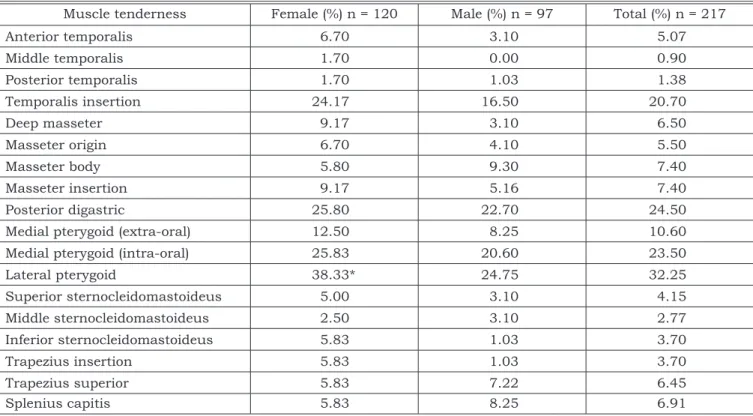
Tables 1 and 2 show the prevalence of the dif-ferent clinical signs of TMD (components of CMI) according to gender. Pterygoid lateral muscle ten-derness was the most frequent sign of palpation index found in 32.25% of the total sample. The most frequent sign of DI was TMJ sounds (mouth opening), occurring in 19.8% of the total sample.
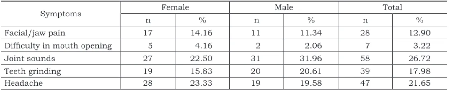
The prevalence of subjective symptoms of TMD according to gender is presented in Table 3. The most prevalent symptom was TMJ sound (26.72%), followed by headache (21.65%). There was no sta-tistical association between genders for clinical signs and symptoms, except for pterygoid lateral muscle tenderness, which presented a higher prev-alence among girls (p < 0.05).
open-94 95
94 95
ing the mouth wide, joint sounds and headache, CMI and PI had significantly higher scores than in adolescents that did not report any symptom (p < 0.05), whereas adolescents who reported TMJ sounds presented a significantly higher mean val-ue for DI (p < 0.05). In addition, individuals who reported teeth grinding presented significantly higher PI scores than those who did not report this symptom (p < 0.05).
DISCUSSION
This study evaluated the prevalence of signs and symptoms of TMD in adolescents through a questionnaire and physical examination. The
de-cision to use a dysfunction index in this study, specifically the CMI, was based on the possibil-ity of objectively measuring the severpossibil-ity of prob-lems in mandibular movements, joint noises, and muscle and joint tenderness, using clearly defined criteria, simple clinical methods and easy scor-ing. In addition, this index had a good intra- and inter-examiner correlation8,9. The symptom
ques-tionnaire proved to be a simple and suitable tool easily understood by the volunteers, thus allowing smaller examiner influence on the individuals and their answers. The application of an anamnestic questionnaire for detecting TMD symptoms has the advantage of being easily used by general practitio-ners or epidemiologists. The CMI scores obtained TABLE 1 - Percentage distribution of clinical signs (muscle tenderness) according to gender.
Muscle tenderness Female (%) n = 120 Male (%) n = 97 Total (%) n = 217
Anterior temporalis 6.70 3.10 5.07
Middle temporalis 1.70 0.00 0.90
Posterior temporalis 1.70 1.03 1.38
Temporalis insertion 24.17 16.50 20.70
Deep masseter 9.17 3.10 6.50
Masseter origin 6.70 4.10 5.50
Masseter body 5.80 9.30 7.40
Masseter insertion 9.17 5.16 7.40
Posterior digastric 25.80 22.70 24.50
Medial pterygoid (extra-oral) 12.50 8.25 10.60
Medial pterygoid (intra-oral) 25.83 20.60 23.50
Lateral pterygoid 38.33* 24.75 32.25
Superior sternocleidomastoideus 5.00 3.10 4.15
Middle sternocleidomastoideus 2.50 3.10 2.77
Inferior sternocleidomastoideus 5.83 1.03 3.70
Trapezius insertion 5.83 1.03 3.70
Trapezius superior 5.83 7.22 6.45
Splenius capitis 5.83 8.25 6.91
*Statistical difference (p < 0.05).
TABLE 2 - Percentage distribution of clinical signs (TMJ sounds and tenderness) according to gender.
Clinical signs Female (%) n = 120 Male (%) n = 97 Total (%) n = 217
Opening click 20.80 18.50 19.80
Closing click 16.60 12.40 14.70
Tenderness in condyle superior region 14.17 6.19 10.60
Tenderness in condyle lateral region 9.17 6.19 7.83
Tenderness in condyle dorsal region 13.33 7.22 10.60
96 97
96 97
in this study were lower than those presented in other studies8,9, probably because this sample
was comprised by adolescents. Studies have re-ported that severe disorder at a young age is rare, supporting the results presented25. Moreover, the
study was carried out in a randomized population and not among people looking for treatment.
The results of muscle tenderness showed that a higher prevalence was observed in the lateral pterygoid muscle (32.25%), but this result must be carefully considered, due to the low specificity of palpation. Nevertheless, this muscle has been part of many current examination schemes4,25,26. It must
be considered that the discomfort or pain observed in response to palpation of the “lateral pterygoid area” may be caused by anatomical structures oth-er than the latoth-eral ptoth-erygoid muscle24. There was
also a high prevalence of tenderness in the pos-terior digastric muscle (24.5%), medial pterygoid muscle (intraoral) (23.5%) and temporalis insertion (20.7%). The scores for intra-oral muscle palpation indicated that the frequency of tenderness of these muscles was higher than at other sites, except for the posterior digastric muscle. Intra-oral palpa-tion may cause pain in normal subjects and thus, false-positives that may lead to a wrong diagnosis, such as myofascial pain8.
As reported in the related literature, the an-terior temporalis region and masseter muscles
have been extensively evaluated. In this research, tenderness of the anterior temporalis region was observed in 5.07% of the adolescents, while for the masseter muscle the correspondent value was 7.4%. These results are similar to those of other studies in adolescents17 and young adults18.
Mas-seter and anterior temporalis area palpation can be considered to be reliable and valuable. This fact supports the belief that pressure pain sensa-tion in these muscles is not derived predominantly from the cutaneous tissues, but from the muscle itself10.
Neck muscle tenderness was also evaluated as part of clinical signs exam in CMI (Table 1). Despite the low prevalence of tenderness in these muscles, their evaluation in TMD patients is im-portant. Moreover, the findings obtained support the theory that a complementary examination of this area should be performed, even when TMD patients do not report any neck problems. Fink et al.7 (2002), corroborating the findings of this study,
mentioned that patients with TMD frequently show symptoms related to the cervical spine region.
Tenderness of the superior and dorsal condyle region occurred in 10.6% of the sample, whereas for the lateral region, it occurred in 7.83%. A num-ber of studies have found a prevalence of TMJ tenderness in adolescents varying from 7.1% to 22.5%2,17,22. Moreover, differences in palpation
TABLE 3 - Percentage of subjective symptoms according to gender.
Symptoms Female Male Total
n % n % n %
Facial/jaw pain 17 14.16 11 11.34 28 12.90
Difficulty in mouth opening 5 4.16 2 2.06 7 3.22
Joint sounds 27 22.50 31 31.96 58 26.72
Teeth grinding 19 15.83 20 20.61 39 17.98
Headache 28 23.33 19 19.58 47 21.65
TABLE 4 - Mean values for CMI, DI and PI among adolescents with and without each subjective symptom.
Symptoms Without CMI DI PI
symptom symptomWith symptomWithout symptomWith symptomWithout symptomWith
Facial/jaw pain 0.075a 0.155b 0.087 0.120 0.055a 0.190b
Difficulty in mouth opening 0.082a 0.186b 0.089 0.168 0.068a 0.204b
Joint sounds 0.069a 0.130b 0.086a 0.108b 0.050a 0.133b
Teeth grinding 0.082 0.098 0.091 0.096 0.067a 0.100b
Headache 0.078a 0.111b 0.090 0.096 0.059a 0.123b
96 97
96 97
techniques and pressure make comparisons very unreliable. In addition, in the present research, TMJ palpation was conducted in three different sites, which could be the cause of disagreement among these results.
The percentage of joint sound upon palpa-tion during mouth opening and closing was 19.8% and 14.7%, respectively, lower than that observed in the study of Nassif et al.18 (2003) (24.7% and
19.5%), performed in young adults. Farsi6 (2003)
found a prevalence of 11.8% for joint sounds in children aged 3 to 15. The small differences among these studies could be due to the fact that the in-cidence of signs and symptoms generally increase and also fluctuate with age5.
The most prevalent subjective symptoms present were TMJ sounds (26.72%), headache (21.65%), tooth grinding (17.98%), and pain in the face or jaw regions (12.9%). Conti et al.2 (2003)
also found TMJ sounds followed by headache as the most commonly reported symptoms, although with lower values. Melis, Abou-Atme16 (2003) and
Conti et al.2 (2003) observed a prevalence rate of
27.2% and 20.5%, respectively, for tooth grinding. Nevertheless, the prevalence of tooth grinding is difficult to estimate, since quite often the subjects are unaware of having the disorder, which can under- or overestimate the amount of people af-fected1. The diversity of TMD prevalence among
different studies has been attributed to the differ-ences in the age groups, the sample sizes and their composition, the number of examiners, as well as the diagnostic criteria used6.
Our findings showed no statistical difference in prevalence of signs and symptoms of TMD be-tween boys and girls. The lateral pterygoid muscle tenderness showed greater prevalence among fe-males, but as mentioned above, the findings for this muscle can be overestimated. Moreover, one problem much found in the literature on TMD is the sparse number of men in comparison with women who seek treatment, resulting in studies that have small numbers of men or studies that limit their investigations to women21. In this
re-search, the lack of statistical differences between genders could be explained by the fact that the sample was comprised of adolescents, some of whom probably have not yet been affected by the effects of puberty. About 90% of the sample was aged 12 to 14 (± 1.43 years). Signs and symptoms of TMD onset tend to occur more frequently in women after puberty and peaks in the reproductive years, and they are smaller in number among chil-dren and adolescents, and among the elderly15.
When CMI, PI and DI values in subjects with or without each subjective symptom were com-pared, there were significantly higher scores for DI in subjects who reported joint sounds. PI and CMI presented significantly higher scores in individu-als with facial/jaw pain, difficulty in opening the mouth wide, joint sounds and headache. Addition-ally, for PI, the same findings were observed when the analysed symptom was teeth grinding. These results are in accordance with those of Kleinknecht
et al.13 (1986) and De Kanter et al.3 (1993), who
found a correspondence between subjective re-ports of TMD symptoms and clinical findings. Katz, Heft12 (2002) found that 53% of subjects with
posi-tive masticatory muscle tenderness also presented positive TMJ sounds and 25% of the subjects with TMJ sounds also presented positive masticatory muscle tenderness. Conti et al.2 (2003) found an
association between joint sounds and joint ten-derness upon palpation. This study supports the proposal that clinical signs and symptoms should be evaluated in combination. However, it is im-portant to note that most of the population-based studies on TMD report a discrepancy between the frequency of symptoms and the frequency of signs of TMJ disturbances. In addition, studies have re-ported varying relationships between subjectively perceived symptoms and signs found upon clinical examination.
Since signs and symptoms of TMD obviously make an early appearance, routine dental exami-nation should include evaluation of these signs and symptoms to identify patients who should be observed more closely. Nevertheless, it must be considered that the signs and symptoms in growing individuals may be due in part to growth changes19. In this phase there are both local and
central factors associated from time to time with TMD developmentand the prediction of single TMD signs for the development of severe disorder later in life is unclear20.
CONCLUSION
98 PB index scores were low, indicating mild disorder,
but these findings do not detract from the impor-tance of early diagnosis, in order to detect factors that can interfere with proper stomatognathic sys-tem growth and development.
ACKNOWLEDGEMENTS
L.R.B. was a CAPES scholarship holder. The authors thank Prof. Glaucia Maria Bovi Ambro-sano for the statistical analysis.
REFERENCES
1. Bader G, Lavigne G. Sleep bruxism; an overview of an oromandibular sleep movement disorder. Sleep Med Rev 2000;4:27-43.
2. Conti A, Freitas M, Conti P, Henriques J, Janson G. Rela-tionship between signs and symptoms of temporomandibu-lar disorders and orthodontic treatment: a cross-sectional study. Angle Orthod 2003;73:411-7.
3. De Kanter RJ, Truin GJ, Burgersdijk RC, Van’t Hof MA, Bat-tistuzzi PG, Kalsbeek H, et al. Prevalence in the Dutch adult population and a meta-analysis of signs and symptoms of temporomandibular disorder. J Dent Res 1993;72:1509-18. 4. Dworkin SF, LeResche L. Research diagnostic criteria for
temporomandibular disorders: review, criteria, examina-tions and specificaexamina-tions, critique. J Craniomandib Disord 1992;6:301-55.
5. Egermark-Eriksson I, Carlsson GE, Magnusson T. A 20-year longitudinal study of subjective symptoms of temporo-mandibular disorders from childhood to adulthood. Acta Odontol Scand 2001;59:40-8.
6. Farsi NM. Symptoms and signs of temporomandibular disorders and oral parafunctions among Saudi children. J Oral Rehabil 2003;30:1200-8.
7. Fink M, Tschernitschek H, Stiesch-Scholz M. Asymptomatic cervical spine dysfunction (CSD) in patients with inter-nal derangement of the temporomandibular joint. Cranio 2002;20:192-7.
8. Fricton JR, Schiffman EL. Reliability of a craniomandibular index. J Dent Res 1986;65:1359-64.
9. Fricton JR, Schiffman EL. The craniomandibular index: validity. J Prosthet Dent 1987;58:222-8.
10. Fujisawa M, Shoji S, Ishibashi K, Clark GT. Pressure pain threshold with and without iontophoretic anesthesia of the masseter muscle in asymptomatic males. J Orofac Pain 1999;13:97-103.
11. Grosfeld O, Czarnecka B. Musculo-articular disorders of the stomatognathic system in school children examined accord-ing to clinical criteria. J Oral Rehabil 1977;4:193-200. 12. Katz J, Heft M. The epidemiology of self-reported TMJ
sounds and pain in young adults in Israel. J Public Health Dent 2002;62:177-9.
13. Kleinknecht RA, Mahoney ER, Alexander LD, Dworkin SF. Correspondence between subjective report of temporo-mandibular disorder symptoms and clinical findings. J Am Dent Assoc 1986;113:257-61.
14. LeResche L. Epidemiology of temporomandibular dis-orders: implications for the investigation of etiologic factors. Crit Rev Oral Biol Med 1997;8:291-305.
15. Meisler JG. Chronic pain conditions in women. J Wom-ens Health 1999;8:313-20.
16. Melis M, Abou-Atme YS. Prevalence of bruxism aware-ness in a Sardinian population. Cranio 2003;21:144-51. 17. Mohlin B, Pilley JR, Shaw WC. A survey of
cranio-mandibular disorders in 1,000 12-year-olds. Study de-sign and baseline data in a follow-up study. Eur J Orthod 1991;13:111-23.
18. Nassif NJ, Al-Salleeh F, Al-Admawi M. The prevalence and treatment needs of symptoms and signs of temporo-mandibular disorders among young adult males. J Oral Rehabil 2003;30:944-50.
19. Pahkala RH, Laine-Alava MT. Changes in TMD signs and in mandibular movements from 10 to 15 years of age in relation to articulatory speech disorders. Acta Odontol Scand 2000;58:272-8.
20. Pahkala R, Qvarnstrom M. Can temporomandibular dysfunction signs be predicted by early morphological or functional variables? Eur J Orthod 2004;26:367-73. 21. Phillips JM, Gatchel RJ, Wesley AL, Ellis E 3rd. Clinical
implications of sex in acute temporomandibular disorders. J Am Dent Assoc 2001;132:49-57.
22. Riolo ML, Brandt D, TenHave TR. Associations be-tween occlusal characteristics and signs and symptoms of TMJ dysfunction in children and young adults. Am J Orthod Dentofacial Orthop 1987;92:467-77.
23. Sonmez H, Sari S, Oksak Oray G, Camdeviren H. Prevalence of temporomandibular dysfunction in Turk-ish children with mixed and permanent dentition. J Oral Rehabil 2001;28:280-5.
24. Stratmann U, Mokrys K, Meyer U, Kleinheinz J, Joos U, Dirksen D, et al. Clinical anatomy and palpability of the inferior lateral pterygoid muscle. J Prosthet Dent 2000;83:548-54.
25. Thilander B, Rubio G, Pena L, de Mayorga C. Preva-lence of temporomandibular dysfunction and its associa-tion with malocclusion in children and adolescents: an epidemiologic study related to specified stages of dental development. Angle Orthod 2002;72:146-54.
26. Wright EF. Referred craniofacial pain patterns in pa-tients with temporomandibular disorder. J Am Dent Assoc 2000;131:1307-15.