Division of Neurosurgery, Hospital das Clínicas (HC), São Paulo University Medical School (FMUSP) São Paulo SP, Brazil: 1Neurosurgery League, FMUSP; 2Department of Neurology and Neurosurgery, FMUSP.
Received 15 September 2003, received in final form 9 March 2004. Accepted 4 May 2004.
Dra. Fernanda Andrade - Rua Rio Grande 678/54 - 04018-001 São Paulo SP - Brasil. E-mail: [email protected]
CLINICAL PRESENTATION, TREATMENT AND OUTCOME
OF PATIENTS WITH CEREBRAL METASTASES
The University of São Paulo series
Fernanda Andrade
1, Paulo Henrique Aguiar
2, Ricardo Bragança de Vasconcellos Fontes
1,2,
Edison Nakagawa
2, Joel Augusto Teixeira
2, Flavio Key Miura
2, Guilherme Lepski
2,
Suely Kazue Nagahashi Marie
2, Raul Marino Jr
2ABSTRACT - Introduction:Secondary neoplasias are the most common tumors affecting the central nerv-ous system and several clinical aspects of this disease are still controversial. Method:Forty-seven consec-utive patients with the diagnosis of cerebral metastases (CM) were retrospectively studied at the Clinical Hospital of São Paulo University Medical School. Mean age was 53.9 years and 25 patients were female.
Results:The most frequent primary sites were breast, lung and skin. Symptoms were related to increased intracranial pressure (ICP) in 48.9%, focal neurological events in 27.7% and both in 17.0%. Single brain metastases were found in 57.4% of those cases, the frontal lobe being most frequently affected. Surgical treatment was performed in 68.1%, radiotherapy in 40.4% and chemotherapy in 17.0%. Conclusion:After statistical analysis, there was a trend towards prolonged survival of female patients, patients with ICP symp-toms and the surgical group. Data from different centers are essential to establish the best management of CM.
KEY WORDS: CNS tumors, secondary neoplasias, cerebral metastases.
Apresentação clínica, tratamento e desfecho de pacientes com metástases cerebrais: a expe-riência da Universidade de São Paulo
RESUMO - Introdução:As neoplasias secundárias são o principal grupo de tumores que afetam o sistema nervoso central. Diversos aspectos da evolução e tratamento desta doença são controversos. Método:
Quarenta e sete pacientes com metástase cerebral foram estudados retrospectivamente no Hospital das Clinicas da Faculdade de Medicina da Universidade de São Paulo. A idade média foi 53,9 anos e 25 pacientes eram do sexo feminino. Resultados: Os sítios primários mais freqüentes foram mama, pulmão e pele. Os sintomas apresentados foram relacionados à hipertensão intracraniana (HIC) em 48,9%, sintomas focais em 27,7% e ambos em 17,0%. Metástases únicas foram encontradas em 57,8% dos casos. O tratamento foi cirurgia em 68,1% dos casos, radioterapia em 40,4% e quimioterapia em 17,0%. Conclusão:Após análise estatística, foram encontradas tendências a maior sobrevida nos pacientes do sexo feminino, no grupo de pacientes que apresentava sintomas de HIC e no grupo cirúrgico. Dados de diferentes centros são essen-ciais para o estabelecer a melhor forma de tratamento para as mestástases cerebrais.
PALAVRAS-CHAVE: tumores SNC, metástases cerebrais, neoplasias secundárias.
The most common group of neoplastic diseases affecting the central nervous system (CNS) is compo-sed of secondary neoplasias, representing a signifi-cant cause of morbidity and mortality. The propor-tion of all cancer patients who develop cerebral metastases (CM) at some time during the course of their disease lies between 20 and 30%1. This
fre-quency is further increased in patient series includ-ing only certain neoplastic diseases such as
mela-noma, lung and breast cancer or series which un-derwent magnetic resonnance to detect CM, reach-ing 50% in an autopsy series of patients with lung cancer2-5. Even though the skull and the meninges
hemisphe-res are the most commonly affected (80%), follo-wed by the cerebellum (15%) and brain stem (5%), reflecting division of blood supply to the brain6.
The usual clinical picture is similar to any other ex-pansive lesion of the CNS, and often includes symp-toms of increased intracranial pressure (ICP) such as headache, nausea, vomiting and seizures, or lo-calized symptoms such as motor and sensitive de-ficits, ataxia, aphasia and simple partial seizures7.
Symptoms are commonly progressive and may ap-pear before, during or after the detection of the primary neoplasm site. Symptom onset, progres-sion and severity depend on several characteristics of the metastatic lesion, such as number, anatom-ic site, size, growth rate and histology8.
Being composed of diverse neoplastic diseases, each with its own characteristics, the clinical man-agement of CM is still controversial on many as-pects. Neurosurgeons continue to debate as to the progression of both the primary and the sec-ondary lesions can be predicted by some form of clinical index or characteristic, so that the treatment of CM could be tailored for each patient. Overly aggressive treatments are often questionable, sin-ce the primary neoplastic disease may not be con-trollable. These aspects are further complicated by the fact that CM present regional and demograph-ic peculiarities whdemograph-ich extrapolate the variations in the individual frequencies of the different neoplas-ms, thus justifying both extensive and regional cli-nical studies.
Therefore, the objective of this study is to report and analyze the clinical data of patients diagnosed with CM and followed by the Brain Tumor Group at the Hospital das Clinicas da Faculdade de Me-dicina da Universidade de Sao Paulo (HC-FMUSP) over the period between November 1999 and May 2002, emphasizing the clinical presentation of the disease and its evolution after treatment.
METHOD
Forty-seven consecutive patients diagnosed with CM and followed by the Brain Tumor Group at the HC-FMUSP from November 1999 until May 2002 were retrospective-ly studied. Medical recorders were searched for infor-mation about age, gender, primary tumor site, clinical pres-entation, evolution and treatment, number and location of the metastases. Demographic data of the 47 patients are shown in Table 1. These data were submitted to sta-tistical analysis employing SPSS 10.0 (SPSS Inc., Chicago, IL, USA). The log-ranktest was used to detect statistical-ly significant differences while the Kaplan-Meier test was utilized to correlate clinical information with patient sur-vival9. Statistical significance was set at 0.05 level.
RESULTS
The most frequent primary neoplasm was breast cancer (23.4%) followed by lung (19.1%) and mela-noma (8.5%). Breast cancer represented 44% of the female cases, as opposed to lung cancer, which was the most frequent primary neoplasm among men (31.8%). Tumors with unknown primary site accoun-ted for 17% of our cases (8 patients); all had the his-tological diagnosis of undifferentiated adenocar-cinoma except for one which had been diagnosed as undifferentiated small cell neoplasm (Table 2).
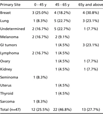
The breast was the most frequent primary site in patients under 45 years of age, while neoplasms with undetermined primary site and lung neo-plasms were found with the same frequency (22.7%) in patients aged 45-65 years. Additionally, older patients presented a high frequency of pri-mary lung cancer (23.1%) followed by pripri-mary gastrointestinal (GI) tumors. Breast cancer was a significant cause of cerebral metastases across all age groups (Table 3).
Table 1. Patient data.
Patients 47
Mean age (range) 53.9 (22-79)
Gender
Female (F) 25 (53.2%)
Male (M) 22 (45.8%)
Table 2. Individual frequency of primary neoplasms.
Primary site Female Male Total (n=25) (n=22) (n=47)
Breast 11 (44.0%) 11 (23.4%)
Lung 2 (8.0%) 7 (31.8%) 9 (19.1%) Undetermined 2 (8.0%) 6 (27.3%) 8 (17.0%) Melanoma 1 (4.0%) 3 (13.6%) 4 (8.5%) GI tumors 1 (4.0%) 3 (13.6%) 4 (8.5%) Lymphoma 2 (8.0%) 1 (4.5%) 3 (6.4%)
Ovary 2 (8.0%) 2 (4.3%)
Kidney 1 (4.0%) 1 (4.5%) 2 (4.3%)
Seminoma 1 (4.5%) 1 (2.1%)
Uterus 1 (4.0%) 1 (2.1%)
Thyroid 1 (4.0%) 1 (2.1%)
The most common symptoms were related to increased ICP (48.9%), especially headache (39.1%) and generalized seizures (30.4%). In contrast, focal neurological events such as motor deficits or sim-ple partial seizures were present in 27.7% of our patients. Symptoms of both localized lesions and increased ICP were reported by 17.0% of our pa-tients and 6.4% (3 papa-tients) related that a bulging mass was their first symptom. Mean time between symptom onset and the first medical evaluation was 5.8 months. Focal manifestations led patients to seek medical attention sooner than increased ICP symptoms (p < 0.05). More than 75% of the patients with focal symptoms sought medical care in less than one month, as opposed to only 5% of those with increased ICP symptoms.
Single brain metastases comprised 57.4% (27 pa-tients) of our cases (Fig 1A/B). One patient (2.1%) had an extra-cranial metastasis only. The frontal lobe was the most affected site (33.3%) followed by the cerebellum (14.8%), the parietal lobe (11.1%), the temporal lobe (3.7%) and the thalamus (3.7%). Furthermore, diffuse single lesions (those involv-ing more than one lobe) were responsible for 33.3% of these cases. Multiple metastases were found in 19 patients (40.4 %) (Fig 1 C/D). Two lesions were identified in 42.1% of these cases, three in 21.1% and more than three in 36.8%. In addition, symp-toms of increased ICP (alone or with focal neurolo-gical events) were more frequent in patients with multiple metastases (78.9% versus59.3% of the
pa-Table 3. Primary site distribution according to age.
Primary Site 0 - 45 y 45 - 65 y 65y and above Breast 3 (25.0%) 4 (18.2%) 4 (30.8%)
Lung 1 (8.3%) 5 (22.7%) 3 (23.1%) Undetermined 2 (16.7%) 5 (22.7%) 1 (7.7%)
Melanoma 2 (16.7%) 2 (9.1%)
GI tumors 1 (4.5%) 3 (23.1%)
Lymphoma 2 (16.7%) 1 (4.5%)
Ovary 1 (4.5%) 1 (7.7%)
Kidney 1 (4.5%) 1 (7.7%)
Seminoma 1 (8.3%)
Uterus 1 (4.5%)
Thyroid 1 (4.5%)
Sarcoma 1 (8.3%)
Total (n=47) 12 (25.5%) 22 (46.8%) 13 (27.7%)
Fig 1. Single brain metastases. (A) Axial T1 weighted MRI after contrast injection showing a hyperintense homogeneous left frontal mass. This mass is a metastatic deposit from a GI tumor in a male patient with focal symptoms. (B) Cerebral comput-ed tomography after contrast injection revealing a hyperdense homogeneous left frontal mass from an ovary metastatic tumor. Patient had ICP symptoms. (C/D) Axial T1 weighted MRI after contrast injection showing two hyperintense heterogeneous mass (left fontal and occipital lobes) from a lung metastatic tumor. Patient had symptoms of ICP.
There was a strong correlation (p< 0.001) bet-ween the number of metastases and supra- or in-fratentorial location. Single tumors were predom-inantly supratentorial (81.5%) while multiple me-tastases were exclusively supratentorial in 52.6%, while being exclusively infratentorial in 5.3% and both supra- and infratentorial in 42.1%.
No correlation was identified between the num-ber of metastases and primary tumor site (p = 0.8). Breast and lung cancer metastases were multiple in 63.6% and 44.4% of cases, respectively, and GI, thyroid, kidney tumors and seminoma cases did pre-sent only single metastases.
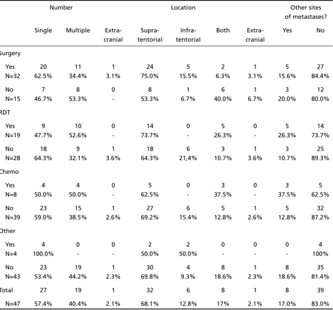
Clinical details according to treatment option are given in Table 4. The treatment category named “Other” in this table refers to 4 patients who un-derwent steroid therapy only. Radiosurgery was
em-ployed in two operated patients after tumor recur-rence. Two patients with metastases in different lobes were operated on (one case with bilateral frontal metastases and one with left cerebellar and parietal tumors). Three other patients had systemic disease with local symptoms (one case each of lymphoma, breast and thyroid cancer). No statistical significance was found between surgi-cal treatment and the number of metastases (p= 0.4), however supratentorial location was found to be a significant factor (p= 0.014).
Sixteen patients (34%) were still being followed as of November 2002. All surviving patients at that time had a Karnofsky Performance Status over 70. The maximum follow-up period was 36 months with a mean survival time after treatment of 8.3 months (Fig 2A). Mean survival time after surgical Table 4. Treatment information.
Number Location Other sites
of metastases? Single Multiple Extra- Supra- Infra- Both Extra- Yes No
cranial tentorial tentorial cranial
Surgery
Yes 20 11 1 24 5 2 1 5 27
N=32 62.5% 34.4% 3.1% 75.0% 15.5% 6.3% 3.1% 15.6% 84.4%
No 7 8 0 8 1 6 1 3 12
N=15 46.7% 53.3% - 53.3% 6.7% 40.0% 6.7% 20.0% 80.0%
RDT
Yes 9 10 0 14 0 5 0 5 14
N=19 47.7% 52.6% - 73.7% - 26.3% - 26.3% 73.7%
No 18 9 1 18 6 3 1 3 25
N=28 64.3% 32.1% 3.6% 64.3% 21,4% 10.7% 3.6% 10.7% 89.3%
Chemo
Yes 4 4 0 5 0 3 0 3 5
N=8 50.0% 50.0% - 62.5% - 37.5% - 37.5% 62.5%
No 23 15 1 27 6 5 1 5 32
N=39 59.0% 38.5% 2.6% 69.2% 15.4% 12.8% 2.6% 12.8% 87.2%
Other
Yes 4 0 0 2 2 0 0 0 4
N=4 100.0% - - 50.0% 50.0% - - - 100%
No 23 19 1 30 4 8 1 8 35
N=43 53.4% 44.2% 2.3% 69.8% 9.3% 18.6% 2.3% 18.6% 81.4%
Total 27 19 1 32 6 8 1 8 39
N=47 57.4% 40.4% 2.1% 68.1% 12.8% 17% 2.1% 17.0% 83.0%
Fig 2. (A) Overall survival. (B/C/D) Kaplan-Meier survival curve (B) according to treatment option (p = 0.2), (C) according to the presence of systemic disease activity (p = 0.68) and (D) for neu-rological symptoms (p= 0.08).
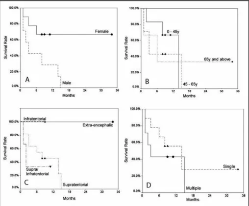
treatment was longer than after other treatment options (14.0 versus 5.0 months), but statistical significance was not reached (p= 0.20). Six-month survival rates after surgery and other treatments were 66.7 and 25%, respectively (Fig 2B). Survival according to the presence of systemic disease activ-ity is shown in Figure 2C. The evidence of extracra-nial disease did not statistically affect survival time (p= 0.68). Patients with focal symptoms had worse survival rates than those with ICP and extra-ence-phalic symptoms, though this was not statistical-ly significant (p= 0.08, Fig 2D). There is a trend to-wards prolonged survival of female patients (p= 0.055 - Fig 3A). Additionally, no association was not-ed between the patient age (p= 0.78), location (p
= 0.30) and the number (p= 0.42) of metastases and survival (Fig 3B, 3C and 3D)
DISCUSSION
Cerebral metastases are the most prevalent of CNS neoplasms, affecting almost 20% of all cancer patients8. Among intracranial diseases, CM are
se-cond only to cerebrovascular disease as the lead-ing cause of mortality7,10. Most studies report that
CM tend to involve male patients more frequent-ly, but this seldom reaches statistical significance. In our series, there is a slight female prevalence, pro-bably owing to the large number of breast neoplasm cases when compared to other patient series11.
Despite single metastases being the most com-mon, the most frequently reported symptom was ICP. This is largely due to the nature of tumor gro-wth and its surrounding edema. Focal symptoms were more frequent in supratentorial lesions con-fined to one lobe only, but a direct relationship bet-ween symptoms and the number of metastases could not be established.
Most patients of this series (68.1%) underwent surgical treatment. Our group adopts classical indi-cations for surgical treatment, taking into account not only the number of metastases, but also the presence of systemic disease, patient age and symp-toms, as well as the tumor site (supra- or infraten-torial)11-17. Lagerwaard, et al. report systemic tumor
activity as an independent, high-impact prognostic factor, and secondary prognostic factors as patient age, number of metastases and histological diag-nosis of the primary tumor11. Sixty-two percent of
the operated patients in our series had a single ce-rebral lesion, while 34.4% had more than one brain lesion and 3.1% had only extra-encephalic symptoms.
Only five of the 32 operated patients had uncon-trolled systemic disease or were treating it. All five patients were under 65 years of age and three of them had single supratentorial metastases. The re-maining two cases had two metastases each, but both could be removed during the same cranioto-my. Surgical treatment was justified in these cases because of their age, evident signs of neurological deterioration and the likelihood that complete resection might improve the survival time12-15,17.
Another nine cases with controlled systemic di-sease and multiple metastases were also operat-ed on. All nine of these patients were under 65 years and the removal of all lesions could be performed via the same craniotomy. Neurological deteriora-tion and controlled systemic disease also played an important role in the decision-making process for these cases. In the study of Bindal et al., the survi-val of patients with controlled systemic disease and multiple CM (which were all removed though one craniotomy) was compared to the survival of those with single metastases15. Both groups were
submitted to surgical treatment and survival rates were similar. Furthermore, complete removal of multiple foci was associated with improved survival rates than suboptimal removal of single metastases. In line with Bindal et al. recommendations, patients in our series who had both multiple CM and were more than 65 years old were not submitted to sur-gical treatment15.
seek-ing of medical attention (p< 0.05). In the present series the data about Karnofsky’s performance scales and/or ECOG (Eastern Cooperative Oncology Group) were not complete; however they have been long recognized as independent, high-impact prognostic factors in the clinical progression of brain metastases11. On the other hand, the results
about the onset symptoms might refine medical decision-making and become a prognostic factor comparable to those scales in the future.
Further investigations into the data presented here should be undertaken before our results can be introduced into clinical practice. In the age of evi-dence-based medicine, the average neurosurgeon is faced with a myriad of guidelines and recommen-dations which are often conflicting. Therefore, criti-cal judgement is necessary to analyze the founda-tions upon which scientific papers are based. Fre-quently, guidelines are established based on inad-equate research designs, which can lead to catas-trophic results. Establishing clinical recommendations based on retrospective, non-randomized studies such as the present study can often be difficult. This study design has several inherent deficiencies, includ-ing selection bias. Treatment options were consid-ered in the light of numerous factors, which may in turn have altered final outcome. On the other hand, it remains doubtful as to whether any form of prospective, randomized study could be conduc-ted in a scientifically correct and ethical manner in the special case of brain metastases. Perhaps the most useful features of our study are the clinical characte-rization of this disease in Brazil and the association of presenting clinical signs and symptoms with oth-er disease charactoth-eristics (such as metastases loca-tion and number) and final outcome. The analysis of treatment results and their relation to final
outco-me should, however, be examined thoroughly and compared to both clinical studies and neurosur-geons’ individual experience.
REFERENCES
1. Aronson SM, Garcia JH, Aronson BE. Metastatic neoplasms of the brain: their frequency in relation to age. Cancer 1964;17:558-563. 2. le Chevalier T, Smith FP, Caille P, et al. Sites of primary malignancies
in patients presenting with cerebral metastases: a review of 120 cases. Cancer 1985;56:880-882.
3. Schellinger PD, Meinck HM, Thron A. Diagnostic accuracy of MRI compared to CT in patients with brain metastases. J Neurooncol 1999;44:275-281.
4. Hochstenbag MMH, Twijnstra A, Wilmink JT, et al. Asymptomatic brain metastases in small cell lung cancer: MR-imaging is useful at ini-tial diagnosis. J Neurooncol 2000;48:243-248.
5. Hirsch FR, Paulson OB, Hansen HH, Vraa-Jensen J. Intracranial metas-tases in small cell carcinoma of lung: correlation of clinical and autop-sy findings. Cancer 1982;50:2433-2437.
6. Subramanian A, Harris A, Piggott K, Shieff C, Bradford R. Metastasis to and from the central nervous system: the “relative protected site”. Lancet Oncol 2002;3:498-507.
7. Victor M, Ropper AH. Intracranial neoplasms and paraneoplastic dis-orders. In Adams and Victor’s. Principles of neurology. Ed 5. McGraw-Hill, 2001;676-733.
8. Crusius P. Metástases intracranianas. In Siqueira MG, Novaes V (eds.). Tumores intracranianos - biologia, diagnóstico e tratamento, Rio de Janeiro: Editora Revinter, 1999.
9. Kaplan EL, Meier P. Nonparametric estimation from incomplete obser-vations. J Am Stat Assoc 1958;53:475-482.
10. Antunes AC, Coutinho MF, Coutinho LM. Hemorrhage in intracranial metastases of melanoma: report of two cases Arq Neuropsiquiatr 1979;37:180-184.
11. Lagerwaard FJ, Levendag PC, Nowak PJCM, et al. Identification of prog-nostic factors in patients with brain metastases: a rewiew of 1292 patients. Int J Radat Oncol Biol Phys 1999;43:795-803.
12. Vecht CJ, Haaxma-Reiche H, Noordiijk EM,et al. Treatment of single brain metastasis: radiotherapy alone or combined with neurosurgery? Ann Neurol 1993;33:583-590.
13. Galicich JH, Sundareasan N, Arbit E, et al. Surgical treatment of single brain metastasis: factor associated with survival. Cancer 1980;45:381-386. 14. Patchell RA, Tibbs PA, Walsh JW, et al. A radomized trial of surgery in the treatment of single metatases to the brain. N Engl J Med 1990;322:494-500. 15. Bindal RJ, Sawaya R, Leavens ME, Lee JJ. Surgical treatment of
multi-ple brain metastases. J Neurosurg 1993;79:210-216.
16. Lang FF, Wildrick DM, Sawaya R. Management of cerebral metastases: the role of surgery. Cancer Control 1998;5:124-129.