ISSN 1806-3713 © 2015 Sociedade Brasileira de Pneumologia e Tisiologia
http://dx.doi.org/10.1590/S1806-37132015000000160 J Bras Pneumol. 2015;41(5):484-484
Continuing EduCation: imaging
484
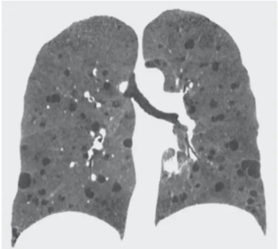
A 57-year-old woman presented with an abdominal mass requiring investigation. She was asymptomatic from a respiratory standpoint. Laboratory test results were unremarkable. A CT scan of the abdomen showed bilateral, fat-containing renal masses and cysts in the lung bases. An HRCT scan showed scattered lung cysts (Figure 1).
Diffuse lung cysts
Edson Marchiori1,2, Gláucia Zanetti2,3, Bruno Hochhegger4,5
Figure 1. HRCT scan showing multiple, round, thin-walled cysts distributed homogeneously throughout the lungs. The remainder of the lung parenchyma is unremarkable. Note that some of the cysts are located in the lung bases.
1. Universidade Federal Fluminense, Niterói, Brasil. 2. Universidade Federal do Rio de Janeiro, Rio de Janeiro, Brasil. 3. Faculdade de Medicina de Petrópolis, Petrópolis, Brasil. 4. Santa Casa de Misericórdia de Porto Alegre, Porto Alegre, Brasil.
5. Universidade Federal de Ciências da Saúde de Porto Alegre, Porto Alegre, Brasil.
The patient basically had diffuse lung cysts on HRCT examination. Cysts are characterized by rounded areas of low attenuation in the lung parenchyma and a well-deined interface with the normal adjacent lung. They are distinguishable from pulmonary emphysema because they have no arterioles at their center and usually have an identiiable wall. The cyst wall is usually thin, but it can vary in thickness. Cysts usually contain air but occasionally contain luid. A cystic pattern is encountered in a number of diseases, the most common being lymphangioleiomyomatosis (LAM), Langerhans cell histiocytosis, lymphocytic interstitial pneumonia (LIP), and
Birt-Hogg-Dubé syndrome (BHDS). Clinically, cysts are usually asymptomatic or accompanied by dyspnea and are often discovered in routine tests or when complications, such as pneumothorax, occur.
Some clinical and tomographic criteria can be useful for the differential diagnosis. In LIP, cysts are less numerous and can be associated with ground-glass opacities. Frequently, LIP occurs in patients with immunological diseases, especially Sjögren’s syndrome. In Langerhans cell histiocytosis, cyst shapes can be more bizarre and, more importantly, cysts predominate in the upper lung ields, sparing the lung bases, especially the costophrenic sulci.
Two syndromic conditions can present with lung cysts and renal masses: tuberous sclerosis and BHDS. In BHDS, cysts are less numerous, are larger, and predominate in the lower lobes. Renal masses, as a rule, correspond to malignant tumors. In tuberous sclerosis, cysts correspond to LAM, are more numerous and diffuse, and also affect the lung bases. Renal masses are benign in nature and are angiomyolipomas.
Tuberous sclerosis is a genetic syndrome, caused by mutations in the TSC1 or TSC2 gene, and is characterized by formation of hamartomas in multiple organs or organ systems. Despite the recent advent of genetic testing for TSC gene mutations, diagnosis continues to be based on clinical criteria. A deinitive diagnosis can be made when patients have at least two of the following indings: cardiac rhabdomyomas; cortical tubers; facial angioibromas; hypermelanotic macules; LAM; renal angiomyolipomas, retinal hamartomas; Shagreen patches; subependymal giant cell astrocytomas; subependymal nodules; or ungual ibromas.
In the case of this patient, taking into account the presence of numerous diffuse cysts in the lung bases, as well as of fat-containing renal masses (angiomyolipomas), the inal diagnosis was tuberous sclerosis-associated LAM.
Recommended Reading