Early m ye lin bre akdo wn fo llo wing
sural ne rve crush: a fre e ze -fracture
study
Departamento de Histologia e Embriologia, Instituto de Ciências Biomédicas, Universidade Federal do Rio de Janeiro, Rio de Janeiro, RJ, Brasil
A.M.B. Martinez and S. Canavarro
Abstract
In this study we describe the early changes of the myelin sheath following surgical nerve crush. We used the freeze-fracture technique to better evaluate myelin alterations during an early stage of Wallerian degeneration. Rat sural nerves were experimentally crushed and ani-mals were sacrificed by transcardiac perfusion 30 h after surgery. Segments of the nerves were processed for routine transmission electron microscopy and freeze-fracture techniques. Our results show that 30 h after the lesion there was asynchrony in the pattern of Wallerian degeneration, with different nerve fibers exhibiting variable degrees of axon disruption. This was observed by both techniques. Careful examination of several replicas revealed early changes in myelin membranes represented by vacuolization and splitting of con-secutive lamellae, rearrangement of intramembranous particles and disappearance of paranodal transverse bands associated or not with retraction of paranodal myelin terminal loops from the axolemma. These alterations are compatible with a direct injury to the myelin sheath following nerve crush. The results are discussed in terms of a similar mechanism underlying both axon and myelin breakdown. Co rre spo nde nce
A.M.B. Martinez
Departamento de Histologia e Embriologia, Bloco F, ICB, UFRJ Av. Brigadeiro Trompowski, s/n 21941-590 Rio de Janeiro, RJ Brasil
E-mail: martinez@ chagas.biof.ufrj.br
Research supported by CAPES, CNPq, FAPERJ and FUJB.
Received April 5, 2000 Accepted September 13, 2000
Ke y wo rds
·Myelin breakdown
·Wallerian degeneration
·Freeze-fracture
·Crush lesion
·Sural nerve
It is generally accepted that axonal cyto-skeleton breakdown during Wallerian de-generation precedes myelin disruption (for a review, see Ref. 1). However, the early changes in myelin sheath following nerve injury are not well understood. We have sometimes observed in fibers undergoing early Wallerian degeneration areas of myelin disruption in the presence of a normal or partially disrupted axoplasm content (data not shown). This finding has also been re-ported by others (2,3). Ludwin (2) described simultaneous degeneration of the axon and myelin after transection of the optic nerve. It is, however, very common to interpret these
findings as artifacts due to poor fixation. If this is not true, it is possible that myelin is directly affected by nerve injury.
using the freeze-fracture technique. Thirty hours after crush was the time chosen for the study because there are fibers in different stages of Wallerian degeneration. At that time, approximately 25% of fibers no longer exhibit cytoskeletal components but the re-maining 75% are either normal or display partial disruption of the axoplasmic cyto-skeleton (4).
Ten Wistar rats were anesthetized and their right sural nerves crushed using a fine forceps and a moderate pressure for 10 s. Left sural nerves were used as controls. Thirty hours after the crush the animals were per-fused intracardially with 2% glutaraldehyde and 2% paraformaldehyde in 0.1 M phos-phate buffer, pH 7.4, and their nerves were dissected out and processed for freeze-frac-ture. Briefly, fragments of the nerves distal stump (the immediately distal 0.5 cm seg-ment was discarded to avoid the traumatic area) were infiltrated in glycerol, mounted on specimen support disks, frozen in liquid nitrogen and fractured at -196oC in Balzers
freeze-fracture equipment, followed by plat-inum shadowing at a 45o angle. Replicas
were obtained after sodium hypochloride digestion, collected on copper grids and ob-served under a Zeiss 900 transmission elec-tron microscope. We also processed tissue for conventional transmission electron mi-croscopy. Briefly, fixed segments of the nerves distal stump were post-fixed in 1% osmium tetroxide, dehydrated in acetone, embedded in PolyBed resin, cut on an RMC ultramicrotome and observed under a Zeiss transmission electron microscope.
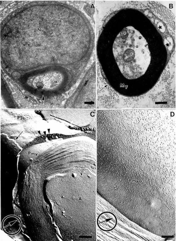
Analysis of ultrathin sections revealed myelinated nerve fibers of normal appear-ance in controls (Figure 1A). In degenerat-ing nerves we could observe normal lookdegenerat-ing myelinated nerve fibers together with fibers exhibiting partial or total granular dissolu-tion of the axoplasm (Figure 1B).
Careful examination of several replicas revealed myelinated nerve fibers fractured at different angles exhibiting several aspects of
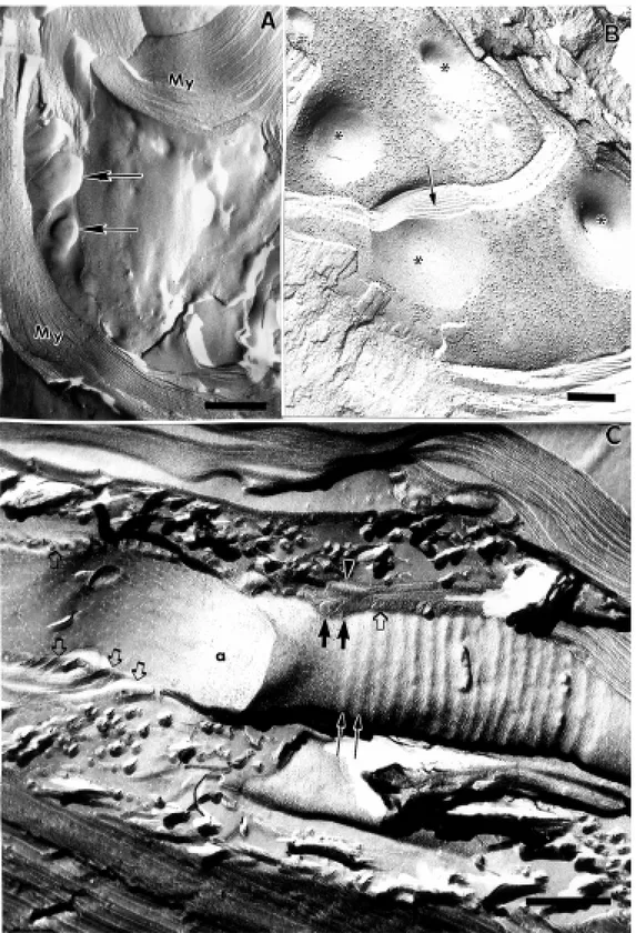
data indicate that myelin alterations can ap-pear at a very early stage of Wallerian degen-eration and are characterized by vacuoliza-tion of myelin lamellae and separavacuoliza-tion and rearrangement of intramembranous particles. Another interesting finding was the
pres-ence, in one replica, of a paranode (Figure 2C) which exhibited the P face of the axolemma with its indentations formed by myelin terminal loops (MTL) which appeared as parallel transverse bands running perpen-dicularly to the long axis of the axon. The
majority of these bands and MTL appeared normal but, as can be observed in Figure 2C, the first paranodal MTL (closest to the node)
is detached from the axolemma and the asso-ciated transverse band is missing. Interest-ingly, the next two MTL are apparently still
attached to the axolemma but their corre-sponding bands have disappeared. However, as can be easily observed in Figure 2C, the intramembranous particles associated with the transverse bands are still present. This data suggest that transverse bands disappear before MTL retraction and that this event is not a consequence of the latter. In agreement with these findings, in a previous study we showed that at 30 h after crush many nodes exhibit an increase in the nodal gap but labeling for sodium channel proteins is still positive (5), indicating that MTL retraction is an early event and might occur before the intramembranous particles degenerate and disappear. Previous works have also shown early myelin detachment from the axolemma (6), but Abrahams et al. (7), who also ad-dressed this issue, did not find this alteration at 24 h after nerve crush. Therefore, we suggest that 30 h is a critical period for the initiation of myelin retraction.
Our results present evidence that in the course of early Wallerian degeneration the myelin sheath suffers alterations in its struc-ture which are represented by splitting and vacuolization of consecutive lamellae, re-distribution of intramembranous particles and disappearance of paranodal parallel trans-verse bands which can appear associated or not with retraction of MTL from the para-nodal axolemma.
The mechanism underlying the initiation of axon degeneration has been a subject of interest for many researchers and, although it is not completely elucidated, many of its aspects have been clarified recently. It has been shown that axonal cytoskeleton degen-eration occurs due to an increase in
intra-axonal calcium concentration (8-16) which activates proteases such as calpains (17). These enzymes are normally present inside axons and are only activated when there is an increase in intra-axonal calcium concentra-tion. Calpains have also been found inside astrocytes and Schwann cell cytoplasm dur-ing Wallerian degeneration (2) and have also been implicated in the degradation of myelin basic protein which is a normal component of the myelin sheath in central and peripher-al myelinated nerve fibers (2). Therefore, it is possible that Schwann cell calpains are also activated by an increase in calcium ions inside the cytoplasm as a general tissue re-sponse to mechanical trauma (nerve crush) and consequently could be responsible for the early myelin alterations as observed in our study and others on nerve degeneration. The involvement of proteolytic enzymes in the mechanism of myelin breakdown has also been described in the course of demyeli-nating diseases (18).
The route of calcium entry into the axon is currently being studied in many research laboratories. Recently it has been postulated that the mechanism of calcium entry is prob-ably due to a reverse operation of the Na+
-Ca+ exchanger (19,20) which pumps
cal-cium into the axon as a response to ATP deprivation and failure of Na+-K+ ATPase.
Although this putative route was described as the mechanism of calcium entry into the axons it is very likely that it can also underlie the mechanism of calcium entry into Schwann cells and therefore be responsible for calpain activation and myelin degradation at an early stage of nerve degeneration.
Re fe re nce s
1. Joseph BS (1973). Somatofugal events in Wallerian degeneration: a conceptual overview . Brain Research,59: 1-18. 2. Ludw in SK (1995). The axon. In: Waxman
SG, Kocsis JD & Stys PK (Editors), Pathol-ogy of the M yelin Sheath. Oxford
Univer-sity Press, Oxford.
3. Cullen M J (1988). Freeze-fracture analy-sis of myelin membrane changes in Wal-lerian degeneration. Journal of Neurocy-tology, 17: 105-115.
4. M albouisson AM B, Ghabriel M N & Allt G
(1984). The non-directional pattern of ax-onal changes in Wallerian degeneration. A computer-aided morphometric analysis. Journal of Anatomy,139: 159-174. 5. M artinez AM B (1999). Distribution of
myelin associated glycoprotein (M AG) during the early stages of Wallerian de-generation. Journal of Submicroscopic Cytology and Pathology,31: 73-81. 6. Ishise J & Rosenbluth J (1987). Nodal and
paranodal structure during Wallerian de-generation in frog spinal nerve. Brain Re-search, 418: 85-97.
7. Abrahams PH, Day A & Allt G (1981). The node of Ranvier in early Wallerian degen-eration: A freeze-fracture study. Acta Neu-ropathologica,54: 95-100.
8. Schlaepfer WW (1971). Experimental al-terations of neurofilaments and neurotu-bules by calcium and other ions. Experi-mental Cell Research,67: 73-80. 9. Schlaepfer WW (1974). Calcium-induced
degeneration of axoplasm in isolated seg-ments of rat peripheral nerve. Brain Re-search,69: 203-215.
10. Schlaepfer WW (1977). Structural alter-ations of peripheral nerve induced by the calcium ionophore A23187. Brain Re-search,136: 1-9.
11. Schlaepfer WW & Hasler M B (1979). Characterization of the calcium-induced disruption of neurofilaments in rat periph-eral nerve. Brain Research,168: 299-309. 12. Kamakura K, Ushiura S, Sugita H & Toyokuura Y (1983). Identification of Ca2+ activated neutral protease in the peripher-al nerves. Journal of Neurochemistry,40: 908-913.
13. M ata M , Staple J & Fink DJ (1986). Changes in intra-axonal calcium distribu-tion follow ing nerve crush. Journal of Neu-robiology,17: 449-467.
14. Glass JD, Schryer BL & Griffin JW (1994). Calcium-mediated degeneration of the axonal cytoskeleton in the Ola mouse. Journal of Neurochemistry, 62: 2472-2475.
15. George EB, Glass JD & Griffin JW (1995). Axotomy-induced axonal degeneration is mediated by calcium influx through ion-specific channels. Journal of Neurosci-ence, 15: 6445-6452.
16. M artinez AM B & Ribeiro LCV (1998).
Ul-trastructural localization of calcium in pe-ripheral nerve fibers undergoing Wallerian degeneration: an oxalate-pyroantimonate and X-ray microanalysis study. Journal of Submicroscopic Cytology and Pathology, 30: 451-458.
17. Bartus RT (1997). The calpain hypothesis of neurodegeneration: evidence for a common cytotoxic pathw ay. Neuroscien-tist, 3: 314-327.
18. Banik NL (1992). Pathogenesis of myelin breakdow n in demyelinating diseases: role of proteolytic enzymes. Critical Re-view on Neurobiology,6: 257-271. 19. Waxman SG, Black JÁ, Ransom BR &
Stys PK (1993). Protection of the axonal cytoskeleton in anoxic optic nerve by de-creased extracellular calcium. Brain Re-search, 614: 137-145.