p38 Mitogen-Activated Protein Kinase
Pathway Regulates Genes during
Proliferation and Differentiation in
Oligodendrocytes
Jeffery D. Haines1, Debra L. Fulton2¤, Stephane Richard3, Guillermina Almazan1,2 *
1Department of Pharmacology and Therapeutics, McGill University, 3655 Sir William Osler Promenade, Montreal, Quebec, Canada, H3G 1Y6,2Department of Neurology and Neurosurgery, Montreal Neurological Institute and Hospital, McGill University, 3801 University St, Montreal, Quebec, Canada, H3A 2B4,3Terry Fox Molecular Oncology Group, Bloomfield Center for Research on Aging, Lady Davis Institute for Medical Research, and Departments of Oncology and Medicine, McGill University, Montreal, Quebec, Canada, H3T 1E2
¤ Current address: Department of Surgery, Urology Division, McGill University, Research Institute McGill University Health Centre, 1001 Decarie Blvd. Montreal, Quebec, Canada, H4A 3A1
Abstract
We have previously shown that p38 mitogen-activated protein kinase (p38 MAPK) is impor-tant for oligodendrocyte (OLG) differentiation and myelination. However, the precise cellular mechanisms by which p38 regulates OLG differentiation remain largely unknown. To deter-mine whether p38 functions in part through transcriptional events in regulating OLG identity, we performed microarray analysis on differentiating oligodendrocyte progenitors (OLPs) treated with a p38 inhibitor. Consistent with a role in OLG differentiation, pharmacological inhibition of p38 down-regulated the transcription of genes that are involved in myelin bio-genesis, transcriptional control and cell cycle. Proliferation assays showed that OLPs treated with the p38 inhibitor retained a proliferative capacity which could be induced upon application of mitogens demonstrating that after two days of p38-inhibition OLGs remained poised to continue mitosis. Together, our results suggest that the p38 pathway regulates gene transcription which can coordinate OLG differentiation. Our microarray dataset will provide a useful resource for future studies investigating the molecular mechanisms by which p38 regulates oligodendrocyte differentiation and myelination.
Introduction
Oligodendrocyte (OLG) maturation involves a complex interplay of cell cycle regulators, tran-scriptional activators and repressors that drive terminal differentiation (extensively reviewed by [1–3]. We have previously shown that p38 mitogen-activated protein kinase (MAPK) regu-lates OLG differentiation and central nervous system (CNS) myelination [4,5].
OPEN ACCESS
Citation:Haines JD, Fulton DL, Richard S, Almazan G (2015) p38 Mitogen-Activated Protein Kinase Pathway Regulates Genes during Proliferation and Differentiation in Oligodendrocytes. PLoS ONE 10 (12): e0145843. doi:10.1371/journal.pone.0145843
Editor:Fatah Kashanchi, George Mason University, UNITED STATES
Received:November 26, 2010
Accepted:December 9, 2015
Published:December 29, 2015
Copyright:© 2015 Haines et al. This is an open access article distributed under the terms of the Creative Commons Attribution License, which permits unrestricted use, distribution, and reproduction in any medium, provided the original author and source are credited.
Funding:This work was supported by operating grants from the Multiple Sclerosis Society of Canada (MSSC) to SR and GA and Canadian Institutes of Health Research (CIHR) to GA. JDH held a studentship from the MSSC. DLF held a postdoctoral fellowship from MSSC. The funders had no role in study design, data collection and analysis, decision to publish, or preparation of the manuscript.
Competing Interests:The authors declare that no competing interests exist.
Pharmacological inhibition of p38 in oligodendrocyte progenitors (OLPs) prevents the accu-mulation of myelin-specific mRNAs and proteins such as myelin basic protein (MBP) and myelin-associated glycoprotein (MAG) [5]. p38 MAPK has also shown to direct myelin-spe-cific gene expression through the differential regulation of myelin-promoter activities [6]. Fur-thermore,in vitrogenetic knock-down of p38αreduces MAG levels, and galactosylceramide (GalC) staining in OLG membrane sheets. Our previous studies also revealed that the down-stream p38 MAPK effector, MK2, is a component of the signaling pathway that promotes OLG differentiation [7]. However, the mechanisms by which p38 MAPK and MK2 regulate OLG differentiation are unknown. Complementing our in vitro work, a recent study has shown that OLG progenitors derived from p38αconditional knockout mice also failed to differentiate in culture. Moreover, electron microscopic analysis showed that the ultrastructure of myelin bun-dles was impaired and the onset of myelination was delayed in the corpus callosum in p38α knockout mice [8].
To further elucidate the mechanisms by which p38 MAPK signaling regulates OLG differ-entiation, we used rat whole genome microarray profiling on oligodendrocyte progenitors (OLPs) treated with the p38α/βisoform inhibitor, PD169316. In addition to the anticipated alterations in myelin gene expression, we identified novel gene targets regulated by the p38 pathway, including transcripts encoding proteins that are involved in vesicular transport, tran-scription factors previously shown to regulate genes in OLGs, and cell cycle regulators. We vali-dated differential expression of several associated gene transcripts by qPCR. Subsequent proliferation assays indicate that OLPs treated with p38 inhibitors are poised in an active cell cycle state before S-phase. Our results suggest that the p38 pathway regulates genes that func-tion to direct OLG identity through cell cycle and eventual arrest to promote terminal differentiation.
Materials and Methods
Reagents and supplies
Ham's F12 medium, PBS, 7.5% BSA fraction V, and penicillin/streptomycin were purchased from Invitrogen (Burlington, ON, Canada). Fetal calf serum and Dulbecco’s Modified Eagle’s Medium (DMEM) were from Wisent Inc (St-Bruno, QC); PDGF-AA and bFGF from Pepro-Tech (Rocky Hill, NJ). PD169316 was from EMD Chemicals (San Diego, CA). Poly-D-lysine, poly-L-ornithine, human transferrin, insulin, HEPES, Triton-X-100, DTT were from Sigma-Aldrich. Western blotting reagents from GE Healthcare Life Sciences (Baie d’Urfe, QC); A2B5 mouse monoclonal antibody from American Type Culture Collection; rabbit polyclonal Ki67 conjugated with FITC from Abcam (Toronto, ON); mouse monoclonal anti-p27kip1 (BD Bio-sciences, Mississauga, ON); rabbit polyclonal p57 (H-91) from Santa Cruz; rabbit monoclonal phopho-CDC2 (TYR15) from Cell Signaling Technology (Danvers, MA); HRP-, FITC-, or Texas Red-conjugated secondary antibodies from Southern Biotechnology, Jackson Immunor-esearch Laboratories (Cedarlane, Hornby, ON), BIO-RAD Canada (Mississauga, ON) or Invi-trogen (Burlington, ON); Hoechst nuclear stain from Molecular Probes Inc. (Eugene, OR). The O4 antibody was a gift (Sommer and Schachner 1981). All other reagents were from Fisher Sci-entific (Whitby, ON), or VWR (Mont-Royal, QC)
Cell cultures
Primary cultures of oligodendrocyte progenitors (OLPs) were prepared from the brains of new-born Sprague-Dawley rats as described previously (McCarthy and de Vellis 1980; Almazan, Afar et al. 1993). All experiments were approved by the McGill Faculty of Medicine Animal Care Committee (permit number 4373) in accordance with Canadian Council on Animal Care
guidelines. OLPs were plated on poly-D-lysine (PDL)-coated culture dishes and grown in serum free media (SFM) consisting of a DMEM-F12 mixture (1:1), 10 mM HEPES, 0.1% bovine serum albumin, 25 mg/mL human transferrin, 30 nM triiodothyronine, 20 nM hydro-cortisone, 20 nM progesterone, 10 nM biotin, 5 mg/mL insulin, 16 mg/mL putrescine, 30 nM selenium and 2.5 ng/mL each of PDGF-AA and bFGF. The OLPs were changed with media that included mitogens every 2d to maintain the cells in a proliferative state. OLPs spontane-ously differentiate upon removal of mitogens. Cultures were characterized immunocytochemi-cally with cell-type-specific antibodies as previously reported (Cohen and Almazan 1994; Radhakrishna and Almazan 1994). On day 0 of differentiation, more than 95% of the cells are positive for gangliosides detected with monoclonal antibody A2B5, a marker for OLPs in
cul-ture while less than 5% were GalC-positive OLGs, GFAP-positive astrocytes or complement type-3-positive microglia. Cells start to express surface sulfatides, recognize with the monoclo-nal antibody O4 on day 1 following growth factor removal, GalC and MAG (myelin-associated protein) on day 2–3 and MBP (myelin basic protein) on days 3–4. The culture media was changed every 2d, and PD169316 was used at 5.0μM for all experiments, unless otherwise
indi-cated. An equivalent DMSO concentration treatment was used for the control.
RNA extraction
Total RNA was extracted from OLGs (~500,000 cells) differentiated in the absence or presence of 5μM PD169316 using the Qiagen RNeasy kit (Qiagen, Mississauga, ON, Canada). Genomic
DNA was eliminated using an on-column DNase digest (DNase set, Qiagen). The RNA was divided into separate aliquots for microarray analysis and quantitative PCR validations. RNA quality was assessed by Génome Québec Innovation Centre (GQIC) using an Agilent RNA BioAnalyzer (Agilent Technologies, Santa Clara, CA), followed by a Qiagen RNA clean-up col-umn, and quantification using a Nanodrop ND-1000 spectrophotometer (Nanodrop Technol-ogies, Wilmington, DE).
Illumina microarray
Gene expression was determined in control and PD169316-treated OLGs using Illumina Rat Whole Genome microarrays in collaboration with GQIC. Complimentary RNA (cRNA) was prepared from 250 ng of total RNA using the Ambion TotalPrep RNA Amplification kit according to the manufacturer's protocol. cRNA (750 ng/array) generated from this kit was hybridized to Rat Illumina Whole Genome Microarrays (RatRef-12 array) according to Illumi-na's protocol. The microarray data is available in the NCBI Gene Expression Omnibus (Refer-ence Number: 15960825). All data are MIAME compliant, and the raw data have been deposited in a MIAME compliant database, as detailed on the MGED Society websitehttp:// www.mged.org/Workgroups/MIAME/miame.html.
FlexArray analysis
Functional Determination using UniProt and literature searches
The "Core Analysis" function included in the Ingenuity Pathway Analysis (Ingenuity Systems, Redwood City, CA, USA) was used to interpret the rat whole genome microarray data in the context of biological processes, pathways and networks that were modulated by
PD169316-treatment. The genes were identified through their unique NCBI Accession num-ber. The biological function of the most up-regulated and down-regulated genes was deter-mined using UniProt Gene Ontology annotations (http://www.uniprot.org/), or through functional annotations provided in published literature available through PubMed (http:// www.ncbi.nlm.nih.gov/pubmed/). The complete table of up- and down-regulated genes classi-fied according to their biological function is available inS1 Table.
Quantitative PCR
One microgram of DNaseI-treated RNA was reverse-transcribed using the AMV reverse tran-scriptase (Roche Diagnostics, Laval, Quebec). When possible, primers were designed to span exon-exon junctions (S2 Table). OneμL of the total cDNA sample was analyzed per reaction,
using the 96 well-block Roche LightCycler 480, and SYBR Green master mix (SABiosciences, Frederick, MD, USA). PCR amplifications were performed as follows: heat inactivation (10 min, 95°C); followed by 35–45 cycles of 94°C, 15 s; 59°C, 30 s; 72°C, 20 s. PCR products were detected by fluorescence at the end of the extension step, and melting curves were analyzed by monitoring the continuous decrease in fluorescence of the SYBR Green signal. PCR products were verified for a single amplification product using melting curve analysis, and the molecular weight of each product was confirmed using PAGE. The fold change in mRNA levels was determined using advanced relative quantification (Pfaffl) method available in the Roche LightCycler 480 software, and data were normalization with 28S rRNA expression levels.
Proliferation assays
Cells were grown to an approximate density of 1.5 × 10⁵cells/cm2in 24-well dishes. OLPs were treated with an increasing dose (1–7.5μM) of PD169316 for 24 or 48 hrs in SFM without
mito-gens, and then incubated with 1μCi/mL3H-thymidine for an additional 24 hr. In experiments
where mitogens were applied after PD169316 treatment, 2.5 ng/mL PDGF-AA and bFGF were added at the same time as the3H-thymidine and incubated for 24 hr, as above. Following the3H-thymidine incorporation period, the medium was aspirated and cultures were rinsed three times with 5% ice-cold trichloroacetic acid and solubilized in 0.2 N NaOH and 0.1% Tri-ton-X-100. Aliquots were mixed with Ecolite liquid scintillation counting fluid and emissions were recorded using aβ-counter.
Time course immunocytochemistry and in vitro quantifications
Primary OPCs were plated on PDL-coated glass coverslips in 24-well plates. When the cells reached 75–80% confluency (denoted Day-0) the cells were treated with 5μM PD169316 in
Results
p38 MAPK regulates diverse cellular targets in OLGs
To further understand how p38 regulates OLG differentiation, we performed an Illumina microarray analysis on primary rat OLPs differentiated in the presence of the p38α/β isoform-specific inhibitor PD169316 to identify gene targets regulated by this signaling pathway. The microarray data was analyzed to determine transcript expression ratio differences as compared to DMSO-treated controls (Fig 1A) and transformed to fold change. Genes that were up- or down-regulated 1.25 fold with a statistical p-value less than or equal to 0.05 were considered to be significantly differentially expressed from control. We classified the fifty most up- and down-regulated genes that were differentially expressed at 48 hrs (2d) after PD169316 treat-ment according to their biological functions using the Gene Ontology (GO) annotations [9] found in the UniProt database [10] or using literature-derived functional annotations. Up-reg-ulated classes included genes involved in cytokinesis, centromere and spindle formation, repli-cation and cell cycle progression. Smaller subsets of genes up-regulated by PD169316
treatment included amino acid transporters, cytoskeletal proteins, vesicular transporters, extra-cellular matrix molecules, and genes involved in oxidative stress. Down-regulated gene tran-scripts encoded proteins belonging to bone morphogenetic signaling pathways, chondroitin sulfate proteoglycans, and cytoskeletal/vesicle trafficking proteins. A prominent set of down-regulated genes remained unclassified with no known function (Fig 1B and 1CandS1 Table).
Levels of myelin gene transcripts were correctly regulated during OLG
differentiation
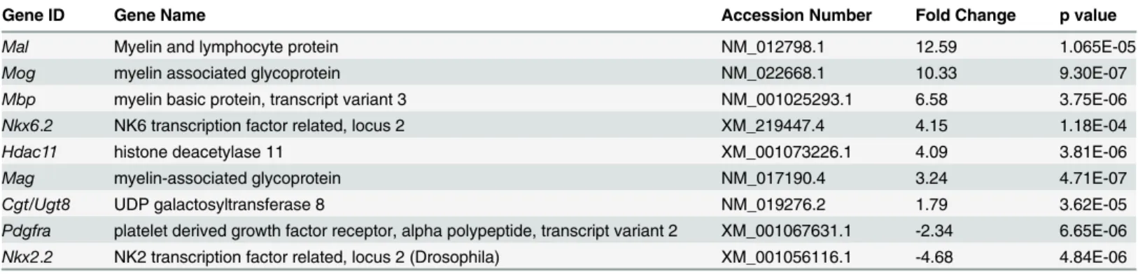
To confirm that the microarray gene expression profiles aligned with previously known gene expression following OLG differentiation, we analyzed transcript levels of typical immature and mature cell-stage specific markers. OLPs continue to proliferate when cultured in the pres-ence of the mitogens PDGF-AA and bFGF and differentiate upon their removal. Consistent with a decreased mitogenic effect upon differentiation, lower expression levels were seen for immature cell-stage markers such as the PDGF receptorα(Pdgfra),nestin, and the early speci-fication marker of the Nkx homeobox gene class family,Nkx2.2. In addition, we observed an upregulation of myelin-specific genes such asMag,Mbp, myelin oligodendrocyte glycoprotein (Mog), and myelin and lymphocyte protein (Mal). The Nkx homeobox geneNkx6.2expression levels were also increased, consistent with its role in OLG differentiation [11]. The expression of another pro-myelin gene activatorHdac11, which promotes the expression ofMbpandPlp during OLP maturation, was also elevated during OLG differentiation [12] (Table 1).
p38 inhibition decreases levels of transcripts encoding myelin-specific
genes
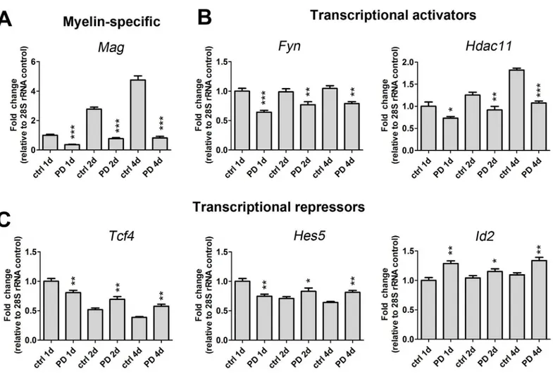
epoxidase, an enzyme involved in a key step in the synthesis of cholesterol that is a necessary component of myelin membrane formation [13].Fyna Src-like tyrosine kinase family member that is important for OLG differentiation and CNS myelination [14], was also down-regulated (Table 2andFig 2B). Further, PD169316 treatment decreased the transcription of genes encod-ingNkx6.2,Hdac11,Sox8and zinc-finger protein 488 (Zfp488), all of which are implicated in activating myelin gene transcription. We employed qRT-PCR analysis to validate the expres-sion level decrease inHdac11and confirmed a decrease at three different time points of differ-entiation (Fig 2B). Additionally, we observed decreased mRNA levels of thyroid hormone receptor,Thra, which binds T3 to activate an intracellular timer controlling OLP differentia-tion [15].
p38 inhibitors increase mRNA levels of OLG transcriptional repressors
and markers associated with early OLPs
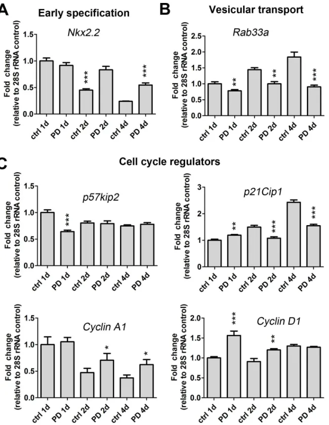
Several transcriptional repressors have been identified that inhibit the progression of OLP mat-uration and function to regulate the onset of OLG differentiation. These include inhibitors of differentiation (Ids), the Wnt/β-catenin target Tcf4, Sox5/6, Notch and one of its downstream effectors Hes5. The expression levels of these repressors decline with OLG maturation to facili-tate the progression of differentiation [16]. In agreement with previously identified roles as inhibitors of differentiation, we observed an up-regulation ofId1andId2following PD19316 treatment (Table 3). We also detected up-regulated levels ofNestinandNkx2.2, which are markers of early OLP development. However, contrary to normal OLP differentiation, the expression level ofPdgfrawas not affected by PD169316 treatment (S1 Table). While there were no appreciable increases in transcription factorsNotch,Hes5andSox6in our microarray results (S1 Table), we did detect a modest, statistically significant up-regulation at day 2 for Hes5, andTcf4by qRT-PCR after p38 inhibition (Fig 2). In agreement with our microarray data, qRT-PCR analyses corroborated up-regulation of the early specification markerNkx2.2 following p38 inhibition (Fig 3).
Fig 1. Gene expression profiling of oligodendrocyte progenitors treated with the p38 MAPK inhibitor, PD169316, reveals gene targets with diverse cellular functions.(A) Schematic diagram of the microarray analysis performed on rat oligodendrocyte (OL) progenitors treated for 1d with the p38 inhibitor, PD169316. Groups of genes were classified by biological function using UniProt and Gene Ontology functional analysis. (B) Functional classes of up-regulated genes included those involved in cytokinesis, spindle formation, replication, cytoskeleton and vesicular transport, p38 target genes, extracellular matrix (ECM), transcription and oxidative stress. (C) down-regulated gene transcript classes included receptors, ligands and transporters, cholesterol and lipid biosynthesis, extracellular matrix (ECM), cytoskeletal remodeling and vesicle trafficking, kinases, phosphatases, cell cycle, bone morphogenetic (BMP) signaling, DNA damage and apoptosis, ubiquitination, transcriptional regulators, and p38 signaling targets.
doi:10.1371/journal.pone.0145843.g001
Table 1. Myelin-specific and other pro-myelin gene transcripts upregulated and/or downregulated during OLG differentiation.OLGs were differenti-ated by removal of PDGF-AA and bFGF for 2d.
Gene ID Gene Name Accession Number Fold Change p value
Mal Myelin and lymphocyte protein NM_012798.1 12.59 1.065E-05
Mog myelin associated glycoprotein NM_022668.1 10.33 9.30E-07
Mbp myelin basic protein, transcript variant 3 NM_001025293.1 6.58 3.75E-06
Nkx6.2 NK6 transcription factor related, locus 2 XM_219447.4 4.15 1.18E-04
Hdac11 histone deacetylase 11 XM_001073226.1 4.09 3.81E-06
Mag myelin-associated glycoprotein NM_017190.4 3.24 4.71E-07
Cgt/Ugt8 UDP galactosyltransferase 8 NM_019276.2 1.79 3.62E-05
Pdgfra platelet derived growth factor receptor, alpha polypeptide, transcript variant 2 XM_001067631.1 -2.34 6.65E-06 Nkx2.2 NK2 transcription factor related, locus 2 (Drosophila) XM_001056116.1 -4.68 4.84E-06
p38 inhibition upregulates the expression of upstream activators of the
p38 MAPK pathway
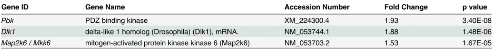
The p38 signaling cascade is initiated by a repertoire of growth stimuli to activate a kinase cas-cade, which includes a number of MAPK kinase kinases (MKKKs). These enzymes, in turn, activate MKK3 or MKK6 (Map2k6) to subsequently activate p38 via phosphorylation of an activation loop domain on p38. Interestingly, we observed an up-regulation of MKK6 following PD169316 treatment suggesting that p38 pathway itself may modulate levels of its upstream activators. In addition, the expression level of PDZ-binding kinase (Pbk), an atypical upstream MKK activator of p38, was also significantly elevated following treatment of OLPs with PD169316. PBK has been shown to specifically activate p38 during cell cycle progression and proliferation [17]. Consistent with a role in regulation of its upstream factors, transcript levels of a p38 MKKK gene,Dlk-1[18,19], was also elevated after p38 inhibition (Table 4).
Fig 2. p38 MAPK regulates the expression of myelin gene transcripts and myelin gene activators and repressors that control oligodendrocyte identity.Gene transcript expression levels of (A) myelin specific (Mag), (B) transcriptional activators (Fyn,Hdac11) and (C) transcriptional repressors (Tcf4, Hes5,Id2) are altered after PD169316 treatment as determined by qRT-PCR. OLPs were treated with 5μM PD169316 for 1d, 2d or 4d and RNA was
harvested, reverse transcribed and analyzed by qRT-PCR. All gene transcripts were normalized to ctrl at 1d all relative to 28S rRNA. Statistical differences were determined using independent t-tests with Bonferroni’s correction (*p<0.05,**p<0.01,***p<0.001 vs same day ctrl).
p38 inhibition modulates the expression of cell cycle regulators, early
growth response proteins and cytokinesis regulators
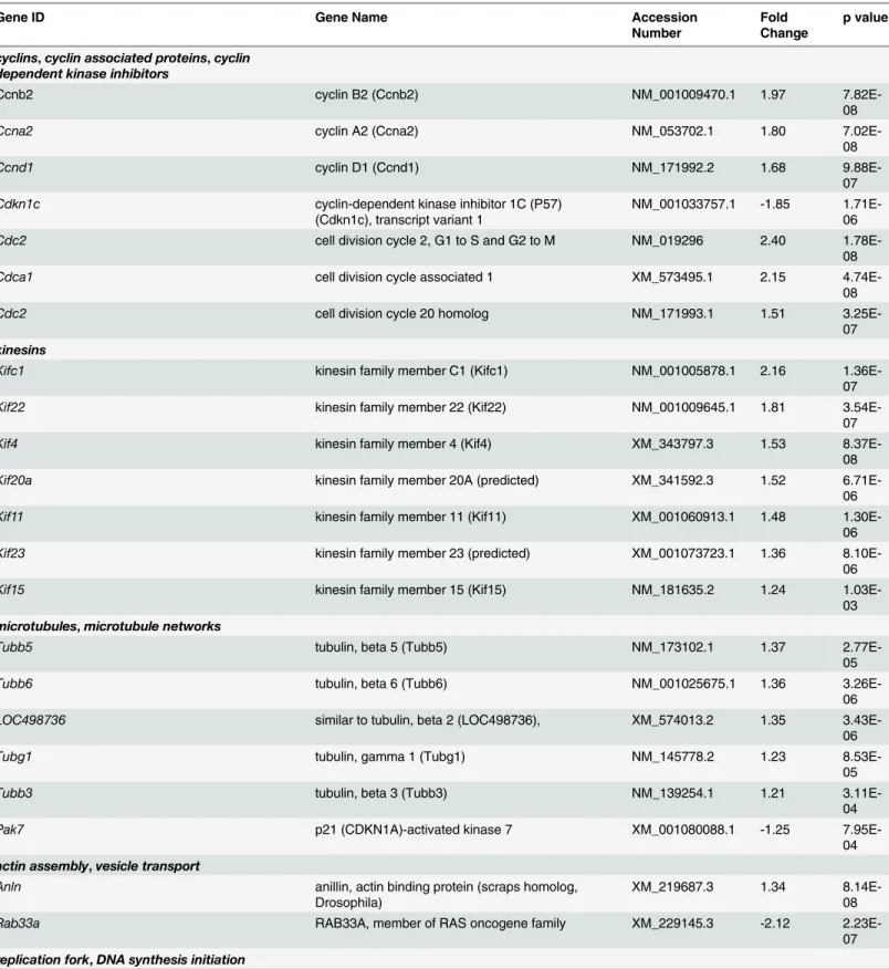
The most abundant group (~45%) of up-regulated genes was composed of transcripts that encode proteins involved in cell cycle progression, spindle and centromere formation, and cytokinesis (Fig 1B). Several cyclins (A2, B2, D1), cyclin associated proteins, and cell division proteins were markedly up-regulated in our microarray and qRT-PCR data following treat-ment of OLPs with PD169316 (Table 5andFig 3) [20]. The p38 pathway also regulates cell cycle checkpoints through phosphorylation of p53 [21]. While we found no changes in mRNA levels ofp53(S1 Table), we did detect decreased levels of p53 target genes includingp21cip1 (Fig 3) andGadd45a(Table 5). We found up-regulated levels for two mitotic checkpoint ser-ine/threonine-protein kinasesBub1 (budding uninhibited by benzimidazoles 1 homolog) and a related isoform,Bub1b. Moreover, PD169316 treatment increased the transcriptional levels of a large group of kinesins involved in microtubule spindle formation during mitosis (Table 5). An up-regulation of cytoskeletal genes involved in mitotic spindle assembly and chromosome separation were also observed, which included: tubulin -b2,-b3,-b5,-b6,-gandanillin. Tran-script levels forRab33a, a protein involved in vesicular transport were downregulated after PD169316 treatment (Table 5andFig 3). Further, transcript expression for p21-activated kinase 7 (Pak7), which regulates Rac/Cdc42 GTPases and cytoskeletal dynamics, was decreased after treatment. We found increased mRNAs levels of a set of genes encoding enzymes with Table 2. Myelin genes and transcriptional activators are decreased by a 24h treatment of OLPs with 5 mM PD169316.
Gene ID Gene Name Accession Number Fold Change p value
Fyn fyn proto-oncogene XM_001062721.1 -1.92 8.44E-09
Mbp myelin basic protein, transcript variant 3 NM_001025293.1 -1.51 6.98E-04
Sox8 SRY-box containing gene 8 (predicted) XM_001060343.1 -1.50 7.26E-07
Cgt/Ugt8 UDP galactosyltransferase 8 NM_019276.2 -1.46 7.48E-07
Sqle squalene epoxidase (Sqle) NM_017136.1 -1.45 1.53E-04
Hdac11 histone deacetylase 11 XM_001073226.1 -1.43 1.27E-06
Thra thyroid hormone receptor alpha (Thra), transcript variant TRalpha2 NM_031134.2 -1.43 6.98E-07
Omgp oligodendrocyte-myelin glycoprotein NM_001005898.2 -1.40 4.55E-06
Nkx6.2 NK6 transcription factor related, locus 2 XM_219447.4 -1.29 0.40 (NS)
Zfp488 zincfinger protein 488 XM_224697.4 -1.29 1.12E-03
Mag myelin-associated glycoprotein NM_017190.4 -1.22 1.96E-03
NS, non-significant.
doi:10.1371/journal.pone.0145843.t002
Table 3. Transcriptional repressors and early OLP markers are upregulated following 24h treatment with 5 mM PD169316.
Gene ID Gene Name Accession Number Fold Change p value
Id1 inhibitor of DNA binding 1 NM_012797.2 1.73 1.33E-06
Nes nestin (Nes) NM_012987.1 1.46 1.56E-06
Jund Jun D proto-oncogene (Jund) XM_001070425.1 1.37 3.80E-04
Id2 inhibitor of DNA binding 2 (Id2) NM_013060.2 1.35 2.01E-05
Nkx2.2 NK2 transcription factor related, locus 2 (Drosophila) (predicted) XM_001056116.1 1.32 0.32 (NS)
Sox6 SRY-box containing gene 6 XM_215016.3 1.22 0.23 (NS)
Pdgfra platelet derived growth factor receptor, alpha polypeptide, transcript variant 2 XM_001067631.1 -1.09 0.22(NS)
Fig 3. p38 MAPK regulates expression of oligodendrocyte specification genes, vesicular transport regulators and cell cycle regulators.Gene transcript expression levels of early (A) specification markers (Nkx2.2), (B) vesicular transport (Rab33a), and (C) cell cycle regulators (p57kip2,p21Cip1, Cyclin A1,Cyclin D1) are altered after PD169316 treatment as determined by qRT-PCR. OLPs were treated with 5μM PD169316 for 1d, 2d or 4d. All gene
transcripts were normalized to ctrl at 1d all relative to 28S rRNA. Statistical differences were determined using independent t-tests with Bonferroni correction (*p<0.05,**p<0.01,***p<0.001 vs. same day ctrl).
roles associated with replication and DNA synthesis, including minichromosome maintenance deficient (MCM), DNA primase and replication factor C. Moreover, the transcription levels of a set of genes involved in spindle body formation and centromere formation were increased after PD169316 treatment, which included aurora kinase B (Aurkb), centromere protein T (Cenpt), nucleolar and spindle associated protein 1 (Nusap1) and polo-like kinase 1 (Plk-1), kinetochore associated proteins and protein regulator of cytokinesis (Prc1) (Table 5).
p38 inhibition suspends OLPs in an active cell cycle state
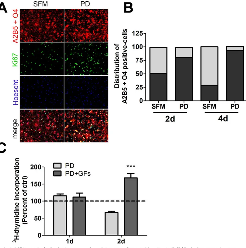
Given the decreased capacity of PD169316-treated cells to incorporate thymidine, we further explored the cell cycle state of p38-inhibited OLPs over a longer time frame by evaluating the presence of nuclear protein Ki67 in cells double-labeled with monoclonal antibodies to OLP markers: A2B5 and O4, as cells positive for either marker can proliferate. Ki67 is expressed dur-ing all active phases of the cell cycle with the exception of G0 restdur-ing stage. OLPs were treated with either PD169316 or DMSO control for 2 or 4d. The populations of Ki67-positive cells and A2B5- and O4-labeled cells were enumerated for each group and compared (Fig 4A and 4B). While fewer than 30% of A2B5 and O4 positive cells treated in DMSO control were Ki67-posi-tive, the significant majority of PD169316-treated A2B5- or O4-positive cells were labeled by the Ki67 marker at 4d. The trend was similar at the 2d time point but the difference in the number of Ki67 labeled cells was smaller. Application of mitogens at the 4d time point to treat-ment and control cells had no significant effect on the proportion of Ki67 labeled cells over those observed in DMSO controls (data not shown). Our results suggest that p38 inhibition over a prolonged duration suspends cells in a non-resting cell state and prevents their differen-tiation, thus providing a potential mechanism for the function of p38 in oligodendrocyte differ-entiation and myelination.
p38 inhibition blocks OLG differentiation, while maintaining OLPs in a
proliferation competent state
Given that our microarray analyses detected an increase in the expression of genes involved in cell cycle and mitosis after p38 inhibition, with accompanying decreases in transcripts encod-ing CDKIs and the thyroid hormone receptor, we explored whether PD169316 treatment per-turbs cell cycle processes in OLPs.3H -thymidine incorporation was performed on cells treated with PD169316 for 1, and 2d (Fig 4). Notably,3H-thymidine incorporation decreased over controls at the 2d time point (Fig 4C). However, when PDGF-AA and bFGF mitogens were applied to PD169316-treated cells for 1d more, a significant increase in thymidine incorpo-ration was observed suggesting that a greater proportion of treated OLPs had the capacity to enter an S-phase cell cycle proliferative state compared to OLPs treated with SFM alone (Fig 4CandS1 Fig). These results combined with the Ki67 assay suggest that p38 may play a role in controlling cell cycle progression in OLPs.
Table 4. PD169316 treatment elevates mRNA levels of p38 upstream activators.
Gene ID Gene Name Accession Number Fold Change p value
Pbk PDZ binding kinase XM_224300.4 1.93 3.40E-08
Dlk1 delta-like 1 homolog (Drosophila) (Dlk1), mRNA. NM_053744.1 1.88 1.48E-06
Map2k6 / Mkk6 mitogen-activated protein kinase kinase 6 (Map2k6) NM_053703.2 1.53 1.67E-05
Table 5. Transcripts encoding cell cycle regulators, kinesins, centromere, spindle and kinetochore associated proteins are upregulated by 5 mM PD169316 treatment.
Gene ID Gene Name Accession
Number
Fold Change
p value
cyclins,cyclin associated proteins,cyclin
dependent kinase inhibitors
Ccnb2 cyclin B2 (Ccnb2) NM_001009470.1 1.97
7.82E-08
Ccna2 cyclin A2 (Ccna2) NM_053702.1 1.80
7.02E-08
Ccnd1 cyclin D1 (Ccnd1) NM_171992.2 1.68
9.88E-07
Cdkn1c cyclin-dependent kinase inhibitor 1C (P57)
(Cdkn1c), transcript variant 1
NM_001033757.1 -1.85 1.71E-06
Cdc2 cell division cycle 2, G1 to S and G2 to M NM_019296 2.40
1.78E-08
Cdca1 cell division cycle associated 1 XM_573495.1 2.15
4.74E-08
Cdc2 cell division cycle 20 homolog NM_171993.1 1.51
3.25E-07
kinesins
Kifc1 kinesin family member C1 (Kifc1) NM_001005878.1 2.16
1.36E-07
Kif22 kinesin family member 22 (Kif22) NM_001009645.1 1.81
3.54E-07
Kif4 kinesin family member 4 (Kif4) XM_343797.3 1.53
8.37E-08
Kif20a kinesin family member 20A (predicted) XM_341592.3 1.52
6.71E-06
Kif11 kinesin family member 11 (Kif11) XM_001060913.1 1.48
1.30E-06
Kif23 kinesin family member 23 (predicted) XM_001073723.1 1.36
8.10E-06
Kif15 kinesin family member 15 (Kif15) NM_181635.2 1.24
1.03E-03
microtubules,microtubule networks
Tubb5 tubulin, beta 5 (Tubb5) NM_173102.1 1.37
2.77E-05
Tubb6 tubulin, beta 6 (Tubb6) NM_001025675.1 1.36
3.26E-06
LOC498736 similar to tubulin, beta 2 (LOC498736), XM_574013.2 1.35
3.43E-06
Tubg1 tubulin, gamma 1 (Tubg1) NM_145778.2 1.23
8.53E-05
Tubb3 tubulin, beta 3 (Tubb3) NM_139254.1 1.21
3.11E-04
Pak7 p21 (CDKN1A)-activated kinase 7 XM_001080088.1 -1.25
7.95E-04
actin assembly,vesicle transport
Anln anillin, actin binding protein (scraps homolog,
Drosophila)
XM_219687.3 1.34
8.14E-08
Rab33a RAB33A, member of RAS oncogene family XM_229145.3 -2.12
2.23E-07
replication fork,DNA synthesis initiation
Discussion
We previously reported that p38 MAPK is required for OLG differentiation and myelination of cultured dorsal root ganglia neurons. To better understand how p38 regulates OLG differentia-tion, we used a rat whole genome microarray analysis to globally survey gene expression Table 5. (Continued)
Gene ID Gene Name Accession
Number
Fold Change
p value
Mcm6 minichromosome maintenance deficient 6 XM_001055953.1 1.99
7.89E-08
Prim1 DNA primase, p49 subunit (Prim1) NM_001008768.1 1.89
2.24E-07
Rfc3 replication factor C (activator 1) 3 NM_001009629.1 1.73
3.57E-07
Mcm7 minichromosome maintenance deficient 7 NM_001004203.1 1.43
9.16E-06
Mcm2 minichromosome maintenance deficient 2 mitotin XM_001072364.1 1.36
3.18E-06
Mcm3 minichromosome maintenance deficient 3 XM_236988.4 1.35
6.18E-06
Mcm10 minichromosome maintenance deficient 10 XM_001071383.1 1.29
4.39E-07
spindle formation,separation
Aurkb aurora kinase B NM_053749.1 2.09
6.10E-09
Prc1 protein regulator of cytokinesis 1 XM_001061201.1 2.06
9.14E-07
Spc25 NDC80 kinetochore complex component, homolog NM_001009654 1.93
2.55E-07
Spbc24 spindle pole body component 24 homolog XM_001077474.1 1.86
4.37E-06
ASPM asp (abnormal spindle)-like, microcephaly
associated (Drosophila)
XM_213891.4 1.82
2.65E-07
Nuf2 Kinetochore protein Nuf2 NM_001012028 1.73
4.72E-10
Nusap1 nucleolar and spindle associated protein 1 XM_001075591.1 1.51
1.19E-05
Kntc1 kinetochore associated 1 XM_001074897.1 1.43
2.48E-07
Kntc2 kinetochore associated 2 (predicted) XM_001055564.1 1.37
3.13E-07
Plk1 polo-like kinase 1 (Drosophila) (Plk1) NM_017100.1 1.34
6.68E-06
Cenpt centromere protein T NM_001024257 1.33
1.11E-04
checkpoints
Ttk Ttk protein kinase (predicted) XM_001062174.1 1.58
2.91E-07
Bub1 budding uninhibited by benzimidazoles 1 homolog XM_215849.4 1.50
1.85E-05 Bub1b budding uninhibited by benzimidazoles 1 homolog,
beta
XM_342494.3 1.33
1.29E-06 Gadd45a growth arrest and DNA-damage-inducible 45 alpha NM_024127.2 -1.37
2.88E-05
Fig 4. p38 inhibitors maintain oligodendrocyte progenitor cells in a non-resting state of the cell cycle.(A, B) Oligodendrocyte progenitors were maintained in serum-free medium in the presence or absence of PD169316 for 2d or 4d and the A2B5 and O4-positive population analyzed for the presence of nuclear Ki67. (A) Representative fluorescent images for the 2d time point, scale bar represents 50μm. (B) Plot of the relative distribution of Ki67+ or
Ki67-A2B5+O4-positive cells. (C) Oligodendrocyte progenitors were treated with 5μM PD169316 for 1d or 2d, followed by3H-thymidine incorporation overnight in
the presence of absence of growth factors (PDGF-AA and bFGF). Statistical differences in (C) were determined by one-way ANOVA followed by Dunnett’s correction (***p<0.001 vs ctrl).
changes in primary OLGs treated with the p38 inhibitor PD169316. Grouping the genes into functional classes using either UniProt or literature searches highlighted an upregulation of groups of genes involved in cytokinesis, centromere and spindle formation, replication and cell cycle progression. Smaller subsets of genes upregulated by PD169316 treatment included amino acid transporters, cytoskeletal proteins, vesicle transporters, extracellular matrix mole-cules, and genes involved in oxidative stress. In contrast, the most prominent group of down-regulated genes was generally unclassified or had no known function. Among other downregu-lated genes were bone morphogenetic proteins, chondroitin sulfate proteoglycans, and cyto-skeletal/vesicle trafficking proteins.
In agreement with other studies, p38 inhibition upregulated myelin-specific gene products associated with OLG differentiation [22,23]. In alignment with our previous study, p38 inhibi-tion downregulated a large number of myelin specific transcripts, includingMag,Mbp,Cnp, PlpandMobp[5]. Novel p38 pathway targets identified in our microarray analysis included the lipid synthesis gene CGT, and squalene epoxidase, a cholesterol biosynthesis enzyme [24]. We also observed decreases in the mRNA levels of pro-myelin gene activators including HDAC11, Fyn and Zfp488. HDAC11 is responsible for activating MBP and PLP promoters, and its knockdown decreases protein levels [12]. The tyrosine kinase Fyn plays multiple roles in oligodendrocyte differentiation, including activation of MBP gene promoter, transport of MBP mRNAs, and phosphorylation of MAG [25,26]. Zfp488 is expressed in differentiating OLGs [27] and it was found to be transcriptionally downregulated in OLG-specific p38α knockout mice at neonatal stages. Furthermore, p38αknockout delays OLP differentiation and causes a number of myelin ultrastructural abnormalities, in addition to a reduction in the num-ber and diameter of axons [8]. Interestingly, our previous results in DRGN/OLG cocultures treated with p38 inhibitors showed a complete and irreversible inhibition of myelination and a lack of organization of the axo-glial adhesion molecule Caspr, while the inhibition of differenti-ation in OLP cultured alone was reversible [5] suggesting that p38 may also play a role in axo-nal integrity.
Another group of gene transcripts that were upregulated in OLPs treated with PD169316 included the upstream enzymes regulating p38 activity such as MKKKs (Dlk1), and PBK, a MKK that specifically phosphorylates p38 during cell cycle [17,28]. These results are in agree-ment with genetic ablation studies of p38αin cardiomyocytes which augmented the expression of p38 upstream activators [29] and suggest that these are compensatory mechanisms in a cel-lular attempt to re-establish basal p38 levels.
Treatment with p38 inhibitor resulted in up-regulation of inhibitors of OLG differentiation or transcriptional repressors:Id1,Id2,Tcf4,and Sox6[16,30–32] as well asNestin, a marker of immature OLG [33]. In contrast, Nkx2.2, which normally decreases with OLG differentiation was increased by PD169316 treatment [34]. However, the p38 pathway appears to differentially regulate immature cell-stage gene targets that are normally downregulated during OLG lineage progression, since no upregulation ofPdgfrawas found with p38 inhibitors. Furthermore, reduced levels of thyroid hormone receptor alpha (Thra) transcripts suggest that p38 inhibition may disrupt the intracellular timer that regulates OLG differentiation.
differentiation by regulating cell cycle state transitions and timing of OLP differentiation [36,
37]. In other cell types, p38 is involved in the regulation of DNA checkpoints including the G2/ M, G1/S and G1/G0 [21]. Examination of Ki67-positive OLPs revealed that the majority of p38 inhibited cells were suspended in an active phase of the cell cycle. Notably, addition of
PDGF-AA and bFGF allowed some OLPs to re-enter S-phase of the cell cycle as shown by thy-midine incorporation. These results are in agreement with those obtained in cardiomyocytes [38–40], and mouse embryonic fibroblasts [41] showing that p38 MAPK pathway regulates genes involved in cellular response to stress, cell division, cell signaling, inflammation and adhesion.
Although the mechanisms by which p38 pathway regulates the cell cycle in OLPs remains to be fully elucidated, our data provides strong support for its role in promoting a transition state between cell proliferation and differentiation through its transcriptional repression of genes involved in cell cycle and inhibitors of differentiation while promoting the expression of pro-myelinating factors. Further studies will be necessary to fully reveal the specific molecular com-ponents involved in this regulatory process. Our microarray dataset will serve as a useful resource for future studies investigating the mechanisms by which p38 regulates oligodendro-cyte proliferation, differentiation and myelination.
Supporting Information
S1 Fig. p38 inhibitors maintain oligodendrocyte progenitor cells in a non-resting state of the cell cycle.OLPs treated for 1d or 2d (A, C) with PD169316 decreased3H-thymidine incor-poration. However, when cultures are maintained for 1d or 2d with PD169316 and then stimu-lated with PDGF-AA and bFGF for an additional 1d, some PD-treated OLPs have the capacity to incorporate thymidine (B, D). Statistical differences were determined by one-way ANOVA followed by Dunnett’s correction (p<0.05,p<0.01,p<0.001 vs ctrl).
(TIF)
S1 Table. List of genes upregulated an downregulated in OLPs following 1d of treatment with the p38 inhibitor PD169316.
(XLSX)
S2 Table. qPCR primers for gene validations.
(DOCX)
Acknowledgments
This work was supported by two operating grants from the Multiple Sclerosis Society of Can-ada (MSSC) to SR and GA and one operating grant from the Canadian Institutes of Health Research (CIHR) to GA. JDH held a doctoral studentship from the MSSC. DLF held a postdoc-toral fellowship from MSSC.
Author Contributions
Conceived and designed the experiments: JDH DLF SR GA. Performed the experiments: JDH DLF. Analyzed the data: JDH DLF. Contributed reagents/materials/analysis tools: SR GA. Wrote the paper: JDH DLF GA.
References
2. Li H, He Y, Richardson WD, Casaccia P. Two-tier transcriptional control of oligodendrocyte differentia-tion. Curr Opin Neurobiol. 2009; 19(5):479–85. doi:10.1016/j.conb.2009.08.004PMID:19740649; PubMed Central PMCID: PMC2826212.
3. Yu Y, Casaccia P, Lu QR. Shaping the oligodendrocyte identity by epigenetic control. Epigenetics. 2010; 5(2):124–8. Epub 2010/02/18. doi: 11160 [pii]. PMID:20160514.
4. Haines JD, Fragoso G, Hossain S, Mushynski WE, Almazan G. p38 Mitogen-activated protein kinase regulates myelination. J Mol Neurosci. 2008; 35(1):23–33. Epub 2007/11/13. doi: 10.1007/s12031-007-9011-0PMID:17994198.
5. Fragoso G, Haines JD, Roberston J, Pedraza L, Mushynski WE, Almazan G. p38 mitogen-activated protein kinase is required for central nervous system myelination. Glia. 2007; 55(15):1531–41. Epub 2007/08/31. doi:10.1002/glia.20567PMID:17729284.
6. Chew LJ, Coley W, Cheng Y, Gallo V. Mechanisms of regulation of oligodendrocyte development by p38 mitogen-activated protein kinase. J Neurosci. 2010; 30(33):11011–27. Epub 2010/08/20. doi: 30/ 33/11011 [pii] doi:10.1523/JNEUROSCI.2546-10.2010PMID:20720108.
7. Haines JD, Fang J, Mushynski WE, Almazan G. Mitogen-activated protein kinase activated protein kinase 2 (MK2) participates in p38 MAPK regulated control of oligodendrocyte differentiation. Glia. 2010; doi:10.1002/glia.21014
8. Chung SH, Biswas S, Selvaraj V, Liu XB, Sohn J, Jiang P, et al. The p38alpha mitogen-activated pro-tein kinase is a key regulator of myelination and remyelination in the CNS. Cell death & disease. 2015; 6:e1748. doi:10.1038/cddis.2015.119PMID:25950478.
9. Ashburner M, Ball CA, Blake JA, Botstein D, Butler H, Cherry JM, et al. Gene ontology: tool for the unifi-cation of biology. The Gene Ontology Consortium. Nat Genet. 2000; 25(1):25–9. doi:10.1038/75556 PMID:10802651; PubMed Central PMCID: PMC3037419.
10. UniProt C. UniProt: a hub for protein information. Nucleic acids research. 2015; 43(Database issue): D204–12. doi:10.1093/nar/gku989PMID:25348405; PubMed Central PMCID: PMC4384041. 11. Liu R, Cai J, Hu X, Tan M, Qi Y, German M, et al. Region-specific and stage-dependent regulation of
Olig gene expression and oligodendrogenesis by Nkx6.1 homeodomain transcription factor. Develop-ment. 2003; 130(25):6221–31. Epub 2003/11/07. doi:10.1242/dev.00868dev.00868[pii]. PMID: 14602683.
12. Liu H, Hu Q, D'Ercole AJ, Ye P. Histone deacetylase 11 regulates oligodendrocyte-specific gene expression and cell development in OL-1 oligodendroglia cells. Glia. 2009; 57(1):1–12. Epub 2008/07/ 16. doi:10.1002/glia.20729PMID:18627006; PubMed Central PMCID: PMC2595137.
13. Saher G, Brugger B, Lappe-Siefke C, Mobius W, Tozawa R, Wehr MC, et al. High cholesterol level is essential for myelin membrane growth. Nat Neurosci. 2005; 8(4):468–75. Epub 2005/03/29. doi: nn1426 [pii] doi:10.1038/nn1426PMID:15793579.
14. Umemori H, Sato S, Yagi T, Aizawa S, Yamamoto T. Initial events of myelination involve Fyn tyrosine kinase signalling. Nature. 1994; 367(6463):572–6. Epub 1994/02/10. doi:10.1038/367572a0PMID: 7509042.
15. Billon N, Jolicoeur C, Tokumoto Y, Vennstrom B, Raff M. Normal timing of oligodendrocyte develop-ment depends on thyroid hormone receptor alpha 1 (TRalpha1). EMBO J. 2002; 21(23):6452–60. Epub 2002/11/29. PMID:12456652; PubMed Central PMCID: PMC136965.
16. Samanta J, Kessler JA. Interactions between ID and OLIG proteins mediate the inhibitory effects of BMP4 on oligodendroglial differentiation. Development. 2004; 131(17):4131–42. doi:10.1242/dev. 01273PMID:15280210.
17. Ayllon V, O'Connor R. PBK/TOPK promotes tumour cell proliferation through p38 MAPK activity and regulation of the DNA damage response. Oncogene. 2007; 26(24):3451–61. Epub 2006/12/13. doi: 1210142 [pii] doi:10.1038/sj.onc.1210142PMID:17160018.
18. Holzman LB, Merritt SE, Fan G. Identification, molecular cloning, and characterization of dual leucine zipper bearing kinase. A novel serine/threonine protein kinase that defines a second subfamily of mixed lineage kinases. J Biol Chem. 1994; 269(49):30808–17. Epub 1994/12/09. PMID:7983011. 19. Nakata K, Abrams B, Grill B, Goncharov A, Huang X, Chisholm AD, et al. Regulation of a DLK-1 and
p38 MAP kinase pathway by the ubiquitin ligase RPM-1 is required for presynaptic development. Cell. 2005; 120(3):407–20. Epub 2005/02/15. doi: S0092-8674(04)01209-7 [pii] doi:10.1016/j.cell.2004.12. 017PMID:15707898.
20. Lavoie JN, Rivard N, L'Allemain G, Pouyssegur J. A temporal and biochemical link between growth fac-tor-activated MAP kinases, cyclin D1 induction and cell cycle entry. Progress in cell cycle research. 1996; 2:49–58. PMID:9552382.
22. Cahoy JD, Emery B, Kaushal A, Foo LC, Zamanian JL, Christopherson KS, et al. A transcriptome data-base for astrocytes, neurons, and oligodendrocytes: a new resource for understanding brain develop-ment and function. J Neurosci. 2008; 28(1):264–78. Epub 2008/01/04. doi: 28/1/264 [pii] doi:10.1523/ JNEUROSCI.4178-07.2008PMID:18171944.
23. Zhang Y, Chen K, Sloan SA, Bennett ML, Scholze AR, O'Keeffe S, et al. An RNA-sequencing transcrip-tome and splicing database of glia, neurons, and vascular cells of the cerebral cortex. J Neurosci. 2014; 34(36):11929–47. doi:10.1523/JNEUROSCI.1860-14.2014PMID:25186741; PubMed Central PMCID: PMC4152602.
24. Abe I, Abe T, Lou W, Masuoka T, Noguchi H. Site-directed mutagenesis of conserved aromatic resi-dues in rat squalene epoxidase. Biochem Biophys Res Commun. 2007; 352(1):259–63. Epub 2006/11/ 23. doi: S0006-291X(06)02482-X [pii] doi:10.1016/j.bbrc.2006.11.014PMID:17112472.
25. Umemori H, Kadowaki Y, Hirosawa K, Yoshida Y, Hironaka K, Okano H, et al. Stimulation of myelin basic protein gene transcription by Fyn tyrosine kinase for myelination. J Neurosci. 1999; 19(4):1393– 7. Epub 1999/02/10. PMID:9952416.
26. White R, Gonsior C, Kramer-Albers EM, Stohr N, Huttelmaier S, Trotter J. Activation of oligodendroglial Fyn kinase enhances translation of mRNAs transported in hnRNP A2-dependent RNA granules. J Cell Biol. 2008; 181(4):579–86. Epub 2008/05/21. doi: jcb.200706164 [pii] doi:10.1083/jcb.200706164 PMID:18490510; PubMed Central PMCID: PMC2386098.
27. Wang SZ, Dulin J, Wu H, Hurlock E, Lee SE, Jansson K, et al. An oligodendrocyte-specific zinc-finger transcription regulator cooperates with Olig2 to promote oligodendrocyte differentiation. Development. 2006; 133(17):3389–98. Epub 2006/08/16. doi: 133/17/3389 [pii] doi:10.1242/dev.02522PMID: 16908628.
28. Dougherty JD, Garcia AD, Nakano I, Livingstone M, Norris B, Polakiewicz R, et al. PBK/TOPK, a prolif-erating neural progenitor-specific mitogen-activated protein kinase kinase. J Neurosci. 2005; 25 (46):10773–85. Epub 2005/11/18. doi: 25/46/10773 [pii] doi:10.1523/JNEUROSCI.3207-05.2005 PMID:16291951.
29. Ambrosino C, Mace G, Galban S, Fritsch C, Vintersten K, Black E, et al. Negative feedback regulation of MKK6 mRNA stability by p38alpha mitogen-activated protein kinase. Mol Cell Biol. 2003; 23(1):370– 81. Epub 2002/12/17. PMID:12482988; PubMed Central PMCID: PMC140674.
30. Stolt CC, Schlierf A, Lommes P, Hillgartner S, Werner T, Kosian T, et al. SoxD proteins influence multi-ple stages of oligodendrocyte development and modulate SoxE protein function. Dev Cell. 2006; 11 (5):697–709. Epub 2006/11/07. doi: S1534-5807(06)00357-1 [pii] doi:10.1016/j.devcel.2006.08.011 PMID:17084361.
31. Hammond E, Lang J, Maeda Y, Pleasure D, Angus-Hill M, Xu J, et al. The Wnt effector transcription fac-tor 7-like 2 positively regulates oligodendrocyte differentiation in a manner independent of Wnt/beta-catenin signaling. J Neurosci. 2015; 35(12):5007–22. doi:10.1523/JNEUROSCI.4787-14.2015PMID: 25810530.
32. Baroti T, Zimmermann Y, Schillinger A, Liu L, Lommes P, Wegner M, et al. Transcription factors Sox5 and Sox6 exert direct and indirect influences on oligodendroglial migration in spinal cord and forebrain. Glia. 2015. doi:10.1002/glia.22919PMID:26345464.
33. Almazan G, Vela JM, Molina-Holgado E, Guaza C. Re-evaluation of nestin as a marker of oligodendro-cyte lineage cells. Microsc Res Tech. 2001; 52(6):753–65. Epub 2001/03/29. doi:10.1002/jemt.1060 [pii] 10.1002/jemt.1060. PMID:11276128.
34. Cai J, Zhu Q, Zheng K, Li H, Qi Y, Cao Q, et al. Co-localization of Nkx6.2 and Nkx2.2 homeodomain proteins in differentiated myelinating oligodendrocytes. Glia. 2010; 58(4):458–68. Epub 2009/09/26. doi:10.1002/glia.20937PMID:19780200; PubMed Central PMCID: PMC2807475.
35. Miki H, Setou M, Kaneshiro K, Hirokawa N. All kinesin superfamily protein, KIF, genes in mouse and human. Proc Natl Acad Sci U S A. 2001; 98(13):7004–11. Epub 2001/06/21. doi:10.1073/pnas. 11114539898/13/7004 [pii]. PMID:11416179; PubMed Central PMCID: PMC34614.
36. Zezula J, Casaccia-Bonnefil P, Ezhevsky SA, Osterhout DJ, Levine JM, Dowdy SF, et al. p21cip1 is required for the differentiation of oligodendrocytes independently of cell cycle withdrawal. EMBO Rep. 2001; 2(1):27–34. Epub 2001/03/17. doi:10.1093/embo-reports/kve008PMID:11252720; PubMed Central PMCID: PMC1083805.
37. Casaccia-Bonnefil P, Hardy RJ, Teng KK, Levine JM, Koff A, Chao MV. Loss of p27Kip1 function results in increased proliferative capacity of oligodendrocyte progenitors but unaltered timing of differ-entiation. Development. 1999; 126(18):4027–37. Epub 1999/08/24. PMID:10457012.
39. Engel FB, Schebesta M, Keating MT. Anillin localization defect in cardiomyocyte binucleation. J Mol Cell Cardiol. 2006; 41(4):601–12. Epub 2006/08/08. doi: S0022-2828(06)00618-3 [pii] doi:10.1016/j. yjmcc.2006.06.012PMID:16889791.
40. Tenhunen O, Rysa J, Ilves M, Soini Y, Ruskoaho H, Leskinen H. Identification of cell cycle regulatory and inflammatory genes as predominant targets of p38 mitogen-activated protein kinase in the heart. Circ Res. 2006; 99(5):485–93. Epub 2006/07/29. doi: 01.RES.0000238387.85144.92 [pii] doi:10.1161/ 01.RES.0000238387.85144.92PMID:16873723.