HISTOCHEMICAL AND ULTRASTRUCTURAL STUDY OF
Caesalpinia
peltophoroides
Benth. (Leguminosae-Caesalpinoideae) SEEDS
1Viviana Borges Corte2, Marília Contin Ventrella3, Eduardo Euclydes de Lima e Borges4, Claudia Aparecida Pontes2 e Daniel Pinho2
ABSTRACT – The objective of this work was to correlate data on light microscopy observations through histochemical analysis and polarized light techniques and investigations in transmission electron microscopy (TEM) to characterize the reserve materials in C. peltophoroides Benth. (Leguminosae-Caesalpinoideae) cotyledons, popularly known as “sibipiruna”, a tropical tree species with wide distribution in Brazil. The cotyledon mesophyll, especially in the abaxial face, is rich in unsaturated neutral lipids contained in numerous lipid bodies dispersed in the cytoplasm. Proteins, more concentrated in the adaxial face of the cotyledons, occur in all the mesophyll and are stored in protein bodies containing globoids, with variable number and size, responsible for accumulation of mineral reserves. Calcium oxalate druses have distribution restricted to the cotyledons adaxial face and are associated with protein bodies. Starch, also distributed all over the cotyledon mesophyll, occurs in small amounts in plastids with developed lamellar system. Secretory cavities rich in phenolic compounds occur among procambial strands.
Keywords: Histochemistry, compartmentalization and reserve materials.
ESTUDO HISTOQUÍMICO E ULTRAESTRUTURAL DE SEMENTES DE
Caesalpinia peltophoroides
Benth. (Leguminosae-Caesalpinoideae)
RESUMO – Este trabalho procurou correlacionar dados de observações em microscopia de luz através de técnicas histoquímicas e de luz polarizada e investigações em microscopia eletrônica de transmissão para caracterizar os materiais de reserva em cotilédones de C. peltophoroides Benth. (Leguminosae-Caesalpinoideae), conhecida popularmente como sibipiruna, ou falso pau-brasil, uma espécie tropical com ampla distribuição no Brasil. O mesofilo cotiledonar, especialmente na face abaxial, apresenta-se rico em lipídios neutros, insaturados, contidos em numerosos corpos lipídicos dispersos no citoplasma. As proteínas, mais concentradas na face adaxial dos cotilédones, ocorrem em todo o mesofilo e são armazenadas em corpos proteicos contendo globoides, com número e tamanho variáveis, responsáveis pelo acúmulo de reservas minerais. Drusas de oxalato de cálcio têm distribuição restrita à face adaxial dos cotilédones e estão associadas aos corpos proteicos. O amido, também distribuído por todo o mesofilo cotiledonar, ocorre em pequena quantidade em plastídios, com sistema lamelar desenvolvido. Entre os cordões procambiais ocorrem cavidades secretoras ricas em compostos fenólicos.
Palavras-chave: Histoquímica, compartimentalização e compostos de reserva.
1 Recebido em 14-12-2007 e aceito para publicação em 23.06.2009.
2 Programa de Pós-Graduação em Ciência Florestal da Universidade Federal de Viçosa (UFV), Viçosa-MG. E-mail:<viviborgescorte@yahoo.com.br> e <pontesac@gmail.com.
3 Departamento de Biologia da Universidade Federal de Viçosa (UFV), Viçosa-MG. E-mail: <ventrella@ufv.br>. 4 Departamento de Engenharia Florestal da UFV. E-mail: <elborges@ufv.br>.
1. INTRODUCTION
Lipids, proteins, carbohydrates and mineral ions represent the main reserve material in seeds. They determine nutritional, industrial and technological
work as an energy source (ATP) to maintain metabolic processes, and as source material for the synthesis of new molecules and tissues that will constitute the seedlings (BEWLEY and BLACK 1983). The content and chemical composition of reserve materials can vary markedly, but they are accumulated in specific organelles that may be present in embryonic or reserve tissues, such as the endosperm and perisperm (BEWLEY and BLACK 1983; BUCKERIDGE et al 2004).
The lipids are synthesized from membranous compartments of the cytoplasm, accumulated in lipid bodies where there is predominance of triacylglycerides that provide energy and structural membrane blocks during the initial germination stages and embryo growth (MURPHY 1990). Under the action of lipases, the fatty acids are liberated and catabolized to produce ATP and acetyl-coA, which is converted to sucrose by the glyoxylate pathway (MURPHY 1990; BUCKERIDGE et al. 2004). Starch, synthesized and accumulated in amyloplasts, is the main carbohydrate reserve in seeds, usually constituted by amylopectin and amylose (MORRISON 1992). During germination, starch is degraded by hydrolysis to maltose, which is later broken down into glucose that can be oxidized or transformed into sucrose. Other reserve carbohydrates can also be found, such as polysaccharides from cell wall, besides sucrose and carbohydrate from raffinose series (BUCKERIDGE et al 2004). Two protein classes are found in seeds, globulins and prolamins, synthesized from the endoplasmic reticulum and the Golgi complex, which are accumulated in protein bodies (MARTY 1999). Proteins serve as the main sulfur nitrogen source and, although not used as ATP sources, they supply carbon skeletons that serve as substratum for the synthesis of others compounds (BUCKERIDGE et al 2004).
Many structural and chemical works have sought to identify reserve compounds in seeds and their cellular compartmentalization (BAGLEY et al. 1963; HORNER and ARNOTT 1965, 1966; SOROKIN 1967; IRWING 1984; PREGO et al. 1998; OTEGUI et al. 1998, 1999; SERRATO-VALENTI 1998). However, few researchers have evaluated tropical species seeds. This work sought to correlate light microscopy data through histochemical and polarized light techniques and investigations under transmission electron microscopy to characterize the reserve materials in C. peltophoroides Benth. (Leguminosae-Caesalpinoideae) cotyledons, popularly known as “sibipiruna”, a tropical tree species with wide
distribution in Brazil, occurring from the Southeast region, in the Atlantic forest, to the Pantanal in Mato-Grosso State (LORENZ I 2002).
2. MATERIALS AND METHODS
This work was accomplished at the Plant Anatomy Laboratory, of the Department of Plant Biology and at the Microscopy and Microanalyses Center of the Universidade Federal de Viçosa (UFV)-MG, Brazil. Ripe Caesalpinia peltophoroides Benth. seeds collected in the municipal district of Viçosa, Minas Gerais, Brazil were used for the study.
2.1. Light microscopy
with Lugol and polarized light were used to show the presence of anisotropic substances, especially crystals and starch (O’BRIEN and McCULLY 1981). All histochemical tests proposed were used in fresh material. To evaluate the extractive or modifying activity of the fixative and the stock solution on different compounds, the tests were also applied in fixed material. For confirmation and location of compounds present in small amounts and comparison of results, it was also used material embedded in methacrylate. Standard control procedures were carried out simultaneously. The images were obtained in light microscope (Olympus AX-70), equipped with polarizing filters, photographic U-photo system, and the Spot-Basic software.
2.2. Transmission electron microscopy
Portions of cotyledons were fixed with 2.5% glutaraldehyde in 0.1M sodium phosphate buffer, pH 7.2, for 1h, at room temperature, and rinsed in the same buffer six times for 10 min. The material was postfixed in 1% sodium phosphate buffered osmium tetroxide (OsO4) for 4h, at 4ºC, and rinsed in the same buffer six times for 10 min. After dehydration in a graded ethanol series, the material was embedded in epoxy resin (Spurr). Ultrathin sections were cut with a diamond knife on a MT2-B ultramicrotome (Du Pont-Sorval), collected on Formvar-coated cupper grids and conventionally stained with uranyl acetate and lead citrate. The sections were observed with a Zeiss EM 109 electron transmission microscope at 80kV.
3. RESULTS
3.1. General features
The Caesalpinia peltophoroides cotyledons show discernible protoderm, ground meristem and procambium, where the ground meristem already presents dorsiventral differentiation, with two to three layers of elongated and juxtaposed cells in the adaxial face, resembling a palisade parenchyma, and several layers of isodiametric cells and compact arrangement on the abaxial face. Among the procambial strands, disposed along the middle region of the cotyledon, large secretory cavities stand out, quite differentiated in the ripe seed. All the cotyledonary cells present thin primary walls, pectocellulosic, stained with PAS, toluidine blue and ruthenium red (Table 1). The Alcian blue test (Table 1) indicates the presence of acid mucopolysaccharides in the external periclinal walls of protoderm cells, probably related to a higher hydrophilia of that region.
3.2. Histochemical tests
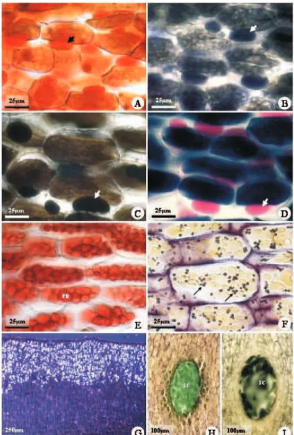
The cotyledon mesophyll is rich in reserve lipids, mainly in the cotyledon abaxial face, being intensely orange stained by Sudan IV (Fig. 1A, Table 1) and black-bluish stained by Sudan Black B (Fig. 1B, Table 1). Lipids are black stained with the OsO4 test (Fig. 1C, Table 1), characterizing unsaturated lipids, while the presence of neutral lipids is confirmed by the characteristic pink staining of Nile blue sulphate (Fig. 1D, Table 1). Free fatty acids were not found in the lipid composition with the rubeanic acid test (Table 1). All tests for lipids demonstrated similar results in fresh and fixed material. The most conclusive results were obtained in fixed material. Reserve lipids were not detected in the material embedded in methacrylate. Because fresh or fixed (without methacrylate embedded) cotyledons were sectioned for the histochemical analyses in light microscopy, lipids coalesce to form one or more oil drops in each cell (Fig. 1A-D) and can occupy over to ¼ of the cellular volume.
P r o t e i n s a r e a b u n d a n t i n a l l c o t y l e d o n mesophyll, especially in the cotyledon adaxial face. Mercury-bromophenol blue and xylidine Ponceau tests (Fig. 1E, Tab.1) confirmed their presence. Similar results were obtained for fresh, fixed or methacrylate embedded material. Among the tests used for protein, xylidine Ponceau presented the most intense and specific coloration, indicating cell compartmentalization in globoid structures, denominated protein bodies.
In the cotyledon adaxial face, the palisade parenchyma shows calcium oxalate crystalline inclusions associated to protein structures, in the form of druses, birefracting under polarized light (Fig. 1G), whose chemical composition is confirmed by its disappearance after treatment with hydrochloric and nitric acid. Fresh, fixed or methacrylate embedded material was suitable to observe crystals under polarization, but the tests were only possible in material without embedding.
COMPOUND TEST COTYLEDON SECRETORY CAVITY Adaxial Abaxial Epithelium Secretion LIPIDS
Sudan III ++ +++ +
-Sudan black B ++ +++ +
-Neutral lipids Nile Blue ++ +++ +
-Fat acids Rubeanic acid - - -
-Insaturated lipids Osmium tetroxide ++ +++ + -PROTEINS
Bromophenol blue ++ + +
-Xylidine Ponceau +++ ++ +
-CARBOHIDRATES
Polysaccharides PAS reagent + + +
-Starch Lugol reagent ++ ++ ++
-Pectins Ruthenium red - - -
-Acid mucopolysaccharides Alcian blue - - - -PHENOLIC COMPOUNDS
Ferric trichloride - - + +++
Toluidine blue - - + +++
Tannins Hydrochloric vanillin - - -
-ALKALOIDS
Wagner reagent - - -
-Dittmar reagent - - -
-Dragendorff reagent - - -
-TERPENOIDS
2,4-dinitrophenylhidrazine - - -
-Essential oils Nadi reagent - - -
-Steroids Anthimonium trichloride - - -
-Sesquiterpene lactones Sulphuric acid - - -
-Abraham reaction - - -
-CALCIUM OXALATE Hydrochloric acid ++ - -
-CRYSTALS Nitric acid ++ - -
-Table 1 – Histochemical tests applied to cotyledons from Caesalpinia peltophoroides Benth. seeds. (+ presence; - absence; * presence only in cell wall; the number of signs expresses the reaction intensity).
Tabela 1 – Testes histoquímicos aplicados em cotilédones de sementes de Caesalpinia peltophoroides Benth. (+ presença;
- ausência; * presente somente na parede celular; o número de sinais expressa a intensidade da reação).
The presence of alkaloids and terpenoids was not detected (Table 1) in the whole cotyledon mesophyll. Phenolic compounds in both fresh and fixed materials were only found in the secretory cavities, indicated by a characteristic greenish color of the material embedded in methacrylate and stained with toluidine blue and with the black color of ferric chloride (Fig. 1H, Table 1). Tannins were not detected with the hydrochloric vanillin test (Table 1). The epithelium of the secretory cavities and adjacent cells are rich in lipids, proteins and starch, positively reacting to the specific tests (Table 1).
Although the usual recommendation for the histochemical analyses is the use of fresh material, the material fixation in FAA50 did not modify qualitative
or quantitatively the reserve materials such as lipid, protein and starch, or even crystals and phenolic compounds, when observed under light microscopy. The embedding in methacrylate was not suitable only for the lipid analysis. However, it was more conclusive for the observation of amyloplasts and crystals, due to their small sizes in this species.
3.3. Ultrastructure of storage cells
Figure 1 – Light micrographs showing the response of Caesalpinia peltophoroides cotyledon cells to histochemical tests. A, characteristic orange color of lipids stained with Sudan IV; large arrows show lipidic drops. B, lipids stained dark blue with the Sudan Black B. C, OsO4 test showing black staining of lipids. D, pink staining of lipids with Nile Blue. E, red staining of protein with xylidine Ponceau; note the large amount of protein bodies in each cotyledon cell. F, small starch grains (narrow arrows) stained black with toluidine blue and lugol reagent. G, calcium oxalate crystals only on the adaxial face, observed under polarized light. These crystals easily dissolved with hydrochloric and nitric acid. H,I, secretory cavities showing phenolic compounds; green staining with toluidine blue and lugol reagent (H); black staining with ferric trichloride test (I).
Figura 1 – Microscopia de luz mostrando a resposta de cotilédones de Caesalpinia peltophoroides aos testes histoquímicos.
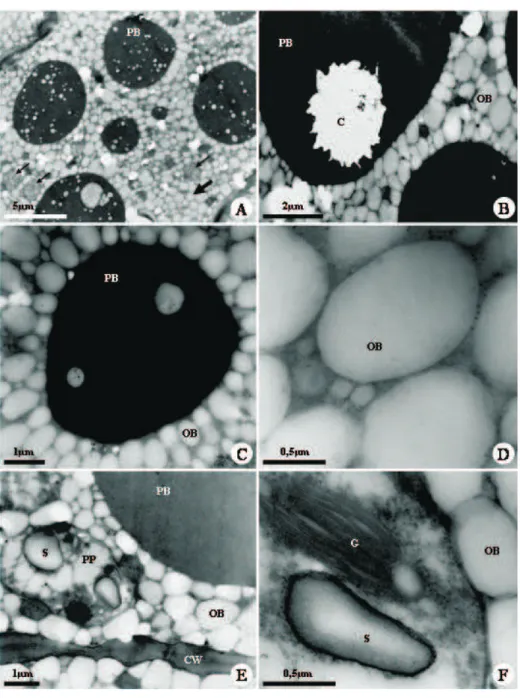
Figure 2 – Transmission electron micrographs of Caesalpinia peltophoroides cotyledon cells, showing the distribution and structure of storage substances. A, cotyledon cell showing protein bodies, lipid bodies (large arrows) and plastids (narrow arrows). B, crystal in the protein body. C, detail of protein body enclosed by lipid bodies. D, detail of lipid bodies. E, plastid with starch grains. F, detail of plastid showing a granun and a starch grain. C, crystal; CW, cell wall; LB, lipid body; PB, protein body; P, plastid; S, starch.
Figura 2 – Micrografia eletrônica de transmissão de células cotiledonares de Caesalpinia peltophoroides mostrando a distribuição
C. peltophoroides protein bodies have a 5-15µm diameter, with electron dense protein matrix, and globoids with variable size and number. Great crystalline inclusions are observed in the protein bodies. However, they are lost during material processing, leaving only their contour in form of druses. Crystals are only observed in the protein bodies located in the palisade parenchyma, and usually only one druse is found in each protein body.
The starch grains have up to 1µm and irregular shape. They are found in 2-4µm plastids, with different stroma and lamellar system, where the thylakoid piling can be seen.
4. DISCUSSION
Among the lipids, oils and fats are the main forms of storage of reduced carbon in seeds, mainly as triacylglycerides, that are neutral lipids in which the fatty acid molecules are linked to the three hydroxyls of the glycerol groups by ester bonds. In the plant lipids, the main saturated fatty acids are lauric, miristic, palm and stearic acids. The main unsaturated fatty acids, oleic, linoleic, and linolenic acids, and their proportion in lipids vary with the species (BUCHANAN et al. 2000). The triacylglycerides are stored in the cotyledon cells cytoplasm or in reserve tissues, organelles known as oleosomes, spherosomes (SOROKIN 1967; FAHN 1985), oil bodies (HORNER and ARNOTT 1965; HUANG 1992; BUCHANAN et al. 2000) or lipid bodies (MURPHY 1990; PREGO et al. 1998; OTEGUI et al. 1999). In lipid bodies, the triacylglycerides are surrounded by a membrane consisting of one layer (monolayer) of phospholipids, stabilized by oleosins, special proteins that recover the surface and hinder the fusion of phospholipids from adjacent lipid bodies (MURPHY 1990; HUANG 1992). Triacylglyceride biosynthesis is attributed to enzymes present in cytoplasm membranous compartments, where the lipid accumulates between two phospholipidic membrane layers, which intumesce and separate as more material is added, releasing a new lipid body that is finally covered by a layer of oleosins formed by the rough endoplasmatic reticulum (MURPHY 1990). In Myrsine laetevirens seed endosperm, the genesis of lipid bodies takes place through the smooth endoplasmatic reticulum secretion, in close association with mitochondrias and plastids, although the rough endoplasmatic reticulum and Golgi are also present (OTEGUI et al. 1999). In C. peltophoroides
cotyledons, the diameter of the lipid bodies varies from 0.5 to 1.5µm. Its uniform distribution over the cytoplasm (Fig. 2) and radiated disposition in relation to the protein bodies is similar to Yucca (HORNER and ARNOTT 1965), Myrsine laetevirens (OTEGUI et al.1999) and Chenopodium quinoa (PREGO et al. 1998) seeds. However, the diameters of these structures measured from transmission electron micrographs are usually underestimated, since most lipid bodies are not viewed at their equatorial planes but rather at planes randomly cut through the organelles.
Crystalline inclusions in the form of calcium oxalate druses are observed in protein bodies, but only in those present in the ground meristem cells designed to be transformed into palisade parenchyma. According to Lott (1981), druse crystals in protein bodies may be common in some species, and their presence can vary depending on the cell and on the tissue. Generally, only one druse is present in each protein body in C. peltophoroides (Fig. 2B), but, in some cells, up to two druses are observed. Observations under light microscopy indicate the chemical nature of the calcium oxalate druses, by the birefraction under polarized light and by the fast dissolution with acids. Although starch, protein, lipid and crystals are generically denominated ergastic or reserve substances (FAHN 1985 DICKISON 2000), commonly found in the endosperm and in the embryo of seeds of several species, crystals present in protein bodies are not actually used during the embryo development or germination process (OTEGUI et al. 1998, 1999; SERRATO-VALENTI et al. 1998; CORTE 2005). Although calcium oxalate crystals represent good taxonomic characteristic (LOTT 1981), their function is still discussed (TILLMAN-SUTELA and KAUPPI 1999; FRANCESCHI and NAKATA 2005). P. tanacetifolia endosperm presents calcium oxalate crystals and globoids rich in calcium in the protein bodies. Nevertheless, during germination only the globoids are digested. In this case, the calcium used by the seedling during the initial development does not come from the calcium oxalate crystals, but only from the globoids (SERRATO-VALENTI et al. 1998).
Starch is a polymer formed by the association of two polysaccharide types, amylose, usually present in the proportion of 25% and amylopectin, in the proportion of 75%. In some cases, this rate can be significantly changed (MORRISON 1992). In chloroplasts, starch grains are temporary because they are quickly degraded after formation, while in the leucoplasts, they are larger, varying from 1 to 150 µm, and stay for a long period in reserve organs (DICKISON 2000). The occurrence of starch grains in Papilionatae seeds is considered abundant, rare in Mimosoidae, and absent in the known Caesalpinioideae (CORNER 1976). In Caesalpinia peltophoroides cotyledons, starch grains are present (Fig. 2A, E-F) in small amounts, with about 1µm and irregular shape, occupying a small plastid area, where
they occur. In these plastids, the presence of piled thylakoids, associated to small starch grains (Fig. 2E-F), can also indicate a transitional form that would result in the conversion to chloroplasts after germination. The radial disposition of the starch around an initial polymerization point, the hilum, confers crystalline properties to the starch grains (FAHN, 1985). Under polarized light, as a result of the double light refraction when crossing the starch grain, an interference cross-shaped figure is usually formed (DICKISON 2000). In C. peltophoroides, due to the irregular shape and reduced size of the starch grains, the birefraction under polarized light is not observed and, consequently, the typical interference figures are not formed. Starch grains are accumulated in plastids during the initial endosperm development of Myrsine laetevirens, and gradually disappear while protein and lipid bodies are formed (OTEGUI et al. 1999). They are absent in the ripe endosperm, representing a temporary form of reserve (OTEGUI et al. 1998).
In general, plant secretions are complex and formed by numerous compounds (ASCENSÃO and PAIS 1987; ASCENSÃO et al. 1997, 1999; SERRATO-VALENTI et al. 1997). Therefore, there can be a predominance of a chemical group, as in the case of the phenolic compounds present in the secretory cavities of C. peltophoroides, which suggests some specificity in the cell secretory activity. Because of the presence of chemical defenses such as the phenolic ones, the secretory cavities could have an ecological role related to the deterrence of predators and fungi and bacterium parasites (FAHN 1985; VAZQUEZ-YANES and OROZCO-SEGOVIA 1993; HARBORNE 1997), assuring the species reproduction.
5. CONCLUSIONS
Concluding, the major reserve materials of C. peltophoroides cotyledons are lipids and proteins, compartmentalized in lipid bodies and protein bodies, respectively. However, small amounts of starch are found in plastids.
6. ACKNOWLEDGEMENTS
7. REFERENCES
ASCENSÃO, L.; PAIS, M. S. S. Glandular trichomes of Artemisia campestris (ssp. maritima): ontogeny and histochemistry of the secretory product. Botanical Gazette, v.148, n.3, p.221-227, 1987.
ASCENSÃO, L.; MARQUES, N.; PAIS, M. S. S. Peltate glandular trichomes of Leonotis leonurus leaves: ultrastructure and histochemical
characterization of secretions. International Journal Plant Science, v.158, n.3, p.249-258, 1997.
ASCENSÃO, L.; MOTA, L.; CASTRO, M. M. Glandular trichomes of the leaves and flowers of Plectranthus ornatus: morphology, distribution and histochemistry. Annals of Botany, v.84, n.4, p.434-447, 1999.
BAGLEY, B. W. et al. A study of protein bodies during germination of peanut (Arachis hipogaea) seed. America Journal of Botany, v.50, n.6, p.523-532, 1963.
BEWLEY, J. D.; BLACK, M. Physiology and biochemistry of seeds in relation to germination. New York: Springer-Verlag, 1983.
BUCHANAN, B. B.; GRUISSEM, W.; JONES, R. Biochemistry & molecular biology of plants. Maryland: American Society of Plant Physiologists, 2000.
BUCKERIDGE, M. S. et al. Acúmulo de reservas. In: FERREIRA, A. G.; BORGUETTI, F. (Eds). Germinação: do básico ao aplicado. Porto Alegre: Artmed, 2004. p.31-50.
CAIN, A. J. The use of Nile Blue in the
examination of lipids. Quarterly Journal of Microscopy Science, v.88, n.1, p.111-116, 1947.
CANIATO, R. et al. Detection of peroxides in intact plant material. Fitoterapia, v.60, n.1, p.549-551, 1989.
CHAMBERLAIN, C. J. Methods in plant histology. 5.ed. Chicago: The University of Chicago Press, 1932.
CORNER, E. J. H. The seeds of dicotyledons. Cambridge: Cambridge University Press, 1976.
CORTE, V. B. Estudos histoquímicos, bioquímicos e fisiológicos em sementes de Caesalpinia peltophoroides Benth.
durante a germinação e crescimento inicial das plântulas. 2005. 70 FOLHAS. Dissertação (Mestrado em Ciência Florestal) -Universidade Federal de Viçosa, Viçosa, MG, 2005.
DAVIDE, R.; CARDE, J.P. Coloration différentielle des inclusions lipidique et terpéniques des pseudophylles du pin maritime au moyen du réactif Nadi. Comptes Rendus Hebdomadaires dês Scéances de I’ Academie dês Sciences Paris, v.258, NUMERO, p.1338-1340, 1964.
DICKISON, W. C. Integrative plant anatomy. San Diego: Academic Press, 2000.
FAHN, A. Plant anatomy. 4.ed. Oxford: Pergamon Press, 1985.
FAHN, A. Secretory tissues in plants. London: Academic Press, 1979.
FEDER, N.; O’BRIEN, T. P. Plant microthecnique: some principles and new methods. America Journal of Botany, v.55, n.1, p.123-142, 1968.
FRANCESCHI, V. R.; NAKATA, P. A. Calcium oxalate in plants: formation and function.
Annual Review Plant Biology, v.56, n.1, p.41-71, 2005.
FURR, M.; MAHLBERG, P. G. Histochemical analyses of laticifers and glandular trichomes in Cannabis sativa. Journal of Natural Products, v.44, n.2, p.153-159, 1981.
GANTER P.; JOLLÉS, G. Histochemic
normale et pathologique. Paris: Gauthier-Villars, 1969/ 1970.
HARBORNE, J. B. Plant secondary metabolism. In: CRAWLEY, M. J. (Ed.) Plant ecology, 2.ed. Oxford: Blackwell Science, 1997. p.132-155.
HORNER, H. T.; ARNOTT, H. J. A hitochemical and ultrastructural study of yucca seed proteins. America Journal of Botany, v.52, n.10, p.1027-1038, 1965.
HORNER, H.T.; ARNOTT, H. J. A hitochemical and ultrastructural study of pré- and post-germinated yucca seeds. America Journal of Botany, v.127, n.10, p.48-64, 1966.
HUANG, A. H. C. Oil bodies and oleosins in seeds. Annual Review of Plant Physiology and Plant Molecular Biology, v.43, p.177-200, 1992.
IRVING, D. W. Seed structure and histochemistry of Prosopis velutina (Leguminosae). Botanical Gazette, v.145, n.3, p.340-345, 1984.
JOHANSEN, D. A. Plant microtechnique. New York: McGraw-Hill, 1940.
KUANG, A. et al. Influence of microgravity on ultrastructure and storage reserves in seeds of Brassica rapa L. Annals of Botany, v.85, n.6, p.851-859, 2000.
LORENZI H. Árvores brasileiras: manual e cultivo de plantas arbóreas nativas do Brasil. Nova Odessa: Plantarum, 2002.
LOTT, J.N.A. Protein body in seeds. Nordic Journal of Botany, v.1, NUMERO, p.423-432, 1981.
MACE, M. E.; HOWELL, C. R. Histochemistry and identification of condensed tannin precursor in roots of cotton seedlings. Canadian Journal of Botany, v.52, n.11, p.2423-2426, 1974.
MACE, M. E.; BELL, A. A.; STIPANOVIC, R. D. Histochemistry and isolation of gossypol and related terpenóides in roots of cotton seedlings. Phytophatology, v.64, n.10, p.1297-1302, 1974.
MARTY, F. Plant vacuoles. Plant Cell, v.11, n.4, p.587-599, 1999.
MAZIA D.; BREWER, P. A.; ALFERT, M. The cytochemistry staining and measurement of protein with mercuric bromophenol blue.
Biological Bulletins, v.104, NUMERO, p.57-67, 1953.
MORRISON, W. R. Analysis of cereal starches. In: LINSKENS, H. F.; JACKSON, J. F. (Eds.) Seed analysis. New York, Springer-Verlag, 1992. p.199-215.
MURPHY, D. J. Storage lipid bodies in plants and other organisms. Progress Lipid Research, v.29, n.4, p.299-324, 1990.
O’BRIEN, T. P.; McCULLY, M. E. The study of plant structure: principles and selected methods. Melburne: Termarcarphy Pty, 1981.
O’BRIEN, T.P.; FEDER, N.; MCCULLY, M. E. Polychromatic staining of plant cell walls by toluidine blue O. Protoplasma, v.59, n.2, p.368-373, 1964.
OTEGUI, M. et al. Histological and chemical characterization of Myrsine laetevirens seed. International Journal Plant Science, v.159, n.5, p.762-772, 1998.
OTEGUI, M. et al. Development of the endosperm of Myrsine laetevirens (Myrcinaceae). II.
Formation of protein and lipid bodies.
International Journal Plant Science, v.160, n.3, p.501-509, 1999.
PEARSE, A. G. E. Histochemistry
theoretical and applied. 4.ed. New York: Longman Group, 1980. v.2.
PREGO, I.; MALDONADO, S.; OTEGUI, M. Seed structure and localization of reserves in
Chenopodium quinoa. Annals of Botany, v.82, n.4, p.481-488, 1998.
SERRATO-VALENTI, G.; MARIOTTI, M.G.; CORNARA, L.; CORALLO, A. A histological and structural study of Phacelia tanacetifolia
endosperm in developing, mature, and germinating seed. International Journal Plant
Science, v.159, n.5, p.753-761, 1998.
SOROKIN, H.P. The spherosomes and the reserve fat in plant cells. America Journal of Botany, v.54, n.8, p.1008-1016, 1967.
SVENDSEN, A. B.; VERPOORTE, R.
Chromatography of alkaloids. New York: Elsevier Scientific Publishing Company, 1983.
TILLMAN-SUTELA, E.; KAUPPI, A. Calcium oxalate crystals in the mature seeds of Norway spruce, Picea abies (L.) Karst. Trees, v.13, n.3, p.131-137, 1999.
VAZQUEZ-YANES, C.; OROZCO-SEGOVIA, A. Patterns of seed longevity and germination in the tropical rainforest. Annual Review
Ecolology of Systems, v.24, p.69-87, 1993.
VIDAL, B. C. Acid glycosaminoglycans and endochondral ossification: