Acta Scientiarum
http://www.uem.br/acta ISSN printed: 1679-9275 ISSN on-line: 1807-8621
Doi: 10.4025/actasciagron.v39i4.32492
Acta Scientiarum. Agronomy Maringá, v. 39, n. 4, p. 535-543, Oct.-Dec., 2017
Histochemical changes during the ontogeny of
malagueta
and
biquinho
pepper seeds
Haynna Fernandes Abud1*, Eduardo Fontes Araújo1, Roberto Fontes Araujo2, Edgard Augusto de Toledo Picoli1 and Maria Izabel Gallão3
1
Universidade Federal de Viçosa, Avenida Peter Henry Rolfs, s/n, 36570-900, Viçosa, Minas Gerais, Brazil. 2Empresa de Pesquisa Agropecuária de Minas Gerais, Viçosa, Minas Gerais, Brazil. 3Universidade Federal do Ceará, Fortaleza, Ceará, Brazil. *Author for correspondence. E-mail: hfabud@gmail.com
ABSTRACT. This study aimed to evaluate the structural changes and reserve deposition of malagueta and
biquinho pepper seeds and to determine their chemical composition during ontogeny. The fruits were collected 25, 40, 55, 70, 85, and 100 days after anthesis (DAA). After collection, the seeds were extracted and fixed in FAA 50 and then stored in 70% ethanol. Subsequently, the seeds were embedded in methacrylate (Historesin) and sectioned with a microtome. The sections were stained with Toluidine blue O for anatomical analysis and Xylidine Ponceau, Periodic acid Schiff (PAS) and Lugol’s iodine solution for histochemical analysis. Sections were made using a cryomicrotome for acid phloroglucinol staining and Sudan III tests. Progressive lignification of the seed coat cells was observed. Proteins and lipids were the main reserve compounds of malagueta and
biquinho pepper seeds stored in the embryo and endosperm. Starch served as a transient reserve during embryo development. The lipid reserves were observed in embryo and endosperm cells from 25 until 100 DAA. At 100 DAA, the protein contents of the malagueta and biquinho pepper seeds were 0.33 and 0.62 g g-1 DM and the lipid
contents were 0.30 and 0.25 g g-1 DM, respectively.
Keywords: Capsicum frutescens, C. chinense, chemical composition, histochemistry, reserves.
Alterações histoquímicas durante a ontogenia de sementes de pimenta malagueta e
biquinho
RESUMO. Objetivou-se acompanhar as alterações estruturais, a deposição de reservas e determinar a composição química de sementes de pimenta malagueta e biquinho durante a sua ontogênese. Frutos foram coletados aos 25, 40, 55, 70, 85 e 100 dias após a antese (DAA). Após a coleta, as sementes foram extraídas e fixadas em FAA 50 e, armazenadas em álcool 70%. Posteriormente, foram incluídas em Historesina Metacrilato e seccionadas em micrótomo. Os cortes foram submetidos à coloração com Azul de Toluidina, para análise anatômica e a análise histoquímica realizada com XylidinePonceau, Periodic acid Schiff (PAS) e Lugol. Para reação com Floroglucina Ácida e Sudam III, foram realizados cortes em criomicrótomo. Verificou-se que houve lignificação progressiva das células do tegumento. As proteínas e os lipídeos constituem os principais compostos de reserva das sementes de pimenta malagueta e biquinho, estocados no embrião e endosperma. O amido constitui uma fonte transitória de reserva para o desenvolvimento do embrião. As reservas lipídicas são observadas nas células do embrião e do endosperma dos 25 aos 100 DAA. Aos 100 DAA, as sementes de pimenta malagueta e biquinho apresentam concentração de proteínas de 0,33 e 0,62 g g-1 M.S. e lipídeos de 0,30 e 0,25 g g-1 M.S.
respectivamente.
Palavras chave:Capsicum frutescens; C. chinense; composição química; histoquímica; reservas.
Introduction
In angiosperms, seed development begins with double fertilization, during which both the zygote and the endosperm are formed. These events also trigger other processes associated with seed development (Berger, Hamamura, Ingouff, Higashiyama, & 2008; Hehenbergeret, Kradolfer, & Köhler, 2012). From fertilization to harvest, morphological, biochemical and physiological changes influence seed formation. Angiosperm seed maturation is a complex process
resulting from the coordinated and concomitant growth and development of distinct structures: tegument, endosperm, and embryo (Ingram, 2010; Kesavan, Song, & Seo, 2013). The ovule is the ontogenetic precursor of the seed. The ovule reaches physiological maturity when the flow of photoassimilates from the plant to the seed ceases (Bewley, Bradford, Hilhorst, & Nonogaki, 2013).
progressive increases in dry matter mass due to the synthesis and deposition of reserves, such as carbohydrates, lipids and proteins (Suda & Giorgini, 2000; Lima et al., 2008; Bewley et al., 2013). These processes occur in specific organelles that may be present in the embryo and/or reserve tissues, such as the endosperm. Some species may also have structural cell wall components, such as hemicellulose, as reserves (Hoch, 2007; Lee et al., 2012). During the intermediate development phase, seeds maintain a high water content and have high germination potential. The end of this process is determined by physiological maturity, defined here as the stage of seed development that presents the maximum germination potential. Later, orthodox seeds dry or dehydrate (Angelovici, Galili, Fernie, & Fait, 2010; Weber, Sreenivasulu, & Weschke, 2010). The orthodox seeds present a spontaneous decline in water content at the end of the developmental program (Bewley et al., 2013).
Knowing the chemical composition and the mechanisms of reserve deposition and mobilization is crucial for seed production technology, as the compounds present in the seeds influence their germination, vigor and storage potential (Bewley et al., 2013). These reserves are commonly found in the form of proteins, lipids and starches stored in specific organelles in plant cells.
The lipids are synthesized in organelles present in the cytoplasm and accumulate in lipid bodies where triacylglycerides predominate and provide energy for initial germination and embryo growth (Murphy, 1990; Graham, 2008). Starch is synthesized and accumulated in amyloplasts, a polymer formed by the association of two polysaccharides: amylose (approximately 25% of the total content) and amylopectin (the remaining 75%), the main component of starch granules (Zeeman, Kossmann, & Smith, 2010). Protein reserves in seeds mainly belong to one of two protein classes, i.e., globulins and prolamins, which are synthesized in the Golgi apparatus and the endoplasmic reticulum and accumulate as protein bodies (Marty, 1999).
During seed germination and seedling development, carbohydrates and lipids are mobilized and used as sources of energy and carbon (Graham, 2008; Bewley et al., 2013). Proteins store mainly nitrogen and sulfur, which are essential for the synthesis of new proteins, nucleic acids, and secondary compounds in the growing seedling (Lima et al., 2008; Bewley et al., 2013).
Previous studies have focused on seed germination and reserve mobilization, in addition to the morphological characterization. Paula et al.
(2016) found that lipids and protein bodies inside the endosperm cells of Morinda citrifolia served as the main reserve components. In Cereus jamacaru, Alencar et al. (2012) discovered that lipids were the main reserve in seeds that were stored in cotyledons while soluble sugar and starch were the minor reserves. In Jatropha curcas seeds, it was determined that the endosperm lipids represent the most abundant reserve and proteins were the second-most important reserve (Alencar et al., 2015).
Little is known of the biochemical and physiological aspects of the chemical composition and reserve deposition in pepper seeds. Plant anatomy and histochemistry have helped to clarify the ontogeny and reserve deposition in different plant organs. The present study aimed to determine the seed composition and cytoplasmatic reserve deposition during the seed ontogeny of the malagueta
(Capsicum frutescens L.) and biquinho peppers (Capsicum chinense Jacq.) at different maturation stages.
Material and methods
Seeds from two pepper species, malagueta (C. frutescens L.) and biquinho (C. chinense Jacq.), were used in this study. The seeds were produced according to methods described by Abud, Araujo, Araujo, Araujo, and Pinto (2013). Pepper fruits from 70 plants were collected randomly 25, 40, 55, 70, 85, and 100 days after anthesis (DAA) for histochemical analyses. For both species, the seeds were manually extracted and washed in running water. Afterwards, the developing seeds were dried at ambient temperature for approximately 24h.
The histochemical analyses were carried out at the Anatomy and Plant Morphogenesis Laboratory of the Department of Plant Biology and Bioagro at the Federal University of Viçosa (UFV). Ten seeds from each maturation period were fixed in FAA 50 [formaldehyde (5): acetic acid (5): ethanol 50% (90)] for 48h and stored in 70% ethanol (Johansen, 1940). Next, the samples were embedded in methacrylate historesin (LEICA) for approximately two months.
Histochemistry during pepper seed ontogeny 537
Acta Scientiarum. Agronomy Maringá, v. 39, n. 4, p. 535-543, Oct.-Dec., 2017
polysaccharides (McManus, 1948); 0,1% Xylidine Ponceau (pH 2.5) for the total proteins (Vidal, 1970); and Lugol’s iodine solution for the starch (Jensen, 1962).
Forty-micrometer cross-sectional cuts of the seeds were made using a cryomicrotome. To obtain the cross sections, the plant material was fixed in FAA 50 and then stored in 70% ethanol until sectioning. To prevent tissue damage during freezing, the samples were transferred and stored overnight in sucrose solution to increase the osmotic potential of the cells. The cuts from each developmental stage were stored in 50% ethanol until the acid phloroglucinol (Jensen, 1962) and Sudan III (Johansen, 1940) reactions.
The plant materials were observed using an optical microscope (AX70RF, Olympus Optical, Tokyo, Japan) equipped with a U-Photo photographic system and a digital camera (AxioCam, Carl Zeiss, Gena, Germany). The images were acquired using Axio Vision LE software.
The chemical analyses were performed at the Laboratory of Post-Harvest Physiology of the Department of Phytotechny at the Federal University of Viçosa.
The proteins were extracted using Tris-HCl buffer 0.1 M pH 7.5 (Alfenas, Peters-Robinson, & Brune, 1991). Four replications were quantified using bovine serum albumin (BSA) as the standard according to methods described by Bradford (1976).
The lipids were chemically extracted using cold hexane (Triebold, 1946). After the plant material was ground and dried, four replications were exposed to hexane for 24h and then centrifuged. The supernatant was removed and placed in test tubes with known weights. The samples were again exposed to hexane for 20 min. and centrifuged. The supernatant was added to the supernatant obtained from the first centrifugation. The determination was carried out using the mass difference after complete hexane evaporation at room temperature (Triebold, 1946).
Statistical analyses were performed using SAS, version 9.3 (SAS Institute, Cary, NC, USA).
Results and discussion
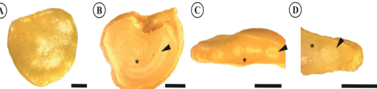
Figure 1 shows the morphological aspects of a completely mature malagueta seed. The malagueta and
biquinho seeds presented similar morphologies; therefore, only one is shown. The pepper seed coat was yellow and homogeneous and had an obovate shape. The seeds of the two species were endospermic. The endosperm was fleshy and consistent and was also yellow. The embryo was peripheral, curved, cylindrical and white. The embryo was easy to identify due to its color difference when compared to the endosperm. Because of the curvature of the embryo, it is possible to visualize the different regions of the embryo, such as the cotyledon and root tissues, in a transverse section (Figure 1C).
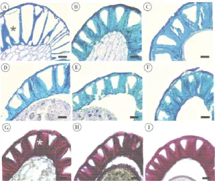
The wall lignification of the integument cells occurred gradually during the development of themalagueta and biquinho pepper seeds. This process can be identified by the thickening of the cell wall at the periclinal and anticlinal divisions of the epidermal cells stained light-blue through the reaction with Toluidine blue O (Figures 1A-F and 2A-F). Positive reaction to acid phloroglucinol confirmed the lignification (Figures 1G-I and 2G-I).
At 25 DAA, there was little lignification of the seed tegument in both species (Figures 2A and 3A). A cross-sectional cut showed the uneven and progressive thickening of the periclinal and anticlinal cell wall divisions towards the inner portion. This led to the formation of a pyramid-shaped lumen in the tegument cells (Figures 2 and 3), visible during early seed formation at 40 DAA (Figures 2B and 3B). As the seeds developed and the cell walls were progressively lignified, the cell lumen became smaller and closer to the outer wall, occupying an increasingly smaller area of the tegument.
Figure 1. Morphological aspects of malagueta seeds (100 DAA). A - The external morphology shown in the whole seed; B - Longitudinal
section showing the internal morphology; C - Transverse section; and D - Transverse section. (Bar – 0.5 mm). Legend: *endosperm; ►
h F D T F 4 D D t 4
The seed t embryo, end earliest stage a histochemical are deposited in which stora Figures 4 and
Figure 2. Cross seeds during diffe DAA; C - 55 DA stained with tolui 100 DAA, reacte Tegma).
Figure 3. Cross seeds during diff 40 DAA; C - 55 DAA, stained w DAA; and I - 100
- 50 μm; *Tegma
Regarding indicated the p the seed tegum 4A and 5A). A
tissues can alr osperm, and analyzed (25 D analyses, the p during seed o age occurs can
5).
sections of the erent developmen AA; D - 70 DAA idine blue O; G - ed with acid phlo
sections of the ferent developme 5 DAA; D - 70 with toluidine blu
0 DAA, reacted w a).
the presence o presence of a sm ment of both sp After 25 DAA,
ready be chara tegument d DAA). Accord protein and lip ontogeny, and n be visualized
tegument in mal
ntal stages: A - 25 A; E - 85 DAA; F 40 DAA; H - 70 oroglucinol. (Bar
tegument in biq
ental stages: A - DAA; E - 85 D ue O; G - 55 D with acid phlorog
of starch, the PA mall number of
pecies at 25 DA i.e., as early a
acterized as during the ding to the pid reserves d the tissue (shown in
lagueta pepper
DAA; B - 40 F - 100 DAA, DAA; and I - r - 50 μm; *
uinho pepper
25 DAA; B - DAA; F - 100 DAA; H - 70 glucinol. (Bar
AS reaction f granules in AA (Figures as 40 DAA,
starch granul components (Figures 4B-(data not sh present until of either pep reserve sou considering reserves, suc were increas suggesting th available in development Protein (Figures 4 evident in gradually d embryo.
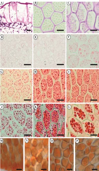
Figure 4. Cro pepper seeds. endosperm, 40 embryo, 25 DA embryo, 70 DA endosperm, 40 100 DAA, XP t O - endosperm with Sudan III.
les were no lon via histochem -C and 5B-C) hown). This re
l the end of see pper species, ind
urce. This i that substan ch as lipids and ses in embryo hat it may con n the seeds
t and germinat (Figures 4D 4M-P and both specie isappeared in
oss sections of d A - tegument, 0 DAA; C - en AA; E - embryo, 4 AA; H - embryo, 8 0 DAA; K - endo
test; M - embryo, m, 40 DAA; and P . (Bar - 20 μm).
nger observed a mical analysis, , or Lugol’s io eserve compon ed developmen
dicating that it is particularly nces common d proteins, wer
and endosper ntribute to the during init ion.
D-L and 5D-L 5M-P) depo s, while star n both the end
ifferent regions o 25 DAA (Bar -dosperm, 100 D 0 DAA; F - embry 85 DAA; I - embry sperm, 70 DAA;
40 DAA; N - em P - endosperm, 10
as seed reserve PAS reaction odine solution nent was not nt in the seeds
is a transitory y important, nly used as re detected, as
rm cell walls, total reserves tial seedling
L) and lipid osition was rch reserves dosperm and
of the malagueta
- 50 μm); B -
Histochemistry during pepper seed ontogeny 539
Acta Scientiarum. Agronomy Maringá, v. 39, n. 4, p. 535-543, Oct.-Dec., 2017
Figure 5. Cross sections of different regions of the
biquinhopepper seeds. A - tegument, 25 DAA; B - endosperm, 40
DAA; C - endosperm, 100 DAA, PAS; D - embryo, 25 DAA; E - embryo, 40 DAA; F - embryo, 55 DAA; G - embryo, 70 DAA; H - embryo, 85 DAA; I - embryo, 100 DAA; J - endosperm, 40 DAA; K - endosperm, 55 DAA; L - endosperm, 85 DAA, XP test; M - embryo, 55 DAA; N - embryo, 100 DAA; O - endosperm, 55 DAA; and P - 100 DAA, reacted with Sudan III. (Bar - 20 μm).
Histochemical tests using XylidinePonceau indicated that the proteins are deposited in the embryo and endosperm during seed formation and that they are an important reserve component of
malagueta and biquinho pepper seeds (Figures 4D-L and 5D-L).
At 25 and 40 DAA (Figures 4D-E and 5D-E), negative reaction to Xylidine Ponceau indicated that the seeds were at a maturation stage in which protein reserves were still absent in the embryo. The accumulation of protein bodies in this seed region began at 55 DAA (Figures 4F and 5F). The amount of stored protein began to increase at this maturation stage (Figures 4G-I and 5G-I). Xylidine Ponceau reaction showed a large amount
of protein in seed embryos at 85 and 100 DAA; the cell cytoplasm was completely filled (Figures 4H-I and 5H-I).
The Xylidine Ponceau test verified the presence of a large accumulation of protein globules in the endosperm of both species during all maturation stages, except at 25 DAA. Figure 3 shows the malagueta pepper seed endosperm at 40, 70, and 100 DAA (Figure 4J-L); Figure 4 shows the biquinho pepper seed endosperm at 40, 55, and 85 DAA (Figure 5L-J).
In the malagueta pepper, the amount of reserve protein deposited in the seed endosperm at 85 and 100 DAA was lower than at previous maturation stages, as evidenced by the empty spaces in the cell cytoplasm. However, this might have been due to cell plasmolysis. Nevertheless, this characteristic was not observed in the embryo.
The protein chemical determination for the
malagueta pepper using methods described by Bradford (1976) showed that the soluble protein stored in the seeds decreased starting at 63 DAA (Figure 6A). The protein content in the malagueta
pepper seeds during development had a quadratic variation, with a peak value of 0.48 g.g-1 dry matter (DM) 55 DDA (Figure 6A). At 25 DDA, the seeds had a protein concentration of 0.28 gg-1 DM, while at 100 DDA, it was 0.33 gg-1 DM (Figure 6A).
However, from 40 to 100 DAA, the amount of soluble protein bodies in the biquinho pepper endosperm followed the same pattern (Figure 5J-L). Figure 6B shows that the accumulation of soluble protein during the development of the biquinho
pepper follows a quadratic behavior. At 25 DAA, the seeds had 0.28 g.g-1 DM with increases in protein content, while at 100 DAA, the protein content reached a maximum of 0.62 gg-1 DM.
The lipid content in the malagueta and biquinho
peppers during seed development is shown in Figure 6. The amount of this compound increased at a rate of 0.0028 g day-1 in the malagueta pepper. At 25 DAA, the seeds had 0.092 g g-1 DM; at 100 DAA, 0.30 gg-1 DM (Figure 6A).
(A) (B)
Figure 6. Soluble Protein content during the development of malagueta (A) and biquinho (B) pepper seeds.
(A) (B)
Figure 7. Lipid content during the development of the malagueta (A) and biquinho (B) pepper seeds.
During malagueta pepper seed development, the protein content initially increased and then decreased after 63 DAA. The lipid reserves increased (Figures 6A and 7A). However, in the biquinho
pepper, protein storage increased up until 100 DDA and lipid accumulation decreased after 74 DAA (Figures 6B and 7B).
The seed development of the malagueta and
biquinho peppers featured similar structural changes and reserve depositions. During each stage of development, peculiarities could be observed in the structural changes of the embryo, endosperm, and tegument cells, and the type and location of the reserve deposition.
The main structural changes in the seed tegument of both species occurred 25 to 40 DAA. At 40 DAA, a pyramid-shaped lumen was observed in the epidermal cells. Several studies on epidermis development in Solanaceae seeds have aimed to investigate changes deriving from the maturity
process, such as those conducted by Edmonds (1983) and Karihaloo and Malik (1996).
In addition to other functions, the tegument plays a major role in endosperm and embryo nutrition during seed development. Phloem discharge occurs in the tegument. The assimilates are then translocated to the embryo and/or endosperm (Weber, Heim, Golombek, Borisjuk, & Wobus
,
1998; Bewley et al., 2013).In a similar study, Karihaloo and Malik (1996) described the tegument structure and development in Solanum melongena and S. violaceum seeds, which had a development pattern similar to the species in the present study. The authors stated that the development of the epidermis in the seeds involved a complex sequence of events, beginning with the thickening of the outer periclinal cell wall face. This characteristic may protect the inner structures of the seeds from the lignified sclerenchyma cells. The results reported by Karihaloo and Malik (1996) and
MALAGUETA
Days after anthesis (DAA)
0 20 40 60 80 100
P ro te in s ( g .g
-1 D.
M ) 0.0 0.1 0.2 0.3 0.4 0.5 0.6 % 83 R x 0001 , 0 x 0166 , 0 0388 , 0 y 2 2 = − + − = BIQUINHO
Days after anthesis (DAA)
0 20 40 60 80 100
Pr o te in s ( g .g
-1 D.
M ) 0.0 0.2 0.4 0.6 0.8 87% R 0,00008x 0,014x 0,0048 y 2 2 = − + − = MALAGUETA
Days after anthesis (DAA)
0 20 40 60 80 100
L
ipi
ds (
g
.g
-1 D.
M ) 0.00 0.05 0.10 0.15 0.20 0.25 0.30 0.35 % 84 R x 0028 , 0 0342 , 0 y 2 = + = BIQUINHO
Days after anthesis (DAA)
0 20 40 60 80 100
Li pi d s ( g .g
-1 D.
Histochemistry during pepper seed ontogeny 541
Acta Scientiarum. Agronomy Maringá, v. 39, n. 4, p. 535-543, Oct.-Dec., 2017
Corner (1976), and the present study in the
malagueta and biquinho peppers demonstrate the structural uniformity of seed tegument epidermis development among some members of the Solanaceae family.
Each seed reserve material is deposited at different rates during maturation. Temporary reserve deposition may occur, which will be used during seed development, or the seeds will be stored and used during germination (Weber, Borisjuk, & Wobus, 1997). Overall, embryo and endosperm storage reserve compositions are similar, except for the polysaccharides stored in the cell wall, which are less abundant in the embryo.
In the malagueta and biquinho pepper, starch was found only at 25 DAA. The absence of starch at later maturation stages indicates that these granules are converted into other components, likely soluble sugars, during the maturation process. Starch may temporarily accumulate in some cells at specific developmental stages (Zeeman et al., 2010). Jordy (2004) mentioned that transitory starch is located near the meristematic tissues and is used as a source of carbon during cell division, expansion, and differentiation. Chen and Lott (1992) reported the absence of starch in mature Capsicum annuum seeds in both the embryo and endosperm.
In both Capsicum seeds analyzed, the proteins were deposited both in the embryo and the endosperm. According to Herman and Larkins (1999), reserve protein synthesis and deposition in seeds are subjected to spatial and temporal regulation, which may vary among species and developmental stages.
Chen and Lott (1992) stated that proteins represented one of the main reserve components in
Capsicum annuum seeds. The authors reported that, in mature seeds, the reserves are deposited inboth the embryo and the endosperm. It can be inferred that the species of this genus must follow the same developmental pattern in terms of protein reserve accumulation. Proteins were also detected as reserve materials in tomato seed embryos and endosperm (Spitzer & Lott, 1980).
Reserve proteins are synthesized in the endoplasmic reticulum and are then transported to their accumulation sites (Herman & Larkins, 1999), i.e., protein bodies or protein vacuoles (Stone & Gifford, 1997). This form of deposition prevents the cleavage of these proteins by proteases present in the cell cytoplasm (Shutov, Baumlein, Blattner, & Muntz, 2003) and enables their storage and later mobilization during seed germination and seedling development (Weber et al., 2010).
Malagueta and biquinho pepper seeds had lipids as reserve components since the early development stages analyzed. Similar to proteins, this reserve is deposited during the seed maturation process in both the embryo and the endosperm. The presence of lipids as reserve material was also observed in seeds of other Solanaceae species, such as C. annuum
(Marion & Dempsey 1964; Chen & Lott, 1992; Pérez-Gálvezet, Garrido-Fernández, & Mínguez-Mosquera, 1999; Embaby & Mokhtar, 2011; Silva, Azevedo, Pereira, Valentão, & Andrade, 2013),
Lycopersicum esculentum (Spitzer & Lott, 1980), C. annuum, C. baccatum, C. chinense, C. frutescens, and C. pubescens (Jarret, Levy, Potter, & Cermak, 2013).
It is important to note that, in the present study, the chemical determination of the reserve components, proteins and lipids were carried out in whole seeds due to the difficulty of separating the storage regions, embryo and endosperm. Therefore, the percentage of accumulated reserves in each tissue during the maturation process could not be determined.
The data on the physiological quality of the
malagueta and biquinho pepper during all of the maturation stages in the present study can be found in Abud et al. (2013).
Conclusion
In conclusion, this study confirmed that proteins and lipids are the main reserve compounds of
malagueta and biquinho pepper seeds. These reserve components are stored during seed development. The accumulation of protein reserves begins during the earliest developmental stages in the cytoplasm of endosperm cells. In embryo cells, the reserves accumulate during the later developmental stages. Starch was only observed as a transient reserve source during initial seed development.
Acknowledgements
The Federal University of Viçosa, the Federal University of Ceará, and the Agricultural Research Corporation Minas Gerais. The Laboratory of Anatomy and Plant Morphogenesis of the Federal University of Viçosa. The Coordination for the Improvement of Higher Education Personnel (CAPES) and the Foundation of Research Support of the State of Minas Gerais (Fapemig) supported this research.
References
ontogênese. Pesquisa Agropecuária Brasileira, 48(12), 1546-1553.
Alencar, N. L. M., Innecco, R. Gomes-Filho, E., Gallão, M. I., Alvarez-Pizarro, J. C., Prisco, J. T., & Oliveira, A. B. (2012). Seed reserve composition and mobilization during germination and early seedling establishment of Cereus jamacaru D.C. ssp. Jamacaru
(Cactaceae). Anais da Academia Brasileira de Ciências,
84(3), 823-832.
Alencar, N. L. M., Gadelha, C. G., Gallão, M. I., Dolder, M. A. H., Prisco, J. T., & Gomes-Filho, E. (2015). Ultrastructural and biochemical changes induced by salt stress in Jatropha curcasseeds during germination and seedling development. Functional Plant Biology,
42(9), 865-874.
Alfenas, A. C., Peters-Robinson, I., & Brune, W. (1991).
Eletroforese de proteínas e isoenzimas de fungos e essências florestais. Viçosa, MG: SIF.
Angelovici, R., Galili, G., Fernie, A. R., & Fait, A. (2010). Seed desiccation: a bridge between maturation and germination. Trends in Plant Science, 15(4), 211-218. Berger, F., Hamamura, Y., Ingouff, M., & Higashiyama, T.
(2008). Double fertilization caught in the act. Trends in Plant Science,13(8), 664-670.
Bewley, J. D., Bradford, K. J., Hilhorst, H. W. M., & Nonogaki, H. (2013). Seeds: physiology of development, germination and dormancy (3rd ed.). New York: Springer. Bradford, M. M. (1976). A rapid and sensitive method for the
quantification of microgram quantities of proteins utilizing the principle of protein-dye binding. Analytical Biochemistry,72, 248-254.
Chen, P., & Lott, J. N. A. (1992). Studies of Capsicum annuum
seeds: structure, storage reserves, and mineral nutrients.
Canadian Journal of Botany,70(3), 518-529.
Corner, E. J. H. (1976). The Seeds of Dicotyledons. Cambridge, UK: Cambridge University Press.
Edmonds, J. M. (1983). Seed coat structure and development in Solanum L. section Solanum (Solanaceae). Botanical Journal of the Linnean Society, 87(3), 229-246.
Embaby, H. E., & Mokhtar, S. M. (2011). Chemical composition and nutritive value of lantana and sweet pepper seeds and nabak seed kernels. Journal of Food Science76(5), 736-741.
Graham, I. A. (2008).Seed storage oil mobilization. Annual Review of Plant Biology, 59, 115-142.
Hehenberger, E., Kradolfer, D., & Köhler, C. (2012). Endosperm cellularization defines an important developmental transition for embryo development.
Development, 139(11), 2031-2039.
Herman, E. M., & Larkins, B. A. (1999). Protein storage bodies and vacuoles. The Plant Cell,11(4), 601-613. Hoch, G. (2007). Cell wall hemicelluloses as mobile carbon
stores in non-reproductive plant tissues. Functional Ecology, 21(5), 823-834.
Ingram, G. C. (2010). Family life at close quarters: communication and constraint in angiosperm seed development. Protoplasma, 247(3), 195-214.
Jarret, R. L., Levy, I. J., Potter, T. L., & Cermak, S. C. (2013) Seed oil and fatty acid composition in Capsicum
spp. Journal of Food Composition and Analysis,30(2), 102-108.
Jensen, W. A. (1962). Botanical histochemistry: principles and practice(1st ed.). Berkley, US: W. H. Freeman and Co. Johansen, D. A. (1940). Plant microtechnique. New York,
USA: McGraw-Hill.
Jordy, M. N. (2004). Seasonal variation of organogenetic activity and reserves allocation in the shoot apex of
Pinuspinaster Ait. Annals of Botany,93(1), 25-37. Karihaloo, J. L., & Malik, S. K. (1996). Seed epidermis
development and histochemistry in Solanum melongena
l. and S. violaceum Ort. Annals of Botany, 77(5), 421-428.
Kesavan, M., Song, J. T., & Seo, H. S. (2013). Seed size: a priority trait in cereal crops. Physiologia Plantarum, 147(2), 113-120.
Lee, K. J. D., Dekkers, B. J. W., Steinbrecher, T., Walsh, C. T., Bacic, A., Bentsink, L., Leubner-Metzger, L., & Knox, J. P. (2012). Distinct cell wall architectures in seed endosperms in representatives of the Brassicaceae and Solanaceae. Plant Physiology, 160(3), 1551-1566. Lima, R. B. S., Gonçalves, J. F. C., Pando, S. C.,
Fernandes, V., & Santos, A. L. W. (2008). Primary metabolite mobilization during germination in rosewood (Anibarosaedora Ducke) seeds. Revista Árvore, 32(1), 19-25.
Marion, J. E., & Dempsey, A. H. (1964). Fatty acids of pimiento pepper seed oil. Journal of the American Oil Chemists’ Society,41(8), 548-549.
Marty, F. (1999). Plant vacuoles. Plant Cell, 11(4), 587-599.
McManus, J. F. A. (1948). Histological and histochemical uses of periodic acid. Stain Technology,23(3), 99-108. Murphy, D. J. (1990). Storage lipid bodies in plants and
other organisms. Progress Lipid Research, 29(4), 299-324.
O’Brien, T. P., Feder, N., & McCully, M. E. (1964). Polychromatic staining of plant cell walls by toluidine blue O. Protoplasma,59(2), 368-373.
Paula, S. O., Sousa, J. A., Brito, E. S., & Gallão, M. I. (2016). The morphological caracterization of the dry seeds and reserve mobilization during germination in
Morinda citrifolia L. Revista Ciência Agronômica, 47(3), 556-563.
Pérez-Gálvez, A., Garrido-Fernández, J., & Mínguez-Mosquera, M. A. (1999). Fatty acid composition of two new pepper varieties (Capsicum annum L. cv. Jaranda and Jariza). Effect of Drying Process and Nutritional Aspects. Journal of the American Oil Chemists’ Society, 76(2), 205-208.
Histochemistry during pepper seed ontogeny 543
Acta Scientiarum. Agronomy Maringá, v. 39, n. 4, p. 535-543, Oct.-Dec., 2017
Silva, L. R., Azevedo, J., Pereira, M. J., Valentão, P., & Andrade, P. B. (2013). Chemical assessment and antioxidant capacity of pepper (Capsicum annuum L.) seeds. Food and Chemical Toxicology, 53, 240-248. Spitzer, E., & Lott, J. N. A. (1980). Thin-section,
freeze-fracture, and energy dispersive x-ray analysis studies of the protein bodies of tomato seeds. Canadian Journal of Botany,58(6), 699-711.
Stone, S. L., & Gifford, D. J. (1997). Structural and biochemical changes in loblolly pine (Pinnus taeda L.) seeds during germination and early-seedling grown. I. Storage protein reserves. International Journal of Plant Science, 158(6), 727-737.
Suda, C. N. K., & Giorgini, J. F. (2000). Seed reserve composition and mobilization during germination and early seedling development of Euphorbia heterophylla.
Brazilian Journal of Plant Physiology,12(3), 226-245. Triebold, H. O. (1946). Quantitative analysis with
applications to agricultural and food products. New York, USA: Van Nostrand.
Vidal, B. C. (1970). Dichroism in collagen bundles stained with xylidine Ponceau 2R. Anal Histochem, 15, 289-296.
Weber, H., Borisjuk, L., & Wobus, U. (1997). Sugar import and metabolism during seed development.
Trends in Plant Science, 2(5), 169-174.
Weber, H., Heim, U., Golombek, S., Borisjuk, L., & Wobus, U. (1998). Assimilate uptake and the regulation of seed development. Seed Science Research, 8(3), 331-345.
Weber, H., Sreenivasulu, N., & Weschke, W. (2010). Molecular physiology of seed maturation and seed storage protein biosynthesis. Plant Developmental Biology, 2, 83-104.
Zeeman, S. C., Kossmann, J., & Smith, A. M. (2010). Starch: Its metabolism, evolution and biotechnological modification in plants. Annual Review of Plant Biology, 61, 209-234.
Received on August 12, 2016. Accepted on December 7, 2016.