Prediction of exposed domains of
envelope glycoprotein in Indian HIV-1
isolates and experimental confirmation
of their immunogenicity in humans
Centre for Biotechnology, Jawaharlal Nehru University, New Delhi, India H. Mohabatkar
and S.K. Kar
Abstract
We describe the impact of subtype differences on the seroreactivity of linear antigenic epitopes in envelope glycoprotein of HIV-1 isolates from different geographical locations. By computer analysis, we predicted potential antigenic sites of envelope glycoprotein (gp120 and gp4l) of this virus. For this purpose, after fetching sequences of proteins of interest from data banks, values of hydrophilicity, flexibil-ity, accessibilflexibil-ity, inverted hydrophobicflexibil-ity, and secondary structure were considered. We identified several potential antigenicepitopes in a B subtype strain of envelope glycoprotein of HIV-1 (IIIB). Solid-phase peptide synthesis methods of Merrifield and Fmoc chemistry were used for synthesizing peptides. These synthetic peptides corre-sponded mainly to the C2, V3 and CD4 binding sites of gp120 and some parts of the ectodomain of gp41. The reactivity of these peptides was tested by ELISA against different HIV-1-positive sera from different locations in India. For two of these predicted epitopes, the corresponding Indian consensus sequences (LAIERYLKQQLLGWG and DIIGDIRQAHCNISEDKWNET) (subtype C) were also synthe-sized and their reactivity was tested by ELISA. These peptides also distinguished HIV-1-positive sera of Indians with C subtype infec-tions from sera from HIV-negative subjects.
Correspondence H. Mohabatkar Centre for Biotechnology Jawaharlal Nehru University New Delhi
India
E-mail: [email protected]
Received August 14, 2003 Accepted November 4, 2003
Key words
•Human immunodeficiency virus
•Glycoprotein •Epitope •Immunogenicity
Introduction
The most effective means of solving the structure of a protein is by using biophysical methods such as X-ray crystallography or nuclear magnetic resonance spectroscopy (1). But both methods require a considerable amount of purified proteins and the use of sophisticated instruments, and methodolo-gies. With the development of DNA sequenc-ing methodology a large number of protein
sequences have become available (2). This sequence information in combination with an ever-increasing database of protein struc-tures, solved via the biophysical methods mentioned above, has led to the populariza-tion of predictive computer algorithms (3).
seg-ments of polypeptide chains have been cor-related with the location of continuous epi-topes in proteins (4,5). Substantial experi-mental and theoretical efforts have been di-rected at understanding the relation between the structure of a protein and its immuno-genic properties.
The first requirement for protein predic-tion is the availability of the amino acid sequence. Protein sequences are available from two different sources, i.e., protein in-formation resource (http:/pir.georgetown. edu), and DNA data banks (http://www.ncbi. nlm.nih.gov), and can be easily converted into primary amino acid structures (6).
The HIV envelope gene codes for a precur-sor polyprotein p88, which is glycosylated to form the envelope precursor protein gp160. gp160 is then cleaved by a cellular protease to form the surface extracellular envelope protein gp120 and the transmembrane glyco-protein gp41 (7). When the gp120 glycopro-teins derived from different HIV-1 isolates were compared, five conserved regions (C1 to C5)and five variable regions (V1 to V5) were identified (8,9). Most of the HIV-neutraliz-ing antibodies fall into two categories: those that recognize a determinant in the V3 loop of gp120 (10,11) and those that block the gp120-CD4 interaction (12) by binding to regions in gp120 conserved between differ-ent HIV strains (4,13,14).
The transmembrane glycoprotein gp41 contains hydrophobic domains which can be arranged into three transmembrane regions and several important epitopes within its ectodomain (15,16).
The HIV strain which predominates in India, is of subtype C (17-19). The first cases of HIV-1 infection in India were reported in Tamil Nadu in 1986, and the first AIDS patient was reported in Bombay in May 1987. The HIV-1 epidemic in India is principally heterosexual (20) with the highest HIV in-fection rates occurring in metropolitan areas in the western part of India (Mumbai) and in the south (Chennai) (21).
In the present study, we predicted ex-posed domains of gp120 and gp41 of Indian isolates of HIV-1 and then we used synthetic peptides mainly corresponding to the C2, V3 and CD4 binding sites of gp120 and some parts of the ectodomain of gp41 against dif-ferent type of sera. Finally, we compared this reactivity with theoretical methods of pre-diction of the immunogenic sites of proteins.
Material and Methods
Peptides
The peptides corresponding to the exposed domains of HIV-1 envelope glycoproteins were synthesized by the solid-phase peptide synthe-sis method of Merrifield (22) and manually by Fmoc chemistry (21,23) or were supplied by the National Institute for Biological Standards and Control, London, UK.
Sera
Venous blood was collected from indi-viduals from different parts of India, kept at room temperature for 2 h, and then left to stand overnight at 4ºC. Blood was then cen-trifuged at 3000 g for 10 min at 4ºC and serum was separated and stored at -20ºC.
For the detection of infection, HIV-1 and HIV-2 sera from different persons were ana-lyzed using four different commercially available kits, i.e., Biochemical Detect HIV™
assay (BioChem ImmunoSystems Inc., Montreal, Quebec, Canada), Innotest HIV-1/HIV-2 antibody assay (Innogenetics N.V., Haven, Zwijndecht, Belgium), Recombigen®
HIV-1/HIV-2 EIA assay (Cambridge Diag-nostics Ireland Ltd., Galway, Ireland), and Inno-LIA HIV-1/HIV-2 antibody assay (In-nogenetics N.V.),according to manufacturer instructions.
Theoretical analysis
was available in the literature (24). The Preditop program provided by Dr. J. Pellequer on a 3½-floppy disk was used to study the exposed domains of the proteins. The scales used in the program to study the structure of a protein are grouped as follows: 9 scales of inverted hydrophobicity (Doolittle, Heijne, Manavala, Prils, Rose, Sweet, Totls, Ges, Zimmermann), 2 scales of hydrophilicity (Hopp, Parker); 4 scales of accessibility (Janin, Chothia, Chothia8, Acrophil), 1 scale of antigenicity (Welling), and 3 scales of secondary structure (Chouf3, grenier3, Levitt). The amino acid sequence of the HIV glycoprotein was read as a moving window of seven residues and of their values corre-sponding to each of the scales used and the mean value was plotted against the fourth residue of the window. In order to compare the profiles obtained by the different meth-ods, various scales were normalized, with the original values of each scale set between +3and -3(25).
Enzyme-linked immunosorbent assay
The peptides were tested for immunoge-nicity using the conventional ELISA de-scribed by Engvall and Perlmann (26). The wells were coated with peptide by incubat-ing 100 µl of peptide solution diluted in carbonate bicarbonate buffer, pH 9.6, at a final amount of 1.2 µl/well in each well of an ELISA plate in a humid chamber overnight at 37ºC. Coated wells were then blocked by incubation with 300 µl of blocking buffer (1 mg/ml bovine serum albumin (BSA) in car-bonate bicarcar-bonate buffer) in each well for at least 6 h in a humid chamber at 37ºC.One hundred microliters of diluted test serum was added to each well and incubated over-night in a humid chamber at 37ºC. The plates were washed four times with phosphate-buffered saline (PBS)-Tween and 100 µl horse radish peroxidase-conjugated goat anti-human antibody was added at 1/1000 dilu-tion in PBS-Tween containing 0.1% BSA.
The plates were washed four times with PBS-Tween and 100 µl of a 2,2'-azinobis (3' ethyl benzothiazoline sulfuric acid) solution (0.5 mg/ml) in citrate phosphate buffer, pH 5.0, containing 1 µl H2O2 per ml was added
to each well, and plates were incubated for 10 min in a humid chamber at 37ºC.After the development of color, absorbance in each well was measured at 405 nm with a micro-plate reader.
Statistical analysis
P values were determined by the rank sum test, with the level of significance set at 0.05.
Results
Prediction of the exposed domains of the HIV-1 IIIB envelope glycoprotein
The amino acid sequence of the HIV-1 IIIB envelope glycoprotein was analyzed with the Preditop program to predict the antigenic determinants of this molecule. The results are shown in Table 1.
Table 1. Amino acid sequences of the envelope glycoprotein of HIV-1 IIIB predicted to be exposed.
Domains predicted to be exposed
74VPTDPNPQEV83
129DTNTNSSSGRM139
219IPIDNDTTSYS229
233KCNNKTFNGTGGPC246
303TRPNNNTRKRIRIQRGPGR321
367GNNKTIIFKQSSGGDPE383
407WSTEGSNNTEGSDTI421
455TRDGGNSNNESE466
469FRLGGGDMRDNWR481
525GAAGSTMGAA534
613ASWSNKSLEQI623
725PRGPDRPE732
Comparison of antigenic domains of HIV IIIB envelope glycoproteins by computational and experimental analysis
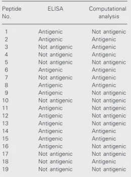
The antigenic domains of HIV IIIB enve-lope glycoproteins were compared by reac-tion with HIV-positive sera and predicted by computational analysis and the results are shown in Table 3. From the results shown in this table, it is clear that peptides Nos. 2, 6, 8, 14 and 15 can be considered to be immuno-genic by both experimental and computa-tional analysis.
Reactivity of synthetic peptides corresponding to the different parts of the envelope glycoprotein of Indian HIV-1 isolates
Two peptides, one corresponding to gp120 (peptide No. 20) and the other
corre-Table 2. Sequences and regions of the peptides used in this study.
Envelope Peptide Region Peptide sequence (percent homology
glycoprotein No. with Indian isolates)
Gp120 1 C2 region 208TQACPKVSFEPIPIHYCAPA227 (90)
2 C2 region 218PIPIHYCAPAGFAILKCNNK237 (95)
3 C2 region 228GFAILKCNNKTFNGTGPCTN247 (90)
4 C2 region 238TFNGTGPCTNVSTVQCTHGI257 (95)
5 C2 region 248VSTVQCTHGIRPVVTQLLL267 (95)
6 V3 region 298VEINCTRPNNNTRKRIRIQ316 (90)
7 V3 region 308NTRKRIRIQRGPGRAFVTIG327 (70)
8 V3 region 318RGPGRAFVTIGKIGNMRQA337 (65)
9 V3 region 328KIGNMRQAHCNISRJAKWNNT347 (70)
10 CD4 binding domain 388NCGGEFFYCNS398 (85)
11 CD4 binding domain 413DTITLPCRIKQIINMWQKVG432 (80)
12 CD4 binding domain 423KQIINMWQKVGKAMYAPPIS442 (85)
13 CD4 binding domain 433KAMYAPPAISGQIRCSSNITG542 (75)
14 CD4 binding domain 443GQIRCSSMTGLLLTRDGGNS462 (70)
Gp41 15 Ectodomain (N-terminal) 520FLGFLGAAGSTMGA533 (100)
16 Ectodomain 582LAVERYLKDQQLLGIWG598 (88)
17 Ectodomain 589DQQLLGIWGCSGKLIGTTAVPWNC623 (96)
18 Ectodomain 729DRPEGIEEEGGERDRS744 (NA)
19 Ectodomain 577LQARILAVERYLKQQL592 (81)
NA indicates that the sequence of Indian isolates was not available at the time of this study.
Antibody-binding properties of synthetic peptides of the different domains of the HIV-1 IIIB envelope glycoproteins
sponding to gp41 of Indian HIV-1 isolates (peptide No. 21) (27,28), were synthesized (Table 4). These peptides were homologous to peptides Nos. 9 and 16, respectively. Both peptides Nos. 9 and 16 had shown negative results by computational analysis and posi-tive results by ELISA with HIV-posiposi-tive sera from Indian individuals. Both peptides Nos. 20 and 21 showed positive results by ELISA with HIV-positive sera from the same indi-viduals.
Discussion
Prediction of secondary structures does not permit definitive conclusions but facili-tates the interpretation of other results and the design of new experiments. The anti-genic domains of a protein molecule can be predicted byanalyzing the protein sequence using a suitable computer program (29). Many such computer programs are available which take into consideration several pa-rameters of adjacent small continuous stretches of amino acids such as inverted hydrophobicity, hydrophilicity, accessibil-ity, antigenicaccessibil-ity, and a tendency to form secondary structures (25). In the present study we used Preditop, one of the more recent programs (version 3.1) wich takes into ac-count 9 scales of inverted hydrophobicity, 2 scales of hydrophilicity, 4 scales of accessi-bility and 1 scale of antigenicity for comput-ing the probability of a given domain of a protein to be exposed on the surface of the molecule.
This program predicted the exposure of several domains of this molecule. According to the prediction, gp120 (a surface glycopro-tein) of the IIIB strain of HIV-1 is supposed to have nine exposed domains, while gp41 (a transmembrane glycoprotein) of this virus is expected to have four exposed domains. The sequences of the predicted domains are shown in Table 1.
Subtype C is the most prevalent subtype of HIV-1 in India. We wanted to know
Table 4. Sequence and region of the peptides corresponding to the Indian consensus sequence envelope glycoprotein of HIV-1.
Envelope Peptide No. Region Peptide sequence
glycoprotein
Gp41 20 Ectodomain 582LAIERYLKEQQLLGIWG598
Gp120 21 V3 region 413DIIGDIRQAHCNISEDKWNET433
Table 3. Comparison of the antigenicity of pep-tides of the envelope glycoproteins of the HIV-1 IIIB strain determined by reactivity with sera (ELISA) and by computational analysis.
Peptide ELISA Computational
No. analysis
1 Antigenic Not antigenic
2 Antigenic Antigenic
3 Not antigenic Antigenic
4 Not antigenic Antigenic
5 Not antigenic Not antigenic
6 Antigenic Antigenic
7 Not antigenic Antigenic
8 Antigenic Antigenic
9 Antigenic Not antigenic
10 Not antigenic Not antigenic
11 Antigenic Not antigenic
12 Antigenic Not antigenic
13 Antigenic Not antigenic
14 Antigenic Antigenic
15 Antigenic Antigenic
16 Antigenic Not antigenic
17 Not antigenic Not antigenic
18 Not antigenic Antigenic
19 Not antigenic Not antigenic
whether predicted domains of the gp120 protein of HIV-1 subtype B react with Indian sera, which were tested with the above kits. Only those sera which were positive in all of the three ELISA tests were selected, and further subjected to the Inno-LIA test. Any serum found to be negative by the Inno-LIA HIV-1/HIV-2 test was discarded. The sera were then classified into the following three groups depending on the reaction pattern found with Inno-LIA HIV-1/HIV-2: a) sera from HIV-1-infected individuals, b) sera from HIV-2-infected individuals, and c) sera from HIV-1 + 2-infected individuals.
referred to as confirmed HIV-positive sera. The synthesized overlapping peptides were then tested for their ability to react with the confirmed HIV-positive sera. Peptides 1, 2, 6, 8, 9, 11, 12, 13, 14, 15, and 16 were found to be reacting with HIV sera. Of the 19 peptides tested, peptides 2, 3, 4, 6, 7, 8, 14, 15 and 18 were predicted to be antigenic in the computational analysis, while peptides 1, 9, 11, 12, 13 and 16 were not, but showed reactivity with Indian HIV-positive sera. In-terestingly, some peptides like 3, 4, 7, and 18 were predicted to be antigenic by computa-tional analysis, but did not show any reactiv-ity with Indian HIV-positive sera by ELISA. On the basis of these results, homologous peptides of 9 and 16 (both showing negative results in computational analysis and posi-tive results in experimental analysis) corre-sponding to the Indian consensus sequence (27) were synthesized (peptides 20 and 21). The reactivity of these peptides with HIV-positive and normal sera was studied by ELISA and both peptides were shown to distinguish normal sera from the HIV-posi-tive ones.
With respect to the difference between the computational and experimental results, it should be mentioned that no method is 100% accurate in predicting the conforma-tion of a protein from its amino acid se-quence (6). In addition, our prediction anal-ysis was performed by using the envelope glycoprotein of HIV-1 IIIB which belongs to
subtype B. However, we had tested peptides corresponding to these parts against the sera of Indian individuals who were infected with HIV-1 subtype C.
Furthermore, It should be noted that anti-bodies directed at gp120 might also bind to conformational, discontinuous epitope(s) quite distinct from the linear sequences de-fined in this study (30,31). An additional factor that should be considered with cau-tion in the interpretacau-tion of these data is the impact of glycosylation on the antibody re-sponse in vivo. Glycosylation may result in the blocking of antibody accessibility to some of the predicted residues (32-34). Further-more, it has been proposed that linear epi-topes may be more exposed in the mono-meric rather than the oligomono-meric form of the envelope glycoprotein (35).
Acknowledgments
We would like to acknowledge the Na-tional Institute for Biological Standards and Control, London, UK, for providing most of the peptides used in this study. We also thank Dr. J.-L. Pellequer (Department of Molecular Biology, The Scripps Research Institute, La Jolla, CA, USA) for sending us a copy of the Preditop program, which was developed by him. We also thank the Na-tional Institute of Communicable Diseases, Delhi, India, for providing HIV-positive sera.
References
1. Gershoni JM, Stern B & Denisova G (1997). Combinatorial libraries, epitope structure and the prediction of protein conformations. Im-munology Today, 18: 108-110.
2. Jameson BA & Wolf H (1988). The antigenic index: a novel algo-rithm for predicting antigenic determinants. Computer Applications in the Biosciences, 4: 181-186.
3. Rooman MJ & Wodak SJ (1998). Identification of predictive se-quence motifs limited by protein structure data base size. Nature, 335: 45-49.
4. Broder CB, Earl PL, Long D, Abedon ST, Moss B & Doms RW (1994). Antigenic implications of human immunodeficiency virus type 1 envelope quaternary structure: oligomeric-specific and
sensi-tive monoclonal antibodies. Proceedings of the National Academy of Sciences, USA, 91: 11699-11703.
5. Kyte J & Doolittle RF (1982). A simple method for displaying the hydropathic characters of a protein. Journal of Molecular Biology, 157: 105-110.
6. Fasman GD (1989). Protein conformational prediction. Trends in Biochemical Sciences, 14: 295-299.
7. Vaishnav YN & Wong-Staal F (1991). The biochemistry of AIDS.
Annual Review of Biochemistry, 60: 577-630.
AIDS or at risk for AIDS. Science, 232:1548-1553.
9. Fung M, Sun C, Gordon W, Lion RS, Chang TW, Daar E & Ho DD (1992). Identification and characterization of a neutralization site within the second variable region of human immunodeficiency virus type 1 gp120. Journal of Virology, 66: 848-856.
10. Moore JP, Trkola A, Korber B, Boots LJ, Kessler JA, McCutchan FE, Moscola J, Ho DD, Robinson J & Conley AJ (1995). A human monoclonal antibody to a complex epitope in the V3 region of gp120 of human immunodeficiency virus type 1 has broad reactivity within and outside clade B. Journal of Virology, 69: 122-130.
11. Casseb J, Katzenstein D, Winters M, Brigido LF, Duarto AJ & Hendry RM (2002). Serotyping HIV-1 with V3 peptides: detection of high avidity antibodies presenting clade-specific reactivity. Brazilian Journal of Medical and Biological Research, 35: 369-375.
12. Lasky LA, Nakarnura G, Smith D, Fennie C, Patzer E, Berman P, Cregory T & Gapon DJ (1987). Delineation of a region of the human immunodeficiency virus type 1 gp120 glycoprotein critical for inter-action with the CD4 receptor. Cell, 50: 975-985.
13. Pestano GA, Hosford KS, Spira AA, Riley J & Xie JM (1995). Seroreactivity of analogous antigenic epitopes in glycoprotein 120 expressed in HIV-1 subtypes A, B, C and D. AIDS Research and Human Retroviruses, 5: 589-596.
14. Xie JM, Pestano GA, Samms M, Lee SK & Guyden J (1996). Immu-nogenic potential of rgp120 from African HIV-1 subtype A. Vaccine, 14: 993-1000.
15. Modrow S, Hahn BA, Shaw GM, Gallo RC, Wong-Staal F & Wolf H (1987). Computer-assisted analysis of envelope protein sequences of seven human immunodeficiency virus isolates: prediction of antigenic epitopes in conserved and variable regions. Journal of Virology, 61: 570-578.
16. Stiegler G, Kunert R, Purtscher M, Wolbank S, Voglauer R, Steindl F & Katinger H (2001). A potent class-clade neutralizing human mono-clonal antibody against a novel epitope on gp41 of human immuno-deficiency virus type 1. Journal of Clinical Microbiology,40: 1010-1022.
17. Weniger BG, Takebe Y, Ou C-Y & Yamazaki S (1994). The molecular epidemiology of HIV in Asia. AIDS, 8 (Suppl 2): S13-S28.
18. Myers G, Korber B, Wain-Hobson S, Smith RF & Pavlakis GN (1993).
Human Retroviruses and AIDS: A Compilation and Analysis of Nucleic Acid and Amino Acid Sequences. Theoretical Biology and Biophysics. Los Alamos National Laboratory, Los Alamos, NM, USA.
19. Delwart EL, Shpaer EG, Louwagie J, McCutchan FE, Grez M, Ruhsaman-Waigmarnn H & Mullins JI (1993). Evolutionary relation-ships determined by a DNA heteroduplex mobility assay: Analysis of HIV-1 env genes. Science, 262: 1257-1261.
20. Tsuchie H, Saraswathy T, Sinniah M, Vijayamalar B, Maniar JK, Monzon OT, Santana RT, Paladin FJ, Wasi C & Thongcharoen P (1995). HIV-1 variants in South and South-East Asia. International Journal of STD and AIDS, 6: 117-120.
21. Charneu P, Borman AW & Quillent C (1994). Isolation and envelope sequence of a highly divergent HIV-1 isolate: definition of a new
HIV-I group. Virology, 205: 247-253.
22. Merrifield RB (1963). Solid phase peptide synthesis. The synthesis of a tetrapeptide. Journal of the American Chemical Society, 85: 2149-2154.
23. Fields GB & Noble RL (1990). Solid phase peptide synthesis utilising 9-fluorenylmethoxy carbonyl amino acids. International Journal of Peptide and Protein Research, 35: 161-214.
24. Ratner L, Haseltine W, Patarka R, Livak KJ, Starcich B, Josephs SF, Doran ER & Rafalsky JA (1985). Complete nucleotide sequence of the AIDS virus, HTLV-III. Nature, 313: 277-285.
25. Pellequer JL, Westh E & Regenmortel MHVV (1991). Predicting location of continuous epitopes in proteins from their primary struc-ture. Methods in Enzymology, 203: 176-201.
26. Engvall E & Perlmann P (1972). Enzyme linked immunosorbent assay, ELISA. 3. Quantitation of specific antibodies by enzyme-labeled anti-immunoglobulin in antigen-coated tubes. Journal of Immunology, 109: 129-135.
27. Dietrich U, Grez M, von Briesen H et al. (1993). HIV-1 strains from India are highly divergent from prototypic African and USA/Euro-pean strains, but are linked to a South African isolate. AIDS, 7: 23-27.
28. Tripathy S, Renjifo B, Wang WK, McLane MF, Bollinger R, Rodriges J, Sterman J, Tripathy S & Essex M (1996). Envelope glycoprotein 120 sequences of primary HIV type 1 isolates from Pune and New Delhi, India. AIDS Research and Human Retroviruses, 12: 199-202. 29. Carmenes RS, Freije JP, Molina MM & Martin JM (1989). Predict 7.
A programme for protein structure prediction. Biochemical and Biophysical Research Communications, 159: 687-693.
30. Benjamin C, Berzofsky JA, East IJ et al. (1984). The antigenic structure of protein: a reappraisal. Annual Review of Immunology, 2: 67-101.
31. Barlow DJ, Edward MS & Thornton IM (1986). Continuous and discontinuous protein antigenic determinants. Nature, 322: 747-748.
32. Agadjanyan M, Luo P, Westerink J, Carey LA, Hutchins W, Steplewski Z, Weiner DB & Kieber-Emmons T (1997). Peptide mim-icry of carbohydrate epitopes on human immunodeficiency virus.
Nature Biotechnology, 15: 547-551.
33. Bernstein HB, Tucker SP, Hunter E, Schutzbach JS & Compans RW (1994). Human immunodeficiency virus type 1 envelope glycopro-tein is modified by O-linked olygosaccharides. Journal of Virology, 68: 463-468.
34. Leonard CK, Spellman MW, Riddle L, Harris RJ, Thomas JN & Gregory TJ (1990). Assignment of intrachain disulfide bonds and characterization of potential glycosylation sites of the type 1 recom-binant human immunodeficiency virus envelope glycoprotein (gp120) expressed in Chinese hamster ovary cells. Journal of Bio-logical Chemistry, 265: 10373-10382.