Reduced plasma levels of angiotensin-(1-7)
and renin activity in preeclamptic patients
are associated with the angiotensin
I-converting enzyme deletion/deletion
genotype
1Departamento de Fisiologia,
2Departamento de Farmacologia, Instituto de Ciências Biológicas, 3Departamento de Ginecologia e Obstetrícia, Faculdade de Medicina,
Universidade Federal de Minas Gerais, Belo Horizonte, MG, Brasil E.P. Velloso1, R. Vieira1,
A.C. Cabral3,
E. Kalapothakis2
and R.A.S. Santos1
Abstract
The relationship between preeclampsia and the renin-angiotensin system (RAS) is poorly understood. Angiotensin I-converting en-zyme (ACE) is a key RAS component and plays an important role in blood pressure homeostasis by generating angiotensin II (Ang II) and inactivating the vasodilator angiotensin-(1-7) (Ang-(1-7)). ACE (I/D) polymorphism is characterized by the insertion (I) or deletion (D) of a 287-bp fragment, leading to changes in ACE activity. In the present study, ACE (I/D) polymorphism was correlated with plasma Ang-(1-7) levels and several RAS components in both preeclamptic (N = 20) and normotensive pregnant women (N = 20). The percentage of the ACE DD genotype (60%) in the preeclamptic group was higher than that for the control group (35%); however, this percentage was not statistically significant (Fisher exact test = 2.86, d.f. = 2, P = 0.260). The highest plasma ACE activity was observed in the ACE DD preeclamptic women (58.1 ± 5.06 vs 27.6 ± 3.25 nmol Hip-His Leu-1
min-1 mL-1 in DD control patients; P = 0.0005). Plasma renin activity
was markedly reduced in preeclampsia (0.81 ± 0.2 vs 3.43 ± 0.8 ng Ang I mL plasma-1 h-1 in DD normotensive patients; P = 0.0012). A
reduced plasma level of Ang-(1-7) was also observed in preeclamptic women (15.6 ± 1.3 vs 22.7 ± 2.5 pg/mL in the DD control group; P = 0.0146). In contrast, plasma Ang II levels were unchanged in pre-eclamptic patients. The selective changes in the RAS described in the present study suggest that the ACE DD genotype may be used as a marker for susceptibility to preeclampsia.
Correspondence R.A.S. Santos
Departamento de Fisiologia e Biofísica
Instituto de Ciências Biológicas UFMG
3127091 Belo Horizonte, MG Brasil
Fax: +55-31-3499-2924 E-mail: [email protected]
Research supported by CNPq, FAPEMIG and PRONEX.
Received November 28, 2005 Accepted January 25, 2007
Key words
•Renin-angiotensin system
•Angiotensin-converting enzyme
•Renin
•Angiotensin-(1-7)
•ACE polymorphism (I/D)
•Preeclampsia
Introduction
Preeclampsia is an idiopathic multisys-tem disorder specific to human pregnancy and puerperium and is associated with
Characteristically, pregnant subjects are nor-motensive or slightly hypotensive (3). Dur-ing pregnancy, there is a progressive in-crease in different renin angiotensin system (RAS) components, such as circulating lev-els of angiotensinogen, renin and angiotensin II (Ang II) (3,4). The physiological conse-quences of the stimulated RAS in normal pregnancy are as yet unclear, as is the issue of how this system may be altered and con-tribute to hypertensive disorders during preg-nancy (3).
In recent years, the recognition of angio-tensin-(1-7) (Ang-(1-7)) as a mediating va-sodilator of the RAS, along with the identifi-cation of the new angiotensin processing enzyme, angiotensin-converting enzyme 2 (ACE 2) (5), and the G protein-coupled re-ceptor MAS as a rere-ceptor for Ang-(1-7) have contributed to the redefinition of the classical concept of the RAS (6). The dipep-tidyl carboxypeptidase ACE is a key RAS component and plays an important role in blood pressure homeostasis by generating the vasoconstrictor peptide Ang II and by inactivating the vasodilator peptides brady-kinin and Ang-(1-7) (7,8). Since Ang-(1-7) is an ACE substrate, clinical conditions in which ACE activity is elevated may result in lower plasma Ang-(1-7) levels (7,9). One such condition is related to ACE insertion/ deletion (I/D) polymorphism (9,10).
The ACE (I/D) polymorphism is charac-terized by the presence (insertion (I) or dele-tion (D)) of a 287-bp fragment and has been identified in intron 16 of this gene (10). The presence of an ACE polymorphism in hu-mans has been postulated from segregation analysis of plasma ACE levels, in which the D allele is associated with higher levels of ACE activity and shorter bradykinin life (9-11). Jalil et al. (9) observed that DD hyper-tensive subjects had higher ACE activity and lower Ang-(1-7) levels. The higher ACE activity in patients presenting the DD geno-type may contribute to the hydrolysis of Ang-(1-7), in which vasodilatory actions
oppose the vasoconstrictor effect of Ang II (8). It has been suggested that DD patients have an increased risk of left ventricular hypertrophy, myocardial infarction, nephrop-athy, and hypertension (9,11). However, data relating ACE polymorphism to preeclamp-sia are scarce. Moreover, the relationship between RAS components and ACE (I/D) polymorphism in preeclampsia has never been investigated, though a significant re-duction in Ang-(1-7) has been observed in preeclamptic women (7).
In the present study, we tested the hypo-thesis that reduced plasma Ang-(1-7) levels in preeclamptic women are related to the DD genotype. For this purpose, ACE (I/D) poly-morphism was correlated with plasma Ang-(1-7) levels and with several RAS compo-nents in both normotensive and preeclamp-tic women.
Subjects, Material and Methods
The study protocol was approved by the Ethics Committee of the Federal University of Minas Gerais and written informed con-sent was obtained from all patients. We in-vestigated 20 hospitalized preeclamptic women and 20 normotensive pregnant women (both in the third trimester of preg-nancy) regarding ACE activity, ACE (I/D) polymorphism, plasma renin activity (PRA), Ang II, and the endogenous ACE substrate, Ang-(1-7). The preeclamptic women (sys-tolic blood pressure 172 ± 5 mmHg, protein-uria ≥+2) had no previous history of hyper-tension or renal, connective-tissue, or meta-bolic diseases. The control patients had no previous history of hypertension or renal, connective-tissue or metabolic diseases, and at time of delivery had normal blood pres-sure (115 ± 1 mmHg). Patients from both groups were on a regular sodium diet and were matched for age, gestational age, race, and laboratory data (Table 1).
sam-pling was performed. Samples were ana-lyzed for PRA, ACE activity, ACE (I/D) polymorphism, Ang II, and Ang-(1-7), as described below.
Angiotensin peptides. Blood was col-lected in a cocktail of protease inhibitors (µL/mL of blood): 50 µL 7.5% EDTA, 10 µL 1 mM phenylmethylsulfonylfluoride, 50 µL 30 mM ortho-phenanthroline, 10 µL 1 mM p-hydroxy-mercury benzoate, and 20 µL 1 mM pepstatin A. Plasma was extracted us-ing C 18 bond-elut phenylsilane cartridge (500 mG/3 mL-varian) pre-activated with a mixture of 20 mL 99.9% acetonitrile (ACN)/ 0.1% heptabutyric acid (HFBA, 0.1%) and 20 mL 0.1% HFBA in ultrapure water. The columns were then washed sequentially with a mixture of 3 mL 0.1% bovine serum albu-min (0.1% BSA/0.1% HFBA), 10 mL 10% ACN/0.1% HFBA and 3 mL 0.1% HFBA. After sample application, the column was washed with 20 mL 0.1% HFBA and 3 mL 20% ACN/0.1% HFBA. The peptides were eluted with 3 mL 99.9% ACN/0.1% HFBA and the solvent was evaporated.
The angiotensins were determined by ra-dioimmunoassay (all samples in the same assay, pg/mL plasma) by the method of Neves et al. (12). For Ang II, samples were recon-stituted with a 0.9% NaCl, 0.1% BSA and 0.03% acetic acid. The Ang II antibody (kindly donated by S. Prennie, Cleveland Clinic Foundation, Cleveland, OH, USA) presented 100% cross-reactivity with Ang-(2-8), Ang-(3-8), and Ang-(4-8). Cross-re-activity of less than 0.001% was observed with Ang I and Ang-(1-7). The inter- and intra-assay levels of variability were 5.2 and 6.8%, respectively (13). Plasma Ang-(1-7) was determined using a polyclonal antibody with less than 0.08% cross-reactivity with Ang-(2-7) and Ang-(3-7), and less than 0.08% cross-reactivity with Ang-(4-7). Cross-reactivity with Ang I, Ang II, and amino-terminal fragments was less than 0.001%. The inter- and intra-assay levels of variability were 8.6and 4.8%, respectively.
Plasma renin activity. PRA is defined as the rate of Ang Igeneration from an endog-enous substrate, and was measured in plas-ma incubated at 37ºC in the presence of EDTA, phenylmethylsulfonyl fluoride and 8-OH quinoleine to prevent the degradation of the generated Ang I. The decapeptide was quantified by radioimmunoassay (12). The polyclonal Ang I antibody presented less than 0.001% cross-reactivity with Ang II and Ang-(1-7). The Ang I antibody cross-reacted 100% with the carboxyl-terminal fragments (2-10), (3-10) and Ang-(4-10). The inter- and intra-assay variability was 7.8 and 3.5%, respectively.
Angiotensin converting-enzyme activity. Plasma ACE activity was measured by a fluorometric method, as described by Santos et al. (14) using 5 mM hippuryl-histidine-leucine as the substrate. Duplicate plasma aliquots (10 µL) were incubated with 500 µL 5 mM Hip-His-Leu in 0.4 M sodium borate buffer, pH 8.3, containing 0.9 mM NaCl, for 15 min at 37ºC. The reaction was stopped by the addition of 1.2 mL 0.34 M NaOH. One
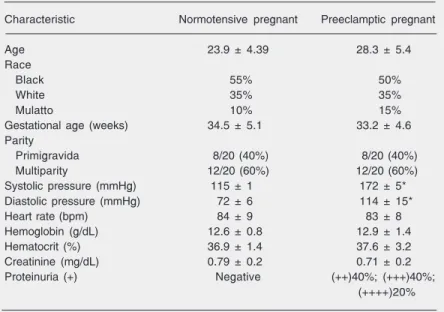
Table 1. Demographic and laboratory data of pregnant normotensive and preeclamptic women.
Characteristic Normotensive pregnant Preeclamptic pregnant
Age 23.9 ± 4.39 28.3 ± 5.4
Race
Black 55% 50%
White 35% 35%
Mulatto 10% 15%
Gestational age (weeks) 34.5 ± 5.1 33.2 ± 4.6 Parity
Primigravida 8/20 (40%) 8/20 (40%)
Multiparity 12/20 (60%) 12/20 (60%)
Systolic pressure (mmHg) 115 ± 1 172 ± 5*
Diastolic pressure (mmHg) 72 ± 6 114 ± 15*
Heart rate (bpm) 84 ± 9 83 ± 8
Hemoglobin (g/dL) 12.6 ± 0.8 12.9 ± 1.4
Hematocrit (%) 36.9 ± 1.4 37.6 ± 3.2
Creatinine (mg/dL) 0.79 ± 0.2 0.71 ± 0.2
Proteinuria (+) Negative (++)40%; (+++)40%;
(++++)20%
Data are reported as means ± SD or percent.
hundred microliters of orthophthaldehyde (20 mg/mL in methanol) was added and after 10 min, 200 µL 3 N HCl was added at room temperature. After centrifugation at 800 g for 5 min, the fluorescence of the supernatant solution (365-nm excitation and 495-nm emission) was measured against water. Blanks were prepared by inverting the order of addition of enzyme and NaOH. A standard curve of 0.5 to 20 nmol His-Leu/ tube was prepared for each assay. Enzyme activity is reported as nmol His-Leu min-1
mL-1. Assays were carried out under
condi-tions that provided constant velocity and constant enzyme-specific activity.
DNA extraction and PCR. Blood was drawn into EDTA-containing tubes and DNA was extracted by a standard phenol-chloro-form isoamilic alcohol method (25:24:1) (15). To determine the ACE genotype, ge-nomic DNA was amplified by PCR, initially using a pair of primers described by Rigat et al. (10) and subsequently, when necessary, with a pair of primers that recognize the insertion of specific sequences for confirma-tion of the specificity of the amplificaconfirma-tion reactions (15). Amplification with the former primer pair resulted in 490- and 190-bp am-plification products, corresponding to the I and D alleles, respectively. PCR amplifica-tion employed 25-µL reacamplifica-tions (50-100 ng genomic DNA), 10 pmol of each primer, 0.5 mM each of deoxy-ATP, GTP, CTP, thymi-dine 5-triphosphate, 50 mM MgCl2, 5 U/µL
Taq DNA polymerase, 500 mM KCl, and 200 mM Tris-HCl buffer, pH 8.4, with 1 min denaturation at 94ºC, followed by 32 cycles of 30 s at 94ºC, 1 min at 58ºC (annealing) and 2 min at 74ºC (extension) in a thermal cycler. The amplification with the latter primer pair resulted in a 335-bp product, corresponding to the I allele and its reaction was performed as described above. The re-action consisted of 30 cycles of amplifica-tion (1 min of initial denaturaamplifica-tion at 94ºC, 30 s of denaturation at 94ºC, 45 s of annealing at 67ºC, and 2 min of extension at 72ºC).
These products were run on 1.5% agarose gel, stained with ethidium bromide and viewed with ultraviolet light.
Statistical analysis
Data are reported as means ± SEM. Sta-tistical analysis was performed using the unpaired Student t-test and the Fisher test for the genotype analyses, with the level of sig-nificance set at P < 0.05 for both tests. Hardy-Weinberg equilibrium was checked by the
χ2 test.
Results
Profile of the renin-angiotensin system in preeclamptic and normotensive pregnant women
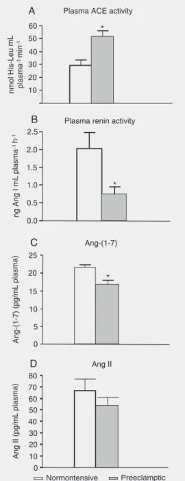
Twenty subjects were studied in each of the two groups. Plasma ACE activity was significantly higher in the preeclamptic group (51.8 ± 4.0 vs 29.3 ± 3.8 nmol Hip-His Leu min-1 mL-1 in the control group; P = 0.0002,
Figure 1A). In contrast, plasma renin activ-ity was markedly reduced in preeclamptic women (0.7 ± 0.1 vs 2.0 ± 0.4 ng Ang I mL plasma-1 h-1 in the control group; P = 0.0071,
Figure 1B). Plasma Ang-(1-7) was signifi-cantly reduced in preeclamptic women (16.9 ± 1.2 vs 21.6 ± 1.1 pg/mL plasma in the control group; P = 0.0063, Figure 1C), whereas plasma Ang II concentration did not differ between groups (54.1 ± 7.0 vs 66.4 ± 10.1 pg/mL plasma in the control group; P = 0.3225, Figure 1D).
Relationship between ACE I/D polymorphism and the renin-angiotensin profile in
preeclamptic and normotensive pregnant women
the preeclamptic group was 5% (1/20) for II, 35% (7/20) for ID and 60% (12/20) for DD. In the control group, gene frequency was 5% (1/20) for II, 60% (12/20) for DI and 35% (7/ 20) for DD. Genotype distribution in pre-eclamptic patients (DD = 0.601; DI = 0.349 and II = 0.051; χ2 = 0.00027; P = 1.0) and the
control group (DD = 0.423; DI = 0.455 and II = 0.123; χ2 = 2.043; P = 0.15) was in
agreement with the Hardy-Weinberg equi-librium. The percentage of the ACE DD genotype (60%) was higher in the preeclamp-tic group than in the control group (35%), but the difference was not statistically sig-nificant (Fisher exact test = 2.86, d.f. = 2, P = 0.260).
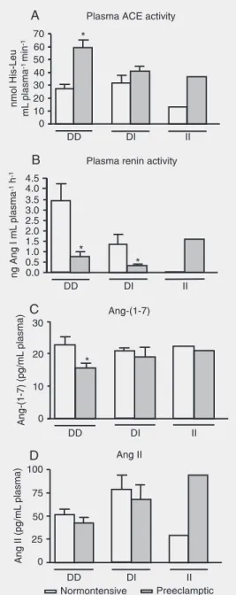
The highest plasma ACE activity was observed in the ACE DD preeclamptic women (58.1 ± 5.06 vs 27.6 ± 3.25 nmol Hip-His Leu min-1 mL-1 in the DD control
patients; P = 0.0005, Figure 2A).
Strikingly, reduced PRA activity was mainly associated with the ACE DD geno-type in preeclamptic women (0.81 ± 0.2 vs 3.43 ± 0.8 ng Ang I mL plasma-1 h-1 in DD
normotensive patients; P = 0.0012, Figure 2B). Low PRA was also observed in pre-eclamptic women with the DI genotype (0.4 ± 0.1 vs 1.4 ± 0.4 ng Ang I mL plasma-1 h-1;
P = 0.0006, Figure 2B).
The reduced plasma level of Ang-(1-7) was also associated with the ACE DD geno-type in preeclamptic women (15.6 ± 1.3 vs 22.7 ± 2.5 pg/mL plasma in the DD control group; P = 0.0146, Figure 2C).
In contrast, plasma Ang II levels were unchanged in preeclamptic patients present-ing the ACE DD genotype (42.77 ± 5.40 vs 51.27 ± 5.98 pg/mL plasma in DD normo-tensive patients; P = 0.3287, Figure 2D).
Discussion
The major finding of the present study was the detection of a significant reduction in plasma Ang-(1-7) levels, significantly higher plasma ACE activity and a marked
Figure 1. Plasma angiotensin-converting enzyme (ACE) activ-ity (A), plasma renin activactiv-ity (B), plasma angiotensin-(1-7) (Ang-(1-7)) levels (C), and plasma an-giotensin II (Ang II) levels (D) of normotensive and preeclamptic pregnant women. *P < 0.05 compared to normotensive preg-nant women (Student t-test). reduction in plasma renin activity in
eclamptic women, especially in patients pre-senting the ACE DD genotype. In contrast, the plasma Ang II levels of preeclamptic women did not differ from those of normo-tensive pregnant subjects.
ship between Ang-(1-7), blood pressure and pregnancy.
There are no studies concerning the in-fluence of Ang-(1-7) on blood pressure lev-els in pregnant women. However, there is considerable experimental evidence of the vasodilator action of this hormone (8). A potent vasodilatory effect of Ang-(1-7) has recently been reported in various vascular beds, including the mesenteric, cutaneous, cerebral, and renal beds of Wistar rats (8,17). In humans, plasma Ang-(1-7) levels have been reported to show a negative correlation with blood pressure (18). Furthermore, a recent study has reported that the control of blood pressure in hypertensive patients treated with the dual ACE and neutral en-dopeptidase inhibitor Omopatrilat was asso-ciated with an increase in urinary Ang-(1-7) levels (19).
As cited above, Merrill et al. (7) demon-strated reduced levels of Ang-(1-7) in pre-eclamptic women when compared to nor-motensive pregnant women. In the present study, we observed for the first time that the greatest reduction of Ang-(1-7) in preeclamp-tic women was associated with the ACE DD genotype. In view of the putative counter-regulatory effects of Ang-(1-7) (8,17) and the diminished vascular reactivity to Ang II in normal pregnancy, a potential role of Ang-(1-7) in both the vasodilation of normal pregnancy and the pathologic vasoconstric-tion observed in preeclampsia should be con-sidered.
Accordingly, Neves et al. (3) observed a significantly increased vasodilator response to Ang-(1-7) in the mesenteric vascular bed of pregnant rats when compared to non-pregnant rats (3). In a previous study, these investigators observed that estrogen replace-ment increased the vasodilatory response of Ang-(1-7) in ovariectomized rats (20). In this regard, estrogen may be a mediator of vascular changes during pregnancy, con-tributing to the vasodilation mediated by nitric oxide-releasing agonists, such as Ang-Figure 2. Plasma
angiotensin-converting enzyme (ACE) activ-ity (A), plasma renin activactiv-ity (B), plasma angiotensin-(1-7) (Ang-(1-7)) levels (C), and plasma an-giotensin II (Ang II) levels (D) in normotensive and preeclamptic pregnant women with DD, DI or II ACE genotypes. D = deletion; I = insertion. *P < 0.05 compared to normotensive pregnant women (Student t-test).
relation-(1-7), through the increase of endothelial nitric oxide synthase expression (21).
Ang-(1-7) is formed by Ang I and Ang II through the effect of tissue peptidases and is rapidly hydrolyzed, mainly by ACE (8). Thus, the decreased plasma Ang-(1-7) levels may be related to the increased ACE activity in pregnant preeclamptic women. However, a direct relationship between ACE activity, plasma Ang-(1-7) levels and preeclampsia is difficult to assume based on currently avail-able data. For example, data related to ACE activity in pregnancy are still controversial. Some studies showed no difference in the plasma ACE activity between normotensive pregnant women and preeclamptic women (22,23). In contrast, other investigators ob-served higher ACE activity in preeclamptic women when compared to normotensive women (7,24), as also observed in our study. Also in keeping with our data, Gurdöl et al. (25) observed that the highest ACE activity in preeclamptic patients was associated with the ACE DD genotype. It is important to highlight that our data suggest that the influ-ence of the RAS in preeclampsia appears to be more related to lower levels of Ang-(1-7) as a consequence of increased ACE activity than to changes in Ang II levels, which were not altered in our study, corroborating find-ings reported by Gordon et al. (4). It remains to be clarified whether changes in ACE ac-tivity and plasma Ang-(1-7) in preeclampsia result in a decreased contribution by other peptides, such as bradykinin, to vasodilator tonus.
Renin is another important RAS compo-nent. Longitudinal studies have demonstrated that PRA increases during pregnancy in nor-motensive women compared to the postpar-tum (26). Langer et al. (22) described sig-nificantly lower PRA in preeclamptic women compared to normotensive pregnant women. Our study corroborated these previous ob-servations, showing that PRA was markedly reduced in preeclamptic women compared
to the control group. A similar finding was reported by Merrill et al. (7). Importantly, our data suggest that this difference is mainly due to the presence of the D allele in pre-eclamptic women. There was also a reduc-tion in PRA among preeclamptic women with the DI genotype, but this reduction was more pronounced among DD patients.
Weir et al. (27) demonstrated a signifi-cant reduction in plasma Ang II in preeclamp-sia, while other studies found no significant difference in plasma Ang II between pre-eclamptic women and pregnant normoten-sive women (4,22). This discrepancy in rela-tion to Ang II could be explained by the blood collection conditions and different assay methodologies employed to measure this peptide (22). In the present study, no alteration in plasma Ang II was observed in preeclamptic women, despite a significant increase in plasma ACE activity. This sug-gests that plasma ACE, and probably tissue ACE as well, may not be a critical factor in determining plasma Ang II levels in such patients (5). Indeed, other enzymes such as chymase and ACE 2 also participate in Ang II metabolism (5).
References
1. Anonymous. Report of the National High Blood Pressure Education Program Working Group on High Blood Pressure in Pregnancy. Am J Obstet Gynecol 2000; 183: S1-S22.
2. Talosi G, Endreffy E, Turi S, Nemeth I. Molecular and genetic aspects of preeclampsia: state of the art. Mol Genet Metab 2000; 71: 565-572.
3. Neves LA, Williams AF, Averill DB, Ferrario CM, Walkup MP, Brosnihan KB. Pregnancy enhances the angiotensin (Ang)-(1-7) vasodilator response in mesenteric arteries and increases the renal concentration and urinary excretion of Ang-(1-7). Endocrinology
2003; 144: 3338-3343.
4. Gordon RD, Symonds EM, Wilmshurst EG, Pawsey CG. Plasma renin activity, plasma angiotensin and plasma and urinary electro-lytes in normal and toxaemic pregnancy, including a prospective study. Clin Sci 1973; 45: 115-127.
5. Crackower MA, Sarao R, Oudit GY, Yagil C, Kozieradzki I, Scanga SE, et al. Angiotensin-converting enzyme 2 is an essential regulator of heart function. Nature 2002; 417: 822-828.
6. Santos RA, Simoes e Silva AC, Maric C, Silva DM, Machado RP, de Buhr I, et al. Angiotensin-(1-7) is an endogenous ligand for the G protein-coupled receptor Mas. Proc Natl Acad Sci USA 2003; 100: 8258-8263.
7. Merrill DC, Karoly M, Chen K, Ferrario CM, Brosnihan KB. Angio-tensin-(1-7) in normal and preeclamptic pregnancy. Endocrine 2002; 18: 239-245.
8. Santos RA, Campagnole-Santos MJ, Andrade SP. Angiotensin-(1-7): an update. Regul Pept 2000; 91: 45-62.
9. Jalil JE, Palomera C, Ocaranza MP, Godoy I, Roman M, Chiong M, et al. Levels of plasma angiotensin-(1-7) in patients with hyperten-sion who have the angiotensin-I-converting enzyme deletion/dele-tion genotype. Am J Cardiol 2003; 92: 749-751.
10. Rigat B, Hubert C, Alhenc-Gelas F, Cambien F, Corvol P, Soubrier F. An insertion/deletion polymorphism in the angiotensin I-convert-ing enzyme gene accountI-convert-ing for half the variance of serum enzyme levels. J Clin Invest 1990; 86: 1343-1346.
11. Cambien F, Costerousse O, Tiret L, Poirier O, Lecerf L, Gonzales MF, et al. Plasma level and gene polymorphism of angiotensin-converting enzyme in relation to myocardial infarction. Circulation
1994; 90: 669-676.
12. Neves LA, Almeida AP, Khosla MC, Santos RA. Metabolism of angiotensin I in isolated rat hearts. Effect of angiotensin converting enzyme inhibitors. Biochem Pharmacol 1995; 50: 1451-1459. 13. Chappell MC, Brosnihan KB, Diz DI, Ferrario CM. Identification of
angiotensin-(1-7) in rat brain. Evidence for differential processing of angiotensin peptides. J Biol Chem 1989; 264: 16518-16523. 14. Santos RA, Krieger EM, Greene LJ. An improved fluorometric assay
of rat serum and plasma converting enzyme. Hypertension 1985; 7:
244-252.
15. Ueda S, Heeley RP, Lees KR, Elliott HL, Connell JM. Mistyping of the human angiotensin-converting enzyme gene polymorphism: fre-quency, causes and possible methods to avoid errors in typing. J Mol Endocrinol 1996; 17: 27-30.
16. Valdes G, Germain AM, Corthorn J, Berrios C, Foradori AC, Ferrario CM, et al. Urinary vasodilator and vasoconstrictor angiotensins during menstrual cycle, pregnancy, and lactation. Endocrine 2001; 16: 117-122.
17. Sampaio WO, Nascimento AA, Santos RA. Systemic and regional hemodynamic effects of angiotensin-(1-7) in rats. Am J Physiol Heart Circ Physiol 2003; 284: H1985-H1994.
18. Luque M, Martin P, Martell N, Fernandez C, Brosnihan KB, Ferrario CM. Effects of captopril related to increased levels of prostacyclin and angiotensin-(1-7) in essential hypertension. J Hypertens 1996; 14: 799-805.
19. Ferrario CM, Smith RD, Brosnihan B, Chappell MC, Campese VM, Vesterqvist O, et al. Effects of omapatrilat on the renin-angiotensin system in salt-sensitive hypertension. Am J Hypertens 2002; 15: 557-564.
20. Brosnihan KB, Neves LA, Joyner J, Averill DB, Chappell MC, Sarao R, et al. Enhanced renal immunocytochemical expression of ANG-(1-7) and ACE2 during pregnancy. Hypertension 2003; 42: 749-753. 21. Weiner CP, Knowles RG, Moncada S. Induction of nitric oxide synthases early in pregnancy. Am J Obstet Gynecol 1994; 171: 838-843.
22. Langer B, Grima M, Coquard C, Bader AM, Schlaeder G, Imbs JL. Plasma active renin, angiotensin I, and angiotensin II during preg-nancy and in preeclampsia. Obstet Gynecol 1998; 91: 196-202. 23. Tamura T, Johanning GL, Goldenberg RL, Johnston KE, DuBard
MB. Effect of angiotensin-converting enzyme gene polymorphism on pregnancy outcome, enzyme activity, and zinc concentration.
Obstet Gynecol 1996; 88: 497-502.
24. Ito M, Itakura A, Ohno Y, Nomura M, Senga T, Nagasaka T, et al. Possible activation of the renin-angiotensin system in the feto-pla-cental unit in preeclampsia. J Clin Endocrinol Metab 2002; 87: 1871-1878.
25. Gurdöl F, Isbilen E, Yilmaz H, Isbir T, Dirican A. The association between preeclampsia and angiotensin-converting enzyme inser-tion/deletion polymorphism. Clin Chim Acta 2004; 341: 127-131. 26. August P, Mueller FB, Sealey JE, Edersheim TG. Role of
renin-angiotensin system in blood pressure regulation in pregnancy. Lan-cet 1995; 345: 896-897.