Identification of
Fusarium oxysporum
f. sp.
lactucae
race 1 as
the causal agent of lettuce wilt in Brazil
Cléia S. Cabral1, Kátia R. Brunelli2, Hélcio Costa3, Maria Esther de N. Fonseca4, Leonardo S. Boiteux4 & Ailton Reis4
1Departamento de Agronomia, Universidade Federal Rural de Pernambuco, 52171-900, Recife, PE, Brazil; 2Sakata Seed
Sudamerica Ltda., Bragança Paulista, 12902-001, SP, Brazil; 3Incaper, Venda Nova do Imigrante, 29375-000, ES, Brazil; 4Embrapa Hortaliças, 70351-970, Brasília, DF, Brazil
Author for correspondence: Ailton Reis, e-mail: ailton.reis@embrapa.br
INTRODUCTION
Fusarium wilt caused by Fusarium oxysporum
Schlechtend.: Fr. f. sp. lactucae Matuo & Motohashi
is one of the most economically important diseases of lettuce (Lactuca sativa L. - Asteraceae) around the
world. Yield losses may vary from 20 to 70% depending on the cultivar, management practices and environmental conditions (Pasquali et al., 2005; 2007). First symptoms of the disease are a poor development and an overall chlorosis. As the disease progresses, older leaves may collapse leading to plant death. An intense brownish color can be seen in the vascular vessels of diseased plants after transversal stem sectioning as a result of the host reaction to the colonization and toxin production by
F. oxysporum f. sp. lactucae (FOLac) (Fujinaga et al.,
2001; Garibaldi et al., 2004b; Scott et al., 2010). Fusarium wilt of lettuce has a wide geographic distribution being reported in Japan (Matuo & Motohashi, 1967), United States (Hubbard & Gerik, 1993), Taiwan (Huang & Lo, 1998), Iran (Millani et al., 1999), Italy (Garibaldi et al., 2002), and Portugal (Pasquali et al., 2007). Three FOLac races have been identified (Fujinaga et al., 2003) and Japan is the only country where all three races are present (Fujinaga et al., 2001; 2003; Yamauchi et al., 2004). Race 1 is the most widespread around the world with reports in Italy, Portugal, Taiwan, Iran, and
the United States (Pasquali et al., 2005) and race 3 has been reported in Taiwan (Chang et al., 2011).
The most commonly used strategy for FOLac race determination is based upon the ability of the isolates to induce (or not) wilt symptoms in a set of differential lettuce cultivars (Ogiso et al., 2002; Fujinaga et al., 2003), which is a cumbersome and time-consuming approach. The development of PCR-based molecular markers for FOLac race determination (Pasquali et al., 2007; Mbofung & Pryor, 2010) is useful for increasing the diagnostic power and conducting large scale survey of races. However, the usefulness of FOLac race-specific PCR primers is yet to be demonstrated for distinct groups of fungal isolates from Neotropical areas of the world.
In Brazil, lettuce vascular wilt caused by F. oxysporum isolates was first reported in Espírito Santo
State (Ventura & Costa, 2008), and later in other lettuce production areas of the country. However, a thorough understanding of the causal agent of the disease in Brazil is lacking, especially the potential presence of distinct FOLac races associated with this disease.
In this context, the objectives of this study were to 1) assess the virulence profile and host specificity of F. oxysporum isolates associated with lettuce vascular wilt,
2) identify FOLac races based on a set of differential lettuce cultivars and 3) assess a PCR protocol for detection and identification of FOLac race 1 isolates. ABSTRACT
The objective of this work was to assess the virulence to lettuce cultivars and host specificity and to identify races of
Fusarium oxysporum f. sp. lactucae (FOLac) isolates associated with lettuce wilt in Brazil. Thirty-one isolates identified based on
morphology were inoculated in a set of lettuce cultivars for race determination. Plantlets of ‘Elisa’, ‘Vera’, and ‘Red Salad Bowl’ cultivars were inoculated with all isolates using a root-dipping method. Isolates were also inoculated in species of Asteraceae and other botanical families. ‘Elisa’ and ‘Vera’ were susceptible to 27 isolates while ‘Red Salad Bowl’ was resistant to all isolates. Plants other than lettuce were not infected by any of the isolates, suggesting their specificity to lettuce. All pathogenic isolates were assigned to race 1 and used to evaluate a PCR protocol with primers targeting race 1. Only amplicons associated with race 1 pattern were observed for all pathogenic isolates but not for one non-pathogenic isolate, thus leading us to conclude that FOLac race 1 is so far the sole causal agent of lettuce wilt in Brazil.
Key words:Lactuca sativa, Fusarium wilt, virulence, pathotypes.
MATERIAL AND METHODS
Collection of isolates
Thirty-one Fusarium isolates associated with lettuce
plants displaying wilt symptoms and intense brownish color of the vascular vessels were collected in seven Brazilian States (Rio Grande do Sul, Santa Catarina, Paraná, São Paulo, Rio de Janeiro, Minas Gerais, and Espírito Santo) and in the Federal District between the years of 2008 to 2012 (Table 1). The isolates were obtained from symptomatic stem segments under aseptic conditions. Affected tissues were washed with tap water for two minutes, disinfested with 70% alcohol for one minute, washed again with sterile distilled water for 30 seconds, and air-dried on the laboratory bench. Five stem segments were allocated into equidistant points within Petri dishes containing Potato Dextrose Agar medium supplemented with 20 mg of tetracycline (PDA-t). The Petri dishes were maintained in a growth chamber for
12 h dark/light cycle at 25oC for three days. Thereafter, the
fungus was transferred to a distinct Petri dish containing PDA-t medium aiming to obtain pure cultures. The isolates were assigned to species based on morphological description in the literature (Nelson et al., 1983; Leslie and Summerell, 2006). The isolates were maintained in Petri dishes containing PDA and in glass vials filled with sterile-distilled water (Castellani, 1963) and deposited in the culture collection of plant pathogenic fungi “Professora Maria Menezes” – CMM (Table 1).
Inoculation procedures
Three mycelial discs (5 mm in diameter each) were obtained from five-day old cultures grown in PDA-t and placed in Erlenmeyer flasks containing 100 mL of Potato + Dextrose liquid medium. These flasks were shaken for ten days (25oC and 12h dark/light cycle) to induce microconidia
production. After this treatment, the suspension was passed
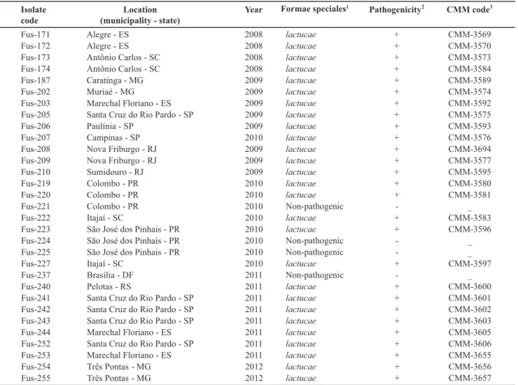
Isolate
code
Location
(municipality - state)
Year Formae specialis 1
Pathogenicity 2
CMM code 3
Fus-171 Alegre - ES 2008 + CMM-3569
Fus-172 Alegre - ES 2008 + CMM-3570
Fus-173 Antônio Carlos - SC 2008 + CMM-3573
Fus-174 Antônio Carlos - SC 2008 + CMM-3584
Fus-187 Caratinga - MG 2009 + CMM-3589
Fus-202 Muriaé - MG 2009 + CMM-3574
Fus-203 Marechal Floriano ES 2009 + CMM-3592
Fus-205
-Santa Cruz do Rio Pardo - SP 2009 + CMM-3575
Fus-206 Paulínia - SP 2009 + CMM-3593
Fus-207 Campinas - SP 2010 + CMM-3576
Fus-208 Nova Friburgo - RJ 2009 + CMM-3694
Fus-209 Nova Friburgo - RJ 2009 + CMM-3577
Fus-210 Sumidouro - RJ 2009 + CMM-3595
Fus-219 Colombo - PR 2010 + CMM-3580
Fus-220 Colombo - PR 2010 + CMM-3581
Fus-221 Colombo - PR 2010 - _
Fus-222 Itajaí - SC 2010 + CMM-3583
Fus-223 São José dos Pinhais - PR 2010 + CMM-3596
Fus-224 São José dos Pinhais - PR 2010 - _
Fus-225 São José dos Pinhais - PR 2010 - _
Fus-227 Itajaí - SC 2010 + CMM-3597
Fus-237 Brasília - DF 2011 - _
Fus-240 Pelotas - RS 2011 + CMM-3600
Fus-241 Santa Cruz do Rio Pardo - SP 2011 + CMM-3601
Fus-242 Santa Cruz do Rio Pardo - SP 2011 + CMM-3602
Fus-243 Santa Cruz do Rio Pardo - SP 2011 + CMM-3603
Fus-244 Marechal Floriano - ES 2011 + CMM-3605
Fus-252 Santa Cruz do Rio Pardo - SP 2011 + CMM-3606
Fus-253 Marechal Floriano - ES 2011 + CMM-3655
Fus-254 Três Pontas - MG 2012 + CMM-3656
Fus-255 Três Pontas - MG 2012
lactucae lactucae lactucae lactucae lactucae lactucae lactucae lactucae lactucae lactucae lactucae lactucae lactucae lactucae lactucae
Non-pathogenic
lactucae lactucae
Non-pathogenic
Non-pathogenic
lactucae
Non-pathogenic
lactucae lactucae lactucae lactucae lactucae lactucae lactucae lactucae
lactucae + CMM-3657
TABLE 1 - Information and attributes of 31 Fusarium oxysporum isolates associated with lettuce (Lactuca sativa L.) wilt in locations
across seven States and in the Federal District, Brazil.
1isolates were assigned to a formae speciales based on pathogenicity and molecular assays.
2Pathogenicity assays were conducted using three lettuce cultivars. (+) = isolate able to induce disease; (-) = no-pathogenic isolate 3Code of the pathogenic isolates deposited in the culture collection of plant pathogenic fungi “Professora Maria Menezes” – CMM.
through a double-layer gauze filter in order to remove mycelium fragments. Spore concentration was determined using an hemocytometer and adjusted to a concentration of 2 x 10⁶ microconidia/mL. All 31 isolates associated with
lettuce wilt were employed in the pathogenicity tests using three commonly grown lettuce cultivars (‘Elisa’, ‘Vera’ and ‘Red Salad Bowl’). The seedlings were produced in polystyrene trays (68 cm x 34 cm) with 128 cells filled
with commercial solid substrate (Bioplant®; Nova Ponte, MG). The trays were kept in a greenhouse free of pest and pathogen infestation with daily irrigation until transplanting and without pesticide applications. Inoculation was performed in 25-day old seedlings by dipping the apical region of their roots (cut approximately 2 cm from the top by sterile scissors) in 50 mL of a conidial suspension for three minutes. Immediately after inoculation, the seedlings were transplanted individually to 1L plastic pots containing sterilized soil. After that, 3 mL of the conidial suspension were added to the collar area of each seedling. The pots were irrigated two hours prior to inoculation until soil saturation (Santos, 1999).
Host range experiment
The isolates were inoculated in a group of six species from the Asteraceae family including lettuce (cv. Elisa),
Cichorium endivia L., Cichorium intybus L., Sonchus oleraceus L., Emilia sonchifolia L., Bidens pilosa L. and Tagetes erecta L. as well as several species from other
botanic families such as Solanaceae (Solanum lycopersicum
L., Capsicum annuum L., Nicotiana tabacum L.), Malvaceae
(Gossypium hirsutum L.), Fabaceae (Phaseolus vulgaris
L.), and Lamiaceae (Ocimum basilicum L.). Inoculation
was made using the root dipping method of the wounded root system essentially as described. Mock-inoculated plants of each species were kept in the same greenhouse conditions to serve as controls. Evaluation was done at 20 to 25 days after inoculation, initially by visual analysis of leaf chlorosis, overall wilt, and vascular browning.
Race determination based on a differential set of lettuce cultivars
The isolates were inoculated in a set of cultivars comprising: ‘Patriot’ (susceptible to all races), ‘Costa Rica No. 4’ (resistance specific to race 1) and ‘Banchu Red Fire’ (resistance specific to race 2). The seedlings were produced in polystyrene trays (68 cm x 34 cm) with 128 cells filled
with commercial solid substrate (Bioplant®; Nova Ponte, MG). The seedlings were removed from the substrate at 25 days after sowing. Inoculation production and inoculation procedures were essentially as described previously. Conidial suspension was adjusted to 2 x 10⁶ microconidia.
mL-1. The evaluation was done 20 days after inoculation
and the plant reaction was assessed using a disease severity ordinal and descriptive scale ranging from 1 to 5 (Santos, 1999). The isolates were classified as pathogenic/virulent to a given cultivar when the average disease severity grade was
above score 2 of the scale (plants without vascular wilt and chlorosis symptoms but with vascular browning; score 1: plant without symptoms). Isolates unable to induce disease in all three cultivars were classified as non-pathogenic or avirulent. Race 1 isolates are pathogenic/virulent only to the cultivars ‘Patriot’ and ‘Banchu Red Fire’, whereas race 2 isolates are pathogenic only to ‘Patriot’ and ‘Costa Rica No. 4’. Isolates able to induce disease in all three accessions would represent race 3, which has been reported only in Japan and Taiwan (Chang et al. 2011).
Molecular FOLac race identification
Twenty-seven isolates characterized as being FOLac race 1 based on the inoculation assays in the set of differentials were tested in additional molecular race phenotyping assays. One isolate of F. oxysporum f. sp. lycopersici (FOL) race 3 (named Fus-191) and one
non-pathogenic F. oxysporum isolate (Fus-237) were also
included. Fungal genomic DNA was extracted using the CTAB protocol with minor modifications (Boiteux et al., 1999). PCR reaction was carried out using the primer par Hani3’ (5’CCC-TCC-AAC-ATT-CAA-CAA-CTG3’) and Hanilatt3rev (5’ATT-CAC-TGT-ACA-CCA-ACC-TTT-T3’), which were designed for specific discrimination of FOLac race 1 isolates (Paquali et al, 2007). PCR reaction was composed of 3 μL of fungal genomic DNA (20 ng/mL), 2 μL 10X buffer (100 mM Tris-HCl, 500 mM KCl, pH 8.3), 0.48 μL MgCl2 (50 mM), 2 μL dNTPs (2.5M each), 0.2 μL
Taq DNA polymerase (5 units/μL), 3 μL of each primer
and 6.32 μL Milli-Q water, with a final reaction volume of 20 µL. Samples were amplified in one My CyclerTM (BIO
RAD) thermocycler. The amplification program employed was carried out as follows: initial cycle (94ºC /1min), ten denaturation cycles (94ºC for 15 seconds), annealing at 66ºC (30 s). The annealing temperature was reduced by 0.5ºC after each cycle, and then the program was shifted to 30 denaturation cycles (94ºC for 15 seconds), 61ºC annealing temperature (30 seconds) and extension of 72ºC (30 seconds). A final extension cycle was carried out at 72ºC for 2 minutes. The introduction of these final 30 cycles was a modification in relation to the original protocol described by Pasquali et al. (2007). Amplicons were analyzed in agarose gel (1% p/v) eletrophoresis, stained with ethidium bromide and visualized under UV light.
RESULTS AND DISCUSSION
The thirty-one purified Fusarium isolates showed the
typical morphological features of the species F. oxysporum
allowed us to assign all pathogenic isolates to F. oxysporum
f. sp. lactucae. The inoculation-based assay is so far the
only available method for distinguishing saprophytic (non-pathogenic) isolates from pathogenic F. oxysporum
isolates, since they are quite similar in terms of cultural and morphological traits (Booth, 1971; Burgess et al., 1994).
Additionally, the 27 pathogenic isolates were not able to induce wilt symptoms in the set of Asteraceae species as well in host species of other botanical families tested in our study, thus suggesting the specificity of these isolates to lettuce. Previously, Ventura & Costa (2008) described the causal agent of lettuce wilt disease in Brazil only to the species level (F. oxysporum).
The reaction of the race differentials to the set of pathogenic isolates was quite similar. ‘Patriot’ and ‘Banchu Red Fire’ were highly susceptible to all isolates (but not to the four nonpathogenic isolates), whereas ‘Costa Rica No. 4’ was found to be resistant, indicating so far the exclusive presence of FOLac race 1 in Brazil, which has been also found in the United States, Japan, Italy, Portugal, Taiwan and Iran (Fujinaga et al., 2001; McCreight et al., 2003; 2005; Garibaldi et al., 2004b; Pasquali et al., 2007).
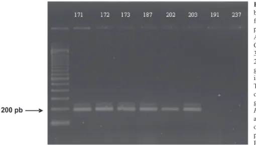
Amplicons corresponding to the region between the clusters of class II transposons (Hua-Van et al., 2000) were amplified via PCR assays for the 27 pathogenic isolates. These amplicons were not present in the control FOL race 3 (isolate Fus. 191) and in one non-pathogenic F. oxysporum (Fus. 237) isolate (Figure 1). The size of the
amplicon was approximately 200 bp. This same primer pair was employed byPasquali et al. (2007) for specific detection of FOLac race 1 using a collection of race 1 isolates from Italy, United States (States of Arizona and California), Portugal, Taiwan, and Japan. In fact, these primers have been previously used as powerful diagnostic tools for FOLac race 1 isolates, but they were not evaluated with isolates from Neotropical areas of
the world. All the results combined suggest that all 27 pathogenic isolates belong to FOLac race 1.
Molecular techniques are important tools to discriminate fungal species with intimate genetic relationships where few morphological diagnostic traits are available (Wuff et al., 2010) as well as to differentiate isolates within the same species (Chandra-Nayaka et al., 2010). In Fusarium, the identification to species
based exclusively upon morphological information is challenging. This genus is especially difficult to fungal taxonomists because either the lack of informative traits or the phenotypic instability of several traits, which expression might be affected by in vitro and in vivo
conditions (Fravel et al., 2003). Our study showed that the PCR primer pair Hani and Hanilatt3rev (Hua-Van et al., 2000) might reliably be used to discriminate FOLac race 1 isolates from Brazil. Robust PCR assays as this one able to differentiate isolates at their formae speciales
and race levels are useful for large-scale surveys of fungal isolates.
It is well known from the literature that distinct
formae speciales of F. oxysporum are very efficiently
seed-transmitted. In fact, the spread of FOLac race 1 isolates into several countries of distinct continents in a short period of time suggests its spread via infested seed, although this has not been conclusively documented (Garibaldi et al., 2004a; Mbofung & Pryor, 2010). Likewise, the presence of FOLac race 1 in several and distant lettuce production regions of Brazil suggests that this pathogen may have been introduced into the country via infected seeds. Further molecular phylogenetic analysis of the Brazilian isolates might allow assessing whether these isolates are either endemic or introduced into the country from abroad.
The control of Fusarium wilt of lettuce via
fungicide and soil desinfestation treatments is usually not economically and ecologically feasible. An effective
FIGURE 1 -PCRamplicons of around 200 bp obtained with the Fusarium oxysporum
f. sp. lactucae (FOLac) race 1-specific
prime pair Hani (5’ GAA-CCC-TCC-AAC-ATT-CAA-CA 3’) and Hanilatt3rev (5’ CCT-CCA-ACA-TTC-AAC-AAC-AAT-G 3’). The lanes 171, 172, 173, 187, 202, and 203 are PCR the amplicons obtained with genomic DNA extracted from six distinct isolates that were pathogenic to lettuce. The lane 191 corresponds to the results obtained with the same prime pair using genomic DNA extracted from one isolateof
F. oxysporum f. sp. lycopersici (FOL) race 3
and the lane 237 corresponds to the results obtained with one F. oxysporum isolate
REFERENCES
Booth C (1971) The genus Fusarium. Kew, England,
Commonwealth Mycological Institute.
Boiteux LS, Fonseca MEN, Simon PW (1999) Effects of plant tissue and DNA purification method on randomly amplified polymorphic DNA based genetic fingerprinting analyses is carrot. Journal of the American Society for Horticultural Science 124:32-38.
Burgess LW, Summerell BA, Bullock S, Gott KP, Backhouse D (1994) Laboratory manual for Fusarium research, 3rd ed., Sidney,
Australia, University of Sidney.
Cabral CS, Reis A (2013) Screening of lettuce accessions for resistance to Fusarium oxysporum f. sp. lactucae race 1. Tropical
Plant Pathology 38:272-281.
Castellani A (1963). The “water cultivation” of pathogenic fungi. Journal of Tropical Medicine and Hygiene 66:283-286.
Chang P, Lin Y, Lai P, Chang T, Wan Y, Huang J (2011) Pathological and molecular race determinations of Fusarium oxysporum f. sp. lactucae from Taiwan. Phytopathology 101(6, supplement):31.
Chandra-Nayaka S, Udaya Shannkar AC, Niranjana SR, Reddy MS, Prakash HS (2010) Detection and quantification of fumonisins from Fusarium verticillioides in maize grown in southern India.
World Journal Microbiology Biotechnology 26:71-78.
Fravel D, Olivain C, Alabouvette C (2003) Fusarium oxysporum
and its biocontrol. New Phytology 157:493-502.
Fujinaga M, Ogiso H, Tsuchiya N, Saito H (2001) Physiological specialization of Fusarium oxysporum f. sp. lactucae, a causal
organism of fusarium root of crisp head lettuce in Japan. Journal of General Plant Pathology 667:205-206.
Fujinaga M, Ogiso H, Tuchiya N, Saito H, Yamanaka S, Nozue M, Kojima M (2003) Race 3, a new race of Fusarium oxysporum f.
sp. lactucae determined by a differential system with commercial
cultivars. Journal of General Plant Pathology 69:23-28.
Garibaldi A, Gilardi G, Gullino ML (2002) First report of Fusarium
oxysporum on lettuce in Europe. Plant Disease 86:1052.
Garibaldi A, Gilardi G, Gullino ML (2004a) Seed transmission of
Fusarium oxysporum f. sp. lactucae. Phytoparasitica 32:61-65.
Garibaldi A, Gilardi G, Gullino ML (2004b)Varietal resistance of lettuce to Fusarium oxysporum f. sp. lactucae. Crop Protection
23:845-851.
Huang JH, Lo CT (1998) Wilt of lettuce caused by Fusarium oxysporum in Taiwan. Plant Pathology Bulletin 7:150-153.
Hua-Van A, Davière JM, Kaper F, Langin, T, Daboussi MJ (2000) Genome organization in Fusarium oxysporum: Clusters of class II
transposons. Current Genetics 37:339-347.
Hubbard JC, Gerik JS (1993) A new disease of lettuce incited by
Fusarium oxysporum f. sp. lactucum forma specialis nov. Plant
Disease 77:750-754.
Leslie JF, Summerell BA (2006) The Fusarium laboratory manual.
Ames, Blackwell.
McCreight JD, Matheron ME, Tickes BR, Platts, B (2003). Occurrence of fusarium root rot race 1 on lettuce in Yuma, Arizona. Hort Science 38:664.
McCreight JD, Matheron ME, Tickes BR, Platts, B (2005) Fusarium wilt race 1 on lettuce. Hort Science 40:529-531. Mbofung GY, Pryor BM (2010) A PCR-based assay for detection of Fusarium oxysporum f. sp. lactucae in lettuce seed. Plant
Disease 94:860-866.
Matuo T, Motohashi S (1967) On Fusarium oxysporum f.
sp. lactucae n. f. causing root rot of lettuce. Transactions of
Mycological Society of Japan 8:13-15.
Millani MJ, Erebarian HR, Alizadeh A (1999) Occurrence of fusarium wilt of lettuce in Shahr-Ray, Varamim and Karaj areas. Iran Journal of Plant Pathology35:44.
Nelson PE, Toussoun TA, Marasas WFO (1983) Fusarium
species: an illustrated manual for identification. University Park, The Pennsylvania State University Press.
Ogiso H, Fujinaga M, Saito H, Takehara T, Yamanaka S (2002) Physiological races and vegetative compatibility groups of
Fusarium oxysporum f. sp. lactucae isolated from crisphead
lettuce in Japan.Journal of General Plant Pathology 68:292-299. Pasquali M, Dematheis F, Gilardi G, Gullino ML, Garibaldi A (2005) Vegetative compatibility groups of Fusarium oxysporum f.
sp. lactucae from lettuce. Plant Disease 89:237-240.
Pasquali M, Dematheis F, Gullino ML, Garibaldi A (2007) Identification of race 1 of Fusarium oxysporum f. sp. lactucae on
lettuce by inter-retrotranposon sequence-characterized amplified region technique. Phytopathology 97:987-996.
Santos JRM (1996) Methodology for screening tomato to Fusarium wilt, Verticillium wilt, gray leaf spot, early blight, and Septoria leaf spot. Pages 164-166. In: Proceedings of the international symposium on tropical tomato diseases, ASHS: IPA. Alexandria, USA.
Scott JC, Kirkpatrick SC, Gordon TR (2010) Variation in susceptibility of lettuce cultivars to fusarium wilt caused by
Fusarium oxysporum f. sp. lactucae. Plant Pathology
59:139-146.
Wulff EG, Sorensen JL, Lübeck M, Nielsen KF, Thrane U, Torp J (2010) Fusarium spp. associated with rice Bakanae: ecology,
genetic diversity, pathogenicity and toxigenicity. Environmental Microbiology 12:649-657.
Yamauchi N, Shimazu J, Horiuchi S, Satou M, Shirakawa T (2004) Physiological races and vegetative compatibility groups of
butterhead lettuce isolates of Fusarium oxysporum f. sp. lactucae
in Japan. Journal of General Plant Pathology 70:308-313. Ventura JA, Costa H (2008) Fusarium wilt caused by Fusarium oxysporum on lettuce in Espirito Santo, Brazil. Plant Disease
92:976.